Introduction
Second branchial cleft anomalies are the commonest of the true branchial cleft abnormalities, and the diagnosis is usually straightforward.Reference Blackham and Lannigan1 Good knowledge of anatomy and embryology are necessary for proper treatment.Reference Barret2 Of the second branchial cleft anomalies, sinuses are more frequent than cysts, and fistulae are extremely rare.Reference Mitroi, Dumitrescu, Simionescu, Popescu, Mogoantă and Cioroianu3, Reference Karabulut, Sönmez, Türkyilmaz, Ozen, Demiroğullari and Güçlü4
Second branchial cleft sinuses may be found along a tract from the anterior border of the sternocleidomastoid muscle to the carotid vessels and the glossopharyngeal and hypoglossal nerves, and can occur anywhere from the tonsillar fossa to the supraclavicular region.Reference Halvorson and Porubsky5, Reference Robson, Kim, Barnes, Kirks and Griscom6 They may be unilateral or bilateral. The classical surgical treatment of second branchial sinus – unilateral or bilateral – is excision of the complete sinus tract via an external approach. With this approach, dissection proceeds from the sinus opening in a cephalad direction until, near the entrance to the tonsillar fossa, the assistant inserts a finger to ‘milk down’ the base, before final excision. Because of the length of sinus tract often encountered, this standard surgical approach is facilitated by a second, ‘step ladder’ incision, originally described by Bailey in 1933.Reference Abhisek, Avik, Biswajit, Utpal and Tapan7
With the classical approach, complete excision must be ensured, but this is doubtful in some cases as recurrence rates following complete excision can be as high as 22 per cent.Reference Calvo, Sancipriano, Diego, Santiago, Rincón and Hermosa8
The current report describes a simple surgical technique that ensures complete excision of a second branchial sinus.
Case presentation
An 11-year-old girl presented to the out-patient clinic with a history of recurrent neck discharge from a persistent hole in the right anterior neck since childhood. The discharge was occasionally purulent and occasionally clear fluid. There was no history of associated pain, swelling or antecedent trauma to the anterior neck.
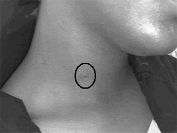
The only significant examination finding was a sinus opening over the anterior border of the right sternocleidomastoid muscle (Figure 1).
The sinus over the anterior border of the sternocleidomastoid muscle (spot in the centre of the black circle), between the junction of the upper two-thirds and lower one-third of the muscle.

A contrast sinugram revealed a blind sinus ending just above the superior border of the hyoid bone.
The patient was prepared for excision of a branchial sinus. The excision was carried out as follows.
The patient was positioned supine, with her face turned away from the surgeon to the left side and the right side of the neck exposed. Surgical cleaning and draping were performed. Methylene blue was instilled into the sinus; only 1.2 ml could be instilled before external spillage occurred. A malleable probe was then inserted to assess the depth of the sinus.
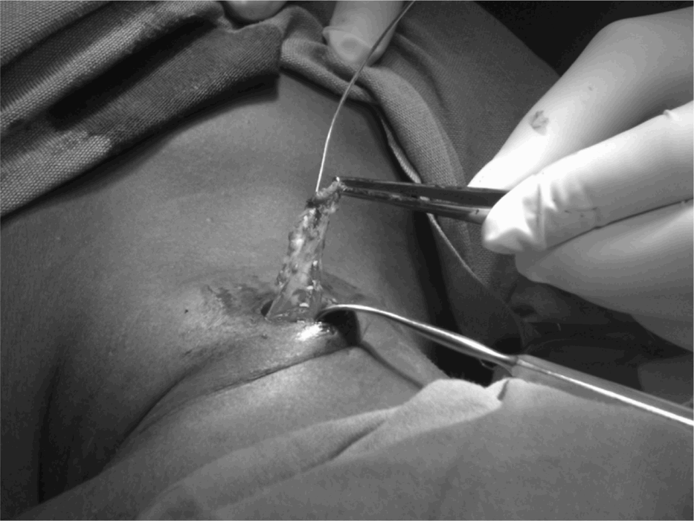
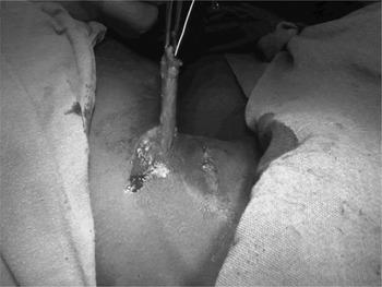
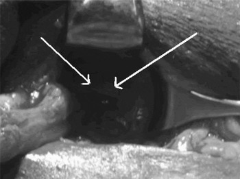
An elliptical skin incision was made encompassing the sinus opening. Dissection of the sinus tract commenced until an approximately 5 cm length of tract had been dissected free, with the malleable probe in situ and clamped to the elliptical skin opening, using lateral traction on the sinus tract to lift it away from the carotid vessels (Figure 2). At this stage, a second, step ladder incision was made, the malleable probe was removed, and the sinus tract was tunnelled through subcutaneous tissue and retrieved through this second incision (Figure 3). The malleable probe was reinserted to guide further dissection of the sinus in a cephalad direction until the hypoglossal nerve was visualised (Figure 4). Dissection proceeded above this level until the tract was noted to turn medially towards the tonsillar fossa.
Initial dissection of the sinus tract, aided by insertion of a malleable metallic probe and retraction of the tract during dissection.
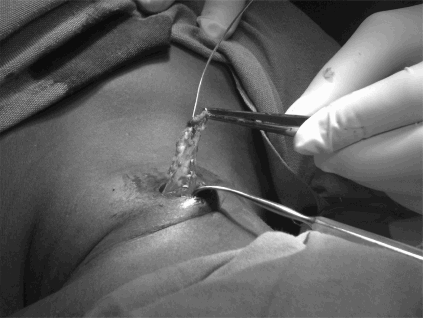
A second, ‘step ladder’ incision was made and the tract tunnelled under the skin, before being retrieved through the second incision as the dissection proceeded in a cephalad direction.

Further cephalad dissection with the sinus tract retracted to the left enabled the hypogossal nerve to be visualised through the dissection bed (arrows).

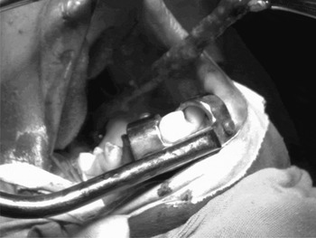
At this stage, the surgeon used digital palpation to assess the depth of dissection and to determine which structures, if any, lay medial to the sinus tract. The assistant exposed the pharynx using a Boyle-Davis mouth gag. Bimanual palpation was then carried out to confirm the attachment of the sinus tract to the tonsillar fossa area. Using McIndoe scissors, the pharyngobasilar fascia was gently opened from the cervical incision behind the posterior faucial pillar, using the surgeon's fingers in the pharynx as a guide. The sinus tract tunnelled into this opening and was retrieved through the pharynx (Figure 5). Attachment of the sinus tract to the postero-superior aspect of the posterior faucial pillar was confirmed, and the complete tract was excised.
The sinus tract was tunnelled through the pharyngobasilar fascia and was retrieved per oris, further confirming attachment of the sinus tract to the superior aspect of the posterior faucial pillar.

The pharyngobasilar fascia opening was closed using interrupted absorbable sutures, and the neck incisions were closed in two layers without a drain.
The patient was commenced on oral feeding from the first post-operative day, and was discharged home after 48 hours.
Neck sutures were removed on the fifth post-operative day. At the time of writing, the patient had been followed up for a year without any recurrence.
Discussion
Surgical management of a second branchial sinus presents a challenge, partly because, unlike the complete branchial fistula, there is difficulty in visualising the cephalad end of the sinus. As such, surgical excision using the classical approach is analogous to a partial amputation, without the benefit of including, within the excised tissue, the blind tonsillar fossa ending. This, in my opinion, may explain the high recurrence rates recorded in some series. The combined approach technique described above overcomes this limitation by ensuring complete excision of the branchial sinus tract.
Previous authors have described the oral approach to management of a second branchial fistula. Stripping of the fistula was first described by Heanley in 1976, but this requires a correct diagnosis and a complete fistula with both internal and external openings, an extremely rare clinical occurrence.Reference Abhisek, Avik, Biswajit, Utpal and Tapan7 In 1995, De and Mikhail described a combined approach to excision of a second branchial fistula followed from the neck to the tonsillar fossa.Reference De and Mikhail9 To my knowledge, the current report represents the first description of a combined approach excision of a unilateral second branchial sinus.
One pertinent issue is whether this technique could be applied to all cases of second branchial sinus. Since there has to date been only one reported case of a branchial cleft anomaly with a tract posterior to the carotid vessels, and since there has been no reported case of a second branchial sinus with a tract posterior to any of the branches of the external carotid vessels, the described technique should be suitable for most second branchial sinuses with tracts anterior to the carotid vessels.Reference Halvorson and Porubsky5 In practice, the technique of lifting up the tract as dissection proceeds would be expected to lateralise the dissection away from the carotid vessels.
The presented patient was followed up for over a year, without any recurrence. Combined approach branchial sinusectomy, as described above, is a simple technique which ensures complete excision of the entire tract of the branchial sinus.