


Introduction
Microsyopidae Osborn and Wortman, Reference Osborn and Wortman1892 is a family of extinct fossil mammals known from the late Paleocene to late Eocene of North America (Gunnell, Reference Gunnell1989; Silcox and Gunnell, Reference Silcox, Gunnell, Janis, Gunnell and Uhen2008; Kihm and Tornow, Reference Kihm and Tornow2014) and the late Paleocene of Europe (Russell, Reference Russell1981; Silcox, Reference Silcox2001; Smith and Smith, Reference Smith, Smith, Wing, Gingerich, Schmitz and Thomas2003; Bloch et al., Reference Bloch, Silcox, Boyer and Sargis2007; but see Hooker et al., Reference Hooker, Russell and Phélizon1999 for an alternative classification of European taxa considered microsyopids here). Three subfamilies are recognized for the North American microsyopids (Gunnell, Reference Gunnell1989). The most primitive and oldest North American genus, Navajovius Matthew and Granger, Reference Matthew and Granger1921 from the early late Paleocene (Tiffanian North American Land Mammal Age [NALMA]; Woodburne, Reference Woodburne2004) is classified in the subfamily Navajoviinae Szalay and Delson, Reference Szalay and Delson1979. The single European member of the family, Berruvius Russell, Reference Russell1964 (including “Sarnacius” sensu Hooker et al., Reference Hooker, Russell and Phélizon1999) from the late Paleocene (Thanetian) of France (Russell, Reference Russell1981; Smith and Smith, Reference Smith, Smith, Wing, Gingerich, Schmitz and Thomas2003), may also pertain to this subfamily (Gunnell, Reference Gunnell1989; Silcox, Reference Silcox2001; but see Hooker et al., Reference Hooker, Russell and Phélizon1999). The later-occurring North American microsyopids can be divided into the smaller-bodied Uintasoricinae Szalay, Reference Szalay1969a (including Uintasorex Matthew, Reference Matthew1909, Niptomomys McKenna, Reference McKenna1960, Choctawius Beard and Dawson, Reference Beard and Dawson2009, Nanomomys Rose et al., Reference Rose, Chew, Dunn, Kraus, Fricke and Zack2012, and Bartelsia Gunnell, Reference Gunnell2012; Alveojunctus Bown, Reference Bown1982 is herein considered a picromomyid rather than a uintasoricine microsyopid following Rose and Bown, Reference Rose and Bown1996; Silcox et al., Reference Silcox, Rose and Walsh2002) and the larger-bodied Microsyopinae Osborn and Wortman, Reference Osborn and Wortman1892 (including Arctodontomys Gunnell, Reference Gunnell1985, Microsyops Leidy, Reference Leidy1872, Megadelphus Gunnell, Reference Gunnell1989, and Craseops Stock, Reference Stock1934). Both Uintasoricinae and Microsyopinae appear in the late Paleocene (Clarkforkian NALMA). Both subfamilies extend to at least the late middle Eocene, and there is also a possible uintasoricine tooth from the late Eocene (Silcox and Gunnell, Reference Silcox, Gunnell, Janis, Gunnell and Uhen2008; Kihm and Tornow, Reference Kihm and Tornow2014). All of the North American members of the family share a distinctive morphology of the lower central incisor, which is enlarged, lanceolate in shape, and oriented so that the expansive, flattened surface is oriented mesially (e.g., see Silcox et al., Reference Silcox, Bloch, Boyer, Chester and López-Torres2017, fig. 3A).
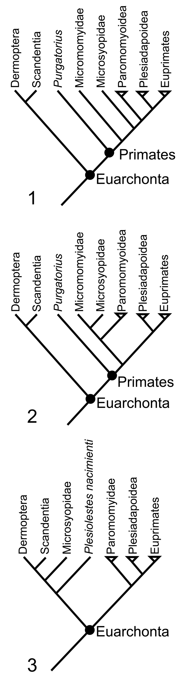
The relationship of Microsyopidae to other mammals has been a matter of some debate. Early workers allied them with either Primates Linnaeus, Reference Linnaeus1758 (e.g., Cope, Reference Cope1884; Osborn and Wortman, Reference Osborn and Wortman1892) or Insectivora Bowditch, Reference Bowditch1821 sensu lato (Matthew, Reference Matthew1915; Simpson, Reference Simpson1945), classifying them alongside Mixodectidae Cope, Reference Cope1883. More recent classifications have allied Microsyopidae (but not Mixodectidae) with Primates (e.g., Beard, Reference Beard1991; McKenna and Bell, Reference McKenna and Bell1997), although in some cases (e.g., Szalay Reference Szalay1969a, b; Gunnell, Reference Gunnell1989, Reference Gunnell2012) with a query (i.e., “?Primates”). Among primates, they have often been included among the plesiadapiforms (e.g., Bown and Gingerich, Reference Bown and Gingerich1973; Bown and Rose, Reference Bown and Rose1976; Gunnell, Reference Gunnell1989; Silcox and Gunnell, Reference Silcox, Gunnell, Janis, Gunnell and Uhen2008; Silcox et al., Reference Silcox, Bloch, Boyer, Chester and López-Torres2017), a likely paraphyletic cluster of 11 families from the Paleocene and Eocene of North America, Europe, and Asia that are similar to each other in having enlarged, procumbent upper and lower central incisors (Silcox and Gunnell, Reference Silcox, Gunnell, Janis, Gunnell and Uhen2008). Within plesiadapiforms, views of microsyopid relationships have varied. Gunnell (Reference Gunnell1989) allied them with the Palaechthonidae (Szalay, Reference Szalay1969b) in the superfamily Microsyopoidea Osborn and Wortman, Reference Osborn and Wortman1892. However, Silcox (Reference Silcox2001, Reference Silcox, Sargis and Dagosto2008; see also Chester et al., Reference Chester, Williamson, Bloch, Silcox and Sargis2017, Reference Chester, Williamson, Bloch, Silcox and Sargis2019) found no support for this grouping, suggesting instead that palaechthonids were a polyphyletic group, some of whose members were more closely allied to paromomyids than to microsyopids. Silcox's (Reference Silcox2001, Reference Silcox, Sargis and Dagosto2008) analyses suggested Microsyopidae might be among the most primitive of stem primates. Subsequent cladistic analyses of plesiadapiform relationships based on dental, cranial, and postcranial data resulted in contradictory placements of Microsyopidae (Bloch et al., Reference Bloch, Silcox, Boyer and Sargis2007; Silcox et al., Reference Silcox, Bloch, Boyer and Houde2010b; Bloch et al., Reference Bloch, Chester and Silcox2016; Chester et al., Reference Chester, Williamson, Bloch, Silcox and Sargis2017, Reference Chester, Williamson, Bloch, Silcox and Sargis2019; Figs. 1.1, 1.2, 2). While results from these analyses all suggest that microsyopids were stem primates, in one analysis (Bloch et al., Reference Bloch, Silcox, Boyer and Sargis2007) they formed a clade with non-micromomyid plesiadapiforms and euprimates, while in others (Silcox et al., Reference Silcox, Bloch, Boyer and Houde2010b; Bloch et al., Reference Bloch, Chester and Silcox2016; Chester et al., Reference Chester, Williamson, Bloch, Silcox and Sargis2017, Reference Chester, Williamson, Bloch, Silcox and Sargis2019) they were the sister taxon to Micromomyidae Szalay, Reference Szalay1974.
Hypotheses of relationships for Euarchonta from (1) Bloch et al. (Reference Bloch, Silcox, Boyer and Sargis2007); (2) Chester et al. (Reference Chester, Williamson, Bloch, Silcox and Sargis2017); (3) Bloch and Silcox (Reference Bloch and Silcox2006). Paromomyoidea in (1, 2) includes Paromomyidae and the analyzed palaechthonids (i.e., Plesiolestes nacimienti in the case of Bloch et al. (Reference Bloch, Silcox, Boyer and Sargis2007); P. nacimienti and Torrejonia wilsoni Gazin, Reference Gazin1968 in the case of Chester et al., Reference Chester, Williamson, Bloch, Silcox and Sargis2017). Note the varying position of Microsyopidae in the three different trees.
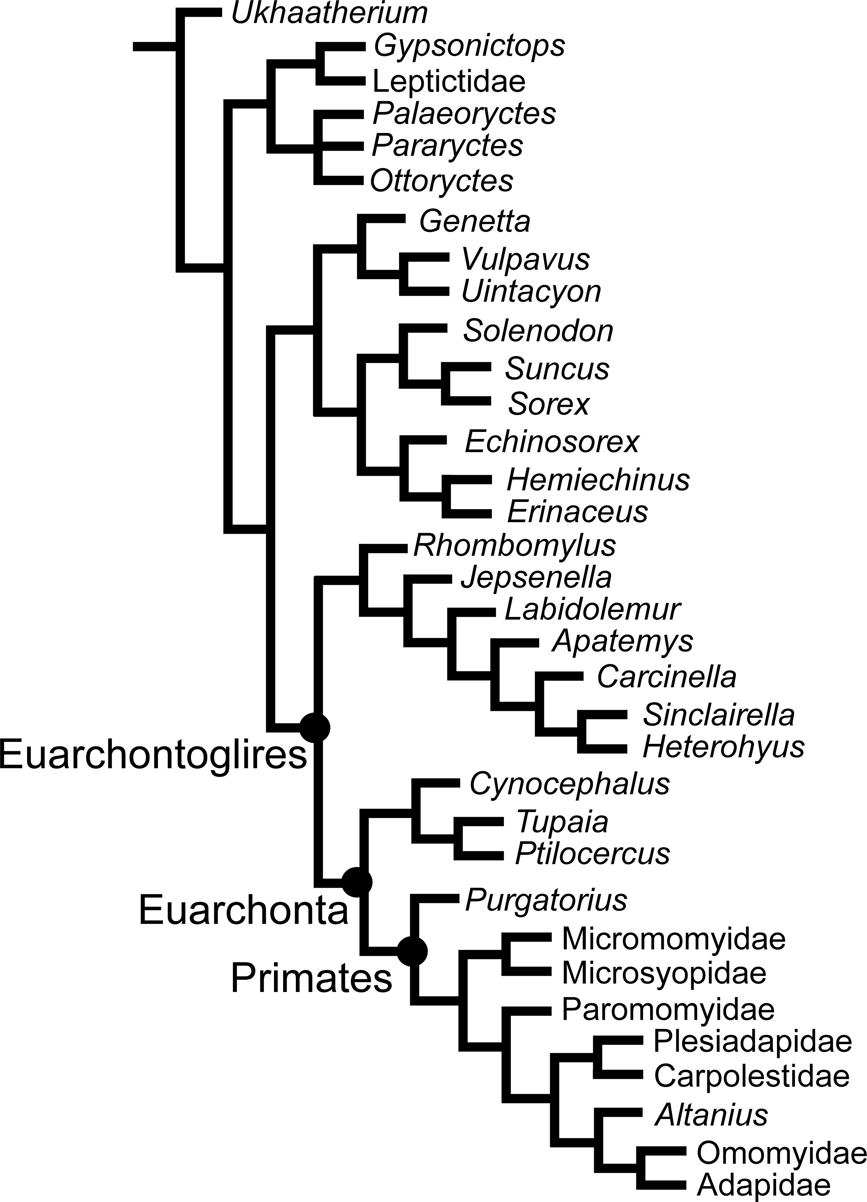
Hypothesis of relationships based on the matrix from Bloch et al. (Reference Bloch, Chester and Silcox2016; analysis with make-up of the bulla scored as “?” for carpolestids and plesiadapids), which includes data from UW 12362 (see also Silcox et al., Reference Silcox, Bloch, Boyer and Houde2010b). Upon evaluating the published codings, it was found that character 77 (Cr, Maxilla, Infraorbital foramen position) needed to be rescored for Microsyopidae (from [0] to [1], above P4). A heuristic search with 1000 replicates using parsimony in PAUP*4.0a165 (Swofford, Reference Swofford2002) found two most parsimonious trees of tree length = 1148; Consistency Index = 0.3328; Retention Index = 0.5227 (all calculated in PAUP 4.0a165). The topology of the resulting strict consensus tree is identical to that published by Bloch et al. (Reference Bloch, Chester and Silcox2016). The corrected data matrix is available on Morphobank (https://morphobank.org/).
It has also been suggested that some or all microsyopids might be more closely allied to Dermoptera llliger, Reference llliger1811 than to Primates (e.g., Szalay and Drawhorn, Reference Szalay, Drawhorn and Luckett1980; Szalay et al., Reference Szalay, Rosenberger and Dagosto1987; Gunnell, Reference Gunnell1989; Ni et al., Reference Ni, Gebo, Dagosto, Meng, Tafforeau, Flynn and Beard2013, Reference Ni, Li, Li and Beard2016). Cranial anatomy plays a critical role in evaluating this hypothesis. In particular, Szalay et al. (Reference Szalay, Rosenberger and Dagosto1987, p. 88) included a footnote in which they discussed possible similarities between the basicranium of dermopterans and microsyopids (see discussion below). Microsyopidae also fell out with Dermoptera + Scandentia Wagner, Reference Wagner1855 in a cladistic analysis of cranial traits (Bloch and Silcox, Reference Bloch and Silcox2006; Fig. 1.3), although with very weak support for that node.
Apart from maxillae, there are no published cranial specimens of either Navajoviinae or Uintasoricinae (but see Silcox and Bloch, Reference Silcox and Bloch2006; White et al., Reference White, Bloch and Silcox2016). However, two very well-preserved specimens are known for the Microsyopinae from the middle Eocene Upper Huerfano Formation of Colorado that were discussed by McKenna (Reference McKenna1966), and subsequently described in detail by Szalay (Reference Szalay1969b). The first of these, AMNH 55286, is a flattened cranium, missing most of the palate rostral to M1 (see Szalay Reference Szalay1969b, pls. 41, 42; Gunnell, Reference Gunnell1989, fig. 75). Originally identified as “Cynodontomys” knightensis Gazin, Reference Gazin1952 (McKenna, Reference McKenna1966), this specimen has been variously referred to Microsyops latidens (Cope, Reference Cope1882) (Szalay, Reference Szalay1969b), “Cynodontomys” latidens (MacPhee et al., Reference MacPhee, Novacek and Storch1988; note that “Cynodontomys” is now generally held to be a junior synonym of Microsyops; see Gunnell, Reference Gunnell1989; Silcox and Gunnell, Reference Silcox, Gunnell, Janis, Gunnell and Uhen2008), and Microsyops knightensis (Gazin, Reference Gazin1952) (Gunnell, Reference Gunnell1989). We follow Gunnell (Reference Gunnell1989) in considering this a specimen of M. knightensis. Although elements of the mesocranium are missing or broken, the posterior basicranium is well preserved, and has formed the primary basis of our previous understanding of the ear region in microsyopids. A second specimen, AMNH 55284, is a partial cranium, originally referred to Microsyops lundeliusi White, Reference White1952 (McKenna, Reference McKenna1966; Szalay, Reference Szalay1969b) that Gunnell (Reference Gunnell1989) referred to a new genus, Megadelphus. This specimen is less compressed than AMNH 55286, preserving a nearly complete palate and orbital region, and partial braincase. Unfortunately, in AMNH 55284 the posterior basicranium is entirely missing. As such, the two specimens provide complementary windows into microsyopine cranial anatomy. However, there are limitations to the information that they can provide on two fronts. First, because they belong to different genera, it is unclear to what extent it is appropriate to generalize from the one to the other. Second, even though each is well preserved in certain regions, there are still elements of anatomy that are unclear or in debate. Perhaps most critically, views vary (e.g., contrast McKenna, Reference McKenna1966 and Szalay, Reference Szalay1969b with Gunnell, Reference Gunnell1989) on the presence or absence of evidence for a bony auditory bulla. Part of the reason for this disagreement is the imperfect preservation of the tympanic roof in AMNH 55286, which makes it difficult to reconstruct its bony mosaic fully, and to determine the extent of bones (e.g., the petrosal) that might contribute to a bony bulla.
Less complete cranial specimens of microsyopine microsyopids include partial braincase roofs of Microsyops annectens (Marsh, Reference Marsh1872) (AMNH 12595; see Szalay Reference Szalay1969b, fig. 20) and Microsyops cf. M. elegans (Marsh, Reference Marsh1871) (UM 99843; see Silcox et al., Reference Silcox, Benham and Bloch2010a, fig. 6). Silcox et al. (Reference Silcox, Benham and Bloch2010a) also described a natural endocast of Microsyops annectens (UW 14559) that was identified based on an associated fragment of basicranium.
This study describes a nearly complete cranium of a microsyopid and the most complete cranium yet recovered for a plesiadapiform. Discovered in 1978 by Jeffery G. Eaton, UW 12362 has previously been mentioned in print (Wible and Covert, Reference Wible and Covert1987; MacPhee et al., Reference MacPhee, Novacek and Storch1988, Reference MacPhee, Cartmill and Rose1989; Silcox et al., Reference Silcox, Bloch, Boyer, Godinot, Ryan, Spoor and Walker2009; Reference Silcox, Benham and Bloch2010a) but never fully described. Silcox et al. (Reference Silcox, Bloch, Boyer, Godinot, Ryan, Spoor and Walker2009) published semicircular canal data from UW 12362, and Silcox et al. (Reference Silcox, Benham and Bloch2010a) detailed the endocranium of this specimen, but neither study provided a description of the external anatomy. The specimen was also used by Silcox et al. (Reference Silcox, Bloch, Boyer and Houde2010b) in coding of characters for phylogenetic analysis, which was subsequently built upon by Bloch et al. (Reference Bloch, Chester and Silcox2016; Fig. 2). However, those authors did not document the cranial anatomy beyond what was reported in the data matrix and character descriptions, and did not specifically consider the impact of the cranial partition for Microsyopidae.
This specimen is significant because it documents, for the first time, all regions of microsyopine cranial anatomy in a single specimen. Also, relative to AMNH 55286, the tympanic roof is better preserved, with some more clearly delineated sutures, allowing for a more thorough description of this critical region. The partial basicranium of UW 14559 is also described; although much less complete, it provides additional information relevant to the anatomy of the tympanic roof. Together, these specimens allow for a comprehensive description of the cranial anatomy of Microsyops annectens, and permit a consideration of the competing hypotheses about the evolutionary relationships of the Microsyopidae.
Materials and methods
X-ray computed tomography scanning
UW 12362 was scanned in June 2007 at the Center for Quantitative Imaging, Penn State University, using the OMNI-X Industrial Scanner. The scan used for this study was performed with energy settings of 170 kV, 0.200 mA, a source-object distance of 170.03 mm, and a field of view of 55.0 mm. Slices were reconstructed using 2400 views with an interslice spacing (z) of 0.0537 mm and an interpixel distance (x, y) of 0.0592 mm. A total of 1394 slices were reconstructed at a matrix size of 1024 x 1024 and stored as 16 bit tiffs. The tiffstack is available from Morphosource (Boyer et al., Reference Boyer, Gunnell, Kaufman and McGeary2016): doi:10.17602/M2/M83384 (please note that the data are mirrored).
UW 14559 was scanned in March 2016 at the Shared Materials Instrumentation Facility, Duke University, using the Nikon XTH 225 ST scanner. The scan was completed in two parts. The isolated petrosal was scanned with energy settings of 175 KV, 0.085 mA, a source-object distance of 54.4 mm and a source-detector distance of 729.47 mm. A total of 794 slices were reconstructed with a matrix size of 1240 x 983 and stored as 16 bit tiffs. Voxels were reconstructed as isotropic with x = y = z = 0.0149139490572667 mm. The tiffstack is available from Morphosource (Boyer et al., Reference Boyer, Gunnell, Kaufman and McGeary2016): doi:10.17602/M2/M83383 (please note that the data are mirrored). The natural endocast and attached ear fragment were scanned with energy settings of 150 KV, 0.100 mA, a source-object distance of 83.2 mm and a source-detector distance of 729.00 mm. A total of 1794 slices were reconstructed with a matrix size of 1397 x 961 and stored as 16 bit tiffs. Voxels were reconstructed as isotropic with x = y = z = 0.0228302486473745 mm. The tiffstack is available from Morphosource (Boyer et al., Reference Boyer, Gunnell, Kaufman and McGeary2016): doi:10.17602/M2/M83384 (data are mirrored).
The data were studied using Image J (Rasband, Reference Rasband1997–2019; Schneider et al., Reference Schneider, Rasband and Eliceiri2012) and Avizo 7 and 9.
Repositories and institutional abbreviations
AMNH, American Museum of Natural History (New York); FMNH, Field Museum of Natural History (Chicago); MNHN-CR, Muséum Nationale d'Histoire Naturelle, Cernay-les-Reims collection (Paris, France), SDSNH, Department of Paleontology, San Diego Natural History Museum (San Diego); UF-M, Florida Museum of Natural History, Mammalogy Collection (Gainesville); UKMNH, University of Kansas Museum of Natural History (Lawrence); UM, University of Michigan Museum of Paleontology (Ann Arbor); UMMZ, University of Michigan Museum of Zoology (Ann Arbor); USNM, United States National Museum (Smithsonian Institutions; Washington DC); UW, University of Wyoming (Laramie); YPM, Yale Peabody Museum (New Haven).
Anatomical terminology follows MacPhee (Reference MacPhee1981) and Wible (Reference Wible2008).
Systematic paleontology
Class Mammalia Linnaeus, Reference Linnaeus1758
Grandorder Euarchonta Waddell, Okada, and Hasegawa, Reference Waddell, Okada and Hasegawa1999
Order ?Primates Linnaeus, Reference Linnaeus1758
Family Microsyopidae Osborn and Wortman, Reference Osborn and Wortman1892
Genus Microsyops Leidy, Reference Leidy1872
Type species
Microsyops elegans (Marsh, Reference Marsh1871) (YPM 11794) from the lower Bridger beds, Bridger Basin, Wyoming.
Microsyops annectens (Marsh, Reference Marsh1872)
Figures 3–19, 20.1
Holotype
Left mandible with M3 (YPM 1171) from Henry's Fork, Bridger Basin, Wyoming.
Occurrence
Both UW 12362 and UW 14559 come from UW locality V-78001 in the Blue Point Marker horizon, Carter Mountain, northwestern Wyoming, which occurs above the Aycross and Wapiti formations and below the Tepee Trail and Wiggins formations (Eaton, Reference Eaton1982). Microsyops annectens is considered to be a diagnostic taxon of Bridgerian 3 (Br3), suggesting an age of ca. 47 Ma (Robinson et al., Reference Robinson, Gunnell, Walsh, Clyde, Storer, Stucky, Froehlich, Ferrusquia-Villafranca, McKenna and Woodburne2004); the published date for the layer from which this specimen derives is 47.9 + 0.5 Ma (Bown, Reference Bown1982; Eaton, Reference Eaton1982).
Description
The following description is based largely on a nearly complete cranium (UW 12362), supplemented by details from a less-completely preserved cranium (UW 14559) when that specimen exhibits differences in morphology or preservation.
Petrosal
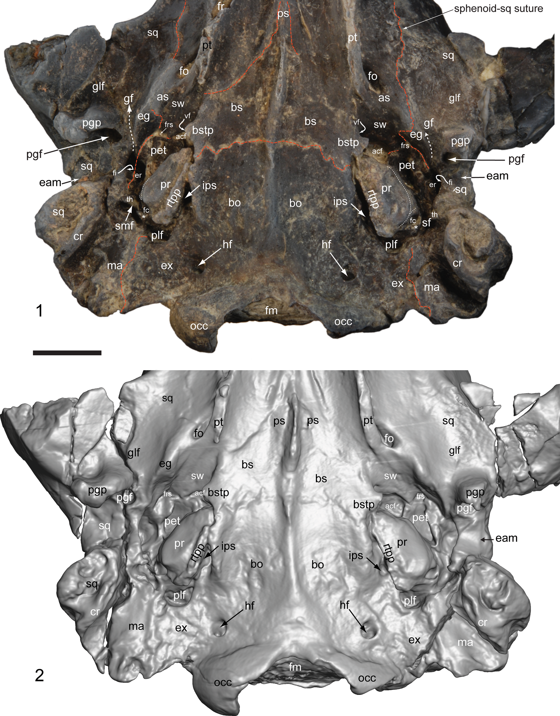
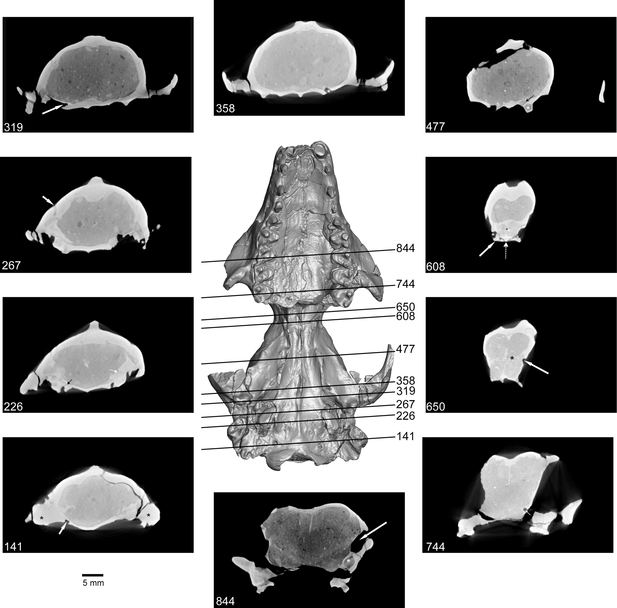
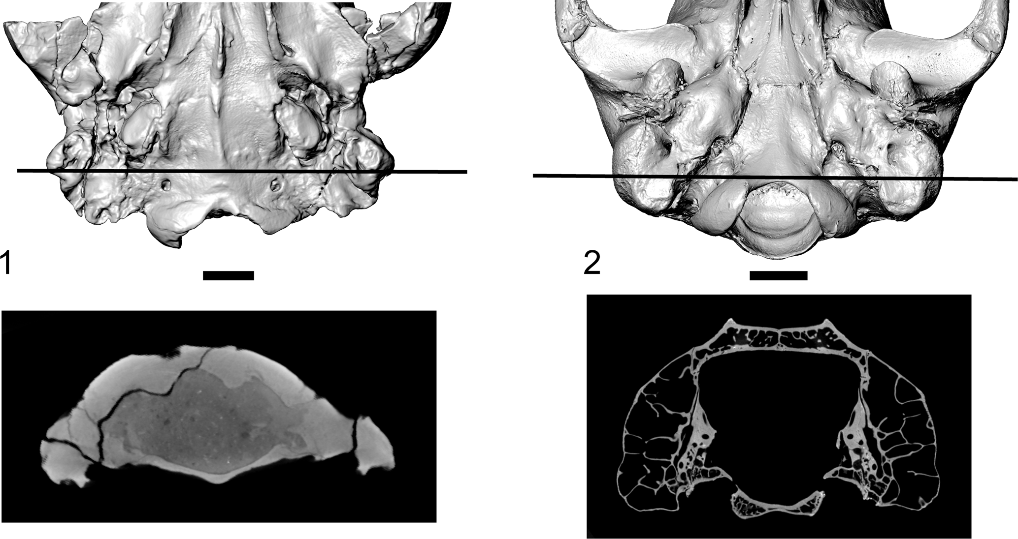
The petrosal contacts the occipital medially, the basisphenoid and epitympanic wing of the sphenoid rostrally, the alisphenoid rostrolaterally, the squamosal laterally (Fig. 5), and the exoccipital and squamosal caudally (Fig. 7, as mastoid [“ma”] on the right side). Analysis of high-resolution CT data suggests that the mastoid process is solid, and not associated with any pneumatisation of the lateral or dorsal walls of the braincase (Fig. 8, slice 141); this structure is formed in part by the mastoid, and in part by the squamosal (Figs. 5, 7). On both the right and left sides of UW 12362, the petrosal appears to be lifted away somewhat from the basioccipital, along the remnant of the basicapsular fissure (i.e., marked in the adult by the inferior petrosal sinus, labelled as “ips” in Fig. 5). This would mean that the rostral tympanic process of the petrosal (rtpp), which extends as a rugose ridge along the medial extent of the promontorium, is oriented more ventrally than would likely have been the case in life (the rtpp is broken away in UW 14559; Fig. 7). Some authors (McKenna, Reference McKenna1966; Szalay, Reference Szalay1969b) have interpreted the corresponding rugose surface in Microsyops knightensis (AMNH 55286) as forming the point of attachment of a bony bulla to the promontorium. However, as Gunnell (Reference Gunnell1989) noted, there is no positive evidence in support of this interpretation—in fact, no currently known specimens (N = 4, including the unpublished SDSNH 47729, see Silcox et al., Reference Silcox, Bloch and Gunnell2012) that preserve the ventral ear region show any clear remnant of a bony bulla, which can be observed in a diversity of fossil mammals with similar preservation. There is no sign of expansion of either the rostral or caudal tympanic processes of the petrosal into a bullar element. Although likely somewhat damaged in UW 12362, the rtpp extends only the length of the promontorium (Fig. 5). The caudal tympanic process of the petrosal (ctpp; asterisk in Figs. 5, 7, 9) is present as a very small ridge of bone caudal and lateral to the fenestra cochleae in both UW 12362 and UW 14559. Neither process shows any evidence of expansion to form significant elements of the floor of the tympanic cavity.
Microsyops annectens (UW 12362). (1) Photograph of the cranium lightly dusted with ammonium chloride and (2) reconstruction from high resolution CT data of a close up ventral view of the basicranium. Both versions are included in this figure because some structures (e.g., sutures) are more easily visualized in the photograph, while others (e.g., foramina) are clearer in the reconstruction. Red dashed lines indicate sutures between bones. White dashed arrows indicate the glaserian fissure (gf). White dotted lines on the promontorium indicate the inferred pathway of the internal carotid artery and promontorial and stapedial branches. The asterisks indicate the very small caudal tympanic process of the petrosal. Scale bar represents 5 mm. Abbreviations: acf = anterior carotid foramen; as = alisphenoid; bo = basioccipital; bs = basisphenoid; bstp = basisphenoid tympanic process; cr = crest on the mastoid; eam = external acoustic meatus; eg = entoglenoid process; er = epitympanic recess; ex = exoccipital; fc = fenestra cochleae; fi = fossa incudis; fm = foramen magnum; fo = foramen ovale; fr = foramen rotundum; frs = foramen for the ramus superior of the stapedial artery; gf = glaserian fissure; glf = glenoid fossa; hf = hypoglossal foramen; ips = inferior petrosal sinus; ma = mastoid; occ = occipital condyle; pet = petrosal; pgf = postglenoid foramen; pgp = postglenoid process; plf = posterior lacerate (jugular) foramen; pr = promontorium; ps = presphenoid; pt = pterygoid; rtpp = rostral tympanic process of the promontorium; sf = stapedius fossa; smf = stylomastoid foramen primitivum; sq = squamosal; sw = epitympanic wing of the sphenoid; th = tympanohyal; vf = vidian foramen.
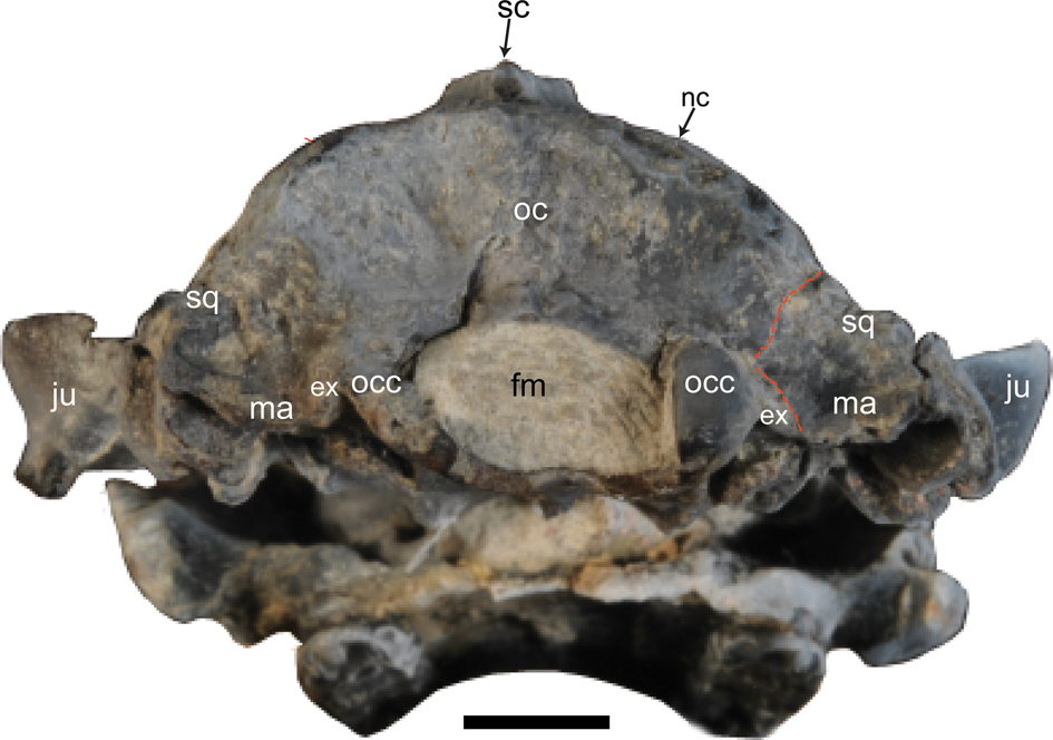
Caudal view of the cranium of Microsyops annectens (UW 12362), lightly dusted with ammonium chloride. The only clear suture (dashed red line) separates the occipital from the mastoid and (presumably) the squamosal. The point of division between the latter two bones is not clear. Scale bar represents 5 mm. Abbreviations: ex = exoccipital; fm = foramen magnum; ju = jugal; ma = mastoid; nc = nuchal crest; oc = occipital complex; occ = occipital condyle; sc = sagittal crest; sq = squamosal.
High resolution X-ray CT images of Microsyops annectens (UW 12362). Numbers correspond to slices in the stack, starting at the caudal extent of the specimen (total = 1,394), as indicated on the reconstruction of the cranium. Dorsal oriented up for CT slices. Brightness and contrast settings were modified for each slice to maximize the visibility of the indicated structures. 141: black asterisks = mastoid processes, white arrow = hypoglossal foramen; 226: white arrow = the facial canal, black arrow = inferior petrosal sinus; 267: white asterisk = the postglenoid foramen, white arrow = foramen on the parietal/squamosal suture; 319: white arrow = vidian foramen, dorsal to the basisphenoid tympanic process; 358: white asterisk = the foramen ovale; 477 black arrow = the canal leading to the sphenorbital fissure, white asterisk = canal for V3 (leading to the foramen rotundum); 608: white solid arrow = palatine, white dotted arrow = vomer, black asterisk = ethmoid, white asterisk = optic foramen; 650: white arrow = the ?ethmoidal foramen, black asterisk = the cribriform plate; 744: white arrow = sphenopalatine foramen; 844 white arrow = lacrimal foramen, white asterisk = infraobital canal.
The petrosal forms most of the caudal portion of the tympanic cavity roof. However, it has a well-demarcated suture with the epitympanic wing of the sphenoid (“sw” in Fig. 5) and alisphenoid (“as” in Fig. 5) in UW 12362, which demonstrates that it does not form the rostral portion of the tympanic cavity roof. Two foramina are evident along the petrosal-sphenoid suture in ventral view (Fig. 5). The more medial one, located near the rostral pole of the promontorium, can be identified based on its position relative to the groove for the promontorial branch of the internal carotid artery as the anterior carotid foramen (“acf”; Figs. 5, 9) for the entry of the promontorial branch of the internal carotid artery to the braincase. The more lateral opening may have conveyed the ramus superior of the stapedial artery (“frs”; Figs. 5, 9), based on the typical course of that artery in eutherians (Wible, Reference Wible1987). Because they sit along the suture between the petrosal and sphenoid, these foramina may have formed from remnants of the piriform fenestra (i.e., the gap between the petrosal and its adjacent bones rostromedially; McDowell, Reference McDowell1958; MacPhee, Reference MacPhee1981; Wible and Martin, Reference Wible, Martin and MacPhee1993), otherwise lost during ontogeny in Microsyops. The ramus superior generally enters the endocranial space through a remnant of that fenestra in eutherians (Wible, Reference Wible1987).
The suture between the petrosal and sphenoid extends laterally into the suture between the petrosal and squamosal, which runs along the roof of the epitympanic recess (“er”; Figs. 5, 9)—as such, this recess was roofed by both of these bones, and the fossa incudis (“fi”; Figs. 5, 9) is located in the squamosal rather than the petrosal. At the caudal pole of the promontorium, the posterior lacerate foramen (“plf” = jugular foramen of Wible, Reference Wible2008; Figs. 5, 9) opens along the suture between the petrosal (mastoid) and the occipital. Just rostral to the plf a very small foramen leading into the structure of the petrosal itself is visible when the cranium is tilted rostrally (“cc” in Fig. 9). Openings similar to this have been variably interpreted in previous discussions of the basicranium of microsyopids. Szalay (Reference Szalay1969b, fig. 17, structure labelled “mec”) considered it to convey a medial entocarotid and the inferior petrosal sinus, while MacPhee and Cartmill (Reference MacPhee, Cartmill, Swisher and Erwin1986, fig. 16) and MacPhee et al. (Reference MacPhee, Novacek and Storch1988, fig. 13) labeled the opening as the cochlear canaliculus. Because there is compelling evidence that eutherian mammals primitively lacked the medial entocarotid (Presley, Reference Presley1979; see also Cartmill and MacPhee, Reference Cartmill, MacPhee and Luckett1980; MacPhee, Reference MacPhee1981; Wible, Reference Wible1983; MacPhee and Cartmill, Reference MacPhee, Cartmill, Swisher and Erwin1986; Gunnell, Reference Gunnell1989), the latter identification seems more likely. This identification is also supported by CT data, since the opening can be observed to lead into the substance of the cochlea, although the density difference between matrix and bone in the cochlea is so slight that it cannot be traced further. A separate opening for the inferior petrosal sinus is present caudally between the petrosal and basioccipital (“ips”; Figs. 5, 9)—this can be traced into the endocranial cavity on the CT data (Fig. 8, slice 226; Silcox et al., Reference Silcox, Benham and Bloch2010a, fig. 3), which extends along the suture between the petrosal and the basioccipital.
Well-demarcated grooves can be identified on the promontorium for the stem of the internal carotid artery, and both the stapedial and promontorial branches (finely dotted lines in Figs. 5, 6, 9). The entry of the internal carotid artery to the middle ear would have been posteromedial. The groove for the stapedial artery extends rostral to the fenestra cochleae, which is oriented posterolaterally, continuing to the fenestra vestibuli, which is oriented more laterally (Figs. 5, 6, 9). From this point, the pathway of the stapedial artery is not clearly indicated by any structures, apart from the above-mentioned opening for the ramus superior, and a groove on the squamosal between the postglenoid process and entoglenoid process (medially continuous with the alisphenoid tympanic process) that can be tentatively identified as the glaserian fissure (“gf”; Figs. 5, 9) based on analogy with Leptictis Leidy, Reference Leidy1868 (Novacek, Reference Novacek1986, fig. 20); as such it could have housed the ramus inferior of the stapedial artery in addition to the chorda tympani (Klaauw, Reference Klaauw1931; Wible, Reference Wible2008). If this interpretation is correct, then the ramus superior followed an endocranial course, while the ramus inferior followed an extracranial one. As noted above, the groove for the promontorial branch of the internal carotid artery leads to the anterior carotid foramen (“acf”) medial to the rostral pole of the promontorium (“pr”; Figs. 5, 9).
Ventral view of the incomplete basicranium in Microsyops annectens (UW 14559) based on a reconstruction from the high resolution X-ray CT data. The black dotted arrow traces the inferred course of the facial nerve. The white dotted lines demarcate the inferred pathway of the internal carotid artery and promontorial and stapedial branches. The asterisks indicate the very small caudal tympanic process of the petrosal. Scale bar represents 1 mm. Abbreviations: cp = crista parotica; cr = crest on the mastoid; er = epitympanic recess; fc = fenestra cochleae; ff = facial foramen (this opening is tucked at the top of the groove for the facial nerve); fv = fenestra vestibuli; ma = mastoid; pr = promontorium; sf = stapedius fossa; th = tympanohyal.
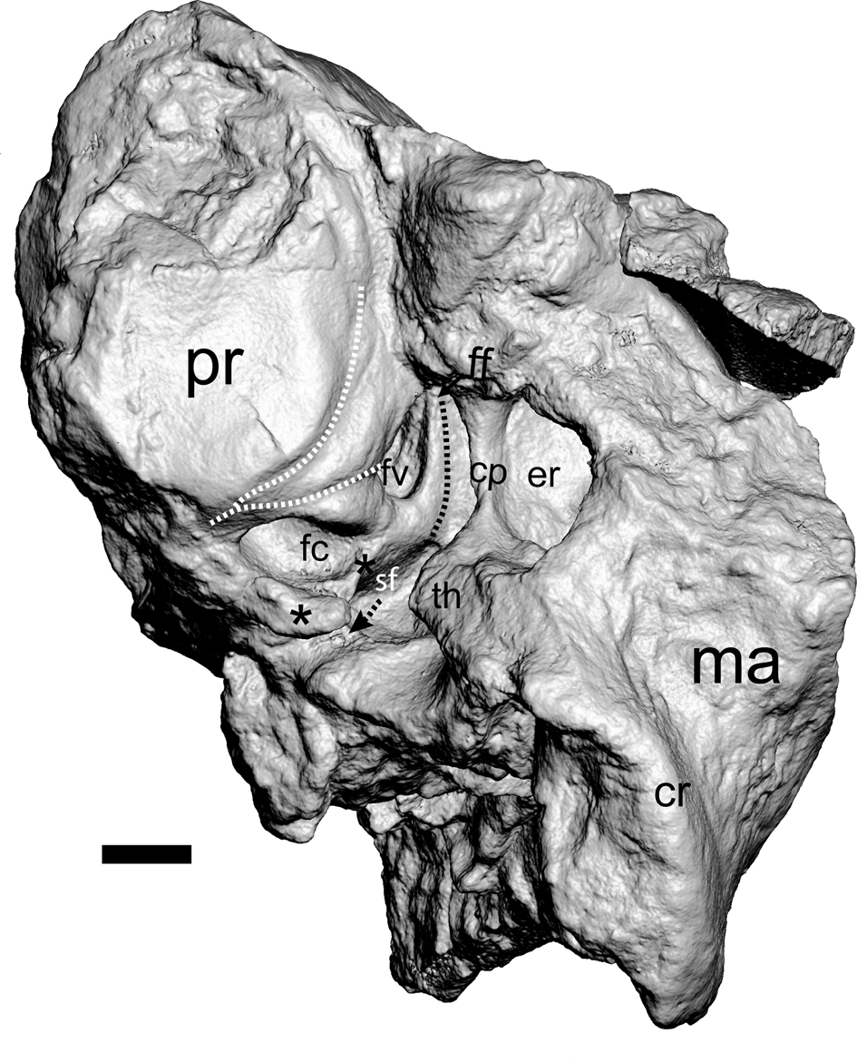
It is difficult to establish the pathway for the facial nerve based on the external anatomy of the basicranium in UW 12362 because the lateral roof of the tympanic cavity is somewhat damaged. However, it is possible to use the high-resolution CT data to trace the course of the facial nerve through the petrosal on the left side of the specimen (Fig. 8, slice 226) to the external surface. Its pathway leads to an opening directly into the middle ear cavity, so there is no bony tube for this nerve. In UW 14559, a groove incised into the lateral floor of the roof of the tympanic cavity provides a likely pathway for this nerve (Fig. 6) because it lies medial to the crista parotica (“cp”; Fig. 6), the attachment point of the tympanohyal, and lateral boundary of the facial nerve in extant mammals (MacPhee, Reference MacPhee1981; Wible et al., Reference Wible, Novacek and Rougier2004). This groove ultimately runs dorsal to the tympanohyal (“th”; Fig. 6). In the absence of a bulla (and associated foramina), exit of the facial nerve from the middle ear would have been through a stylomastoid foramen primitivum (sensu MacPhee, Reference MacPhee1981), between the tympanohyal and caudal tympanic process of the petrosal (Figs. 5, 6). UW 12362 lacks a well-defined groove on the lateral tympanic roof, but shares with UW 14559 the gap (labelled “smf” in Fig. 5) between the th and ctpp, representing the bony portion of the opening for this nerve out of the middle ear cavity. On the left side of UW 12362 there is a well-defined fossa for the stapedius muscle (“sf”; Fig. 6) located medial to the tympanohyal—this space is obscured by matrix on the right side of UW 12362, but is well defined in UW 14559 (Fig. 9). The location of this fossa indicates that the origin of the stapedius muscle would have been near the outer margin of the tympanic cavity.
Occipital complex (=basioccipital, exoccipital, supraoccipital)
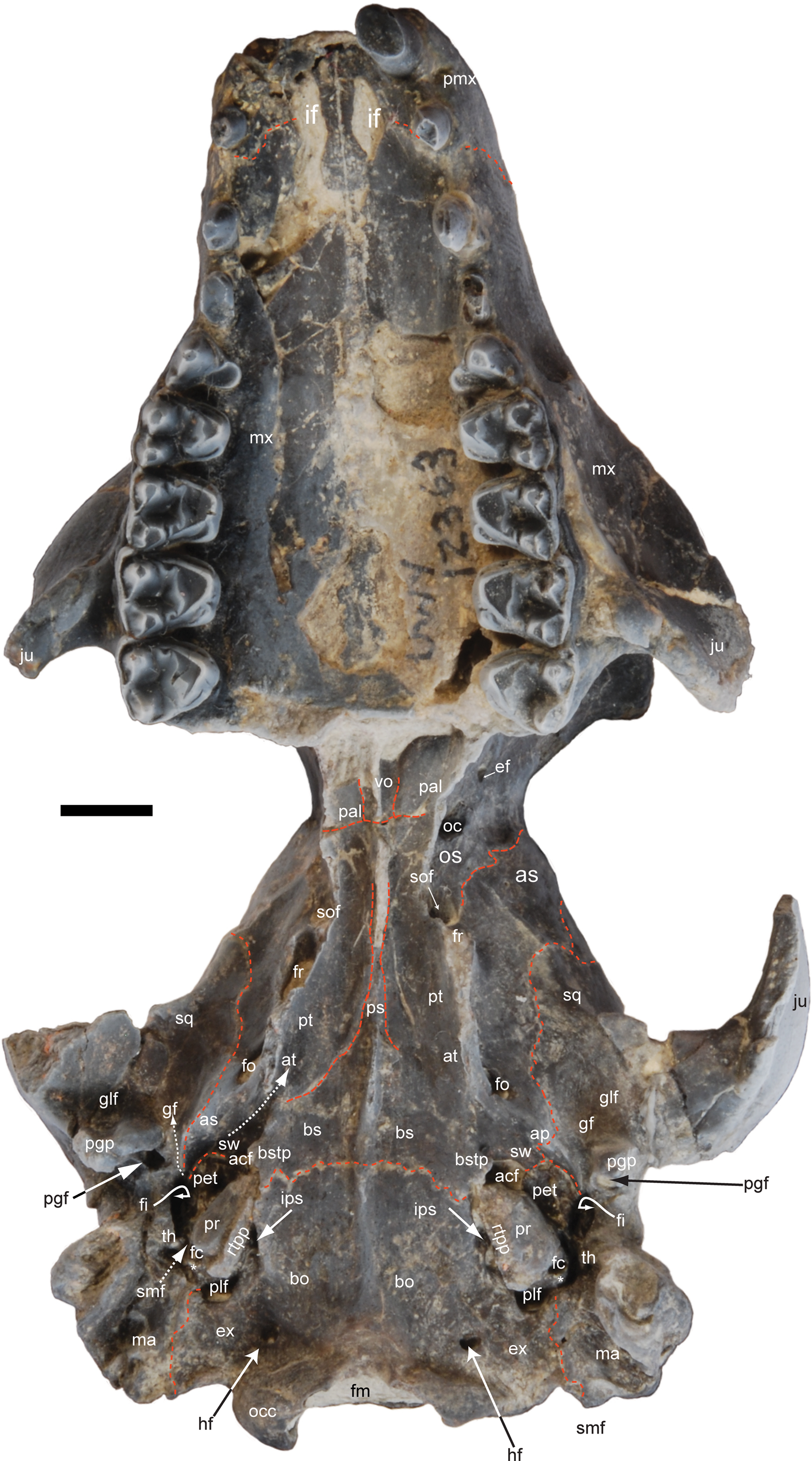
Clear sutures are lacking between the various elements of the occipital, so the term “occipital complex” is used when a more precise identification of the relevant element is ambiguous based on patterns in extant mammals. On the basicranium the occipital complex forms much of the ventral surface of the neurocranium. Laterally it contacts the petrosal at the remnant of the basicapsular fissure (i.e., location of the inferior petrosal sinus; Figs. 5, 9) and along a suture with the mastoid region that is well defined bilaterally in UW 12362 (Figs. 5, 9). Rostrally it contacts the basisphenoid near the rostral pole of the petrosal (Figs. 5, 9). There is a strong midline crest starting ~3.5 mm from the rostroventral edge of the foramen magnum, extending to the level of the suture with the basisphenoid, and continuing on the basiphenoid/presphenoid (i.e., between the right and left “ps” labels in Fig. 10). An opening for the inferior petrosal sinus is present between the petrosal and the basioccipital (Figs. 5, 9, 10). The lateral edge of the basioccipital on both sides of the cranium is unbroken (Fig. 5). This edge is deflected ventrally to a very slight degree along its length, which could be interpreted as a very low basioccipital tympanic process. Based on these observations, if there were a bony bulla, it was not composed of the basioccipital to any significant degree.
Labeled ventral view of Microsyops annectens (UW 12362), lightly dusted with ammonium chloride. Red dashed lines indicate sutures between bones. White dotted arrows represent passage of soft-tissue structures. Scale bar represents 5 mm. Abbreviations: acf = anterior carotid foramen; ap = tympanic process of the alisphenoid; as = alisphenoid; at = auditory tube; bo = basioccipital; bs = basisphenoid; bstp = basisphenoid tympanic process; ef = ethmoidal foramen; ex = exoccipital; fc = fenestra cochleae; fi = fossa incudis; fm = foramen magnum; fo = foramen ovale; fr = foramen rotundum; gf = glaserian fissure; glf = glenoid fossa; hf = hypoglossal foramen; if = incisive foramen; ips = inferior petrosal sinus; ju = jugal; ma = mastoid; mx = maxilla; oc = occipital; occ = occipital condyle; os = orbitosphenoid; pal = palatine; pet = petrosal; pgf = postglenoid foramen; pgp = postglenoid process; plf = posterior lacerate (jugular) foramen; pmx = premaxilla; pr = promontorium; ps = presphenoid; pt = pterygoid; rtpp = rostral tympanic process of the promontorium; smf = stylomastoid foramen primitivum; sof = sphenorbital fissure; sq = squamosal; sw = epitympanic wing of the sphenoid; th = tympanohyal; vo = vomer.
The exoccipital forms the occipital condyles and has a horizontal component on the cranial base that contacts the mastoid portion of the petrosal ventrally (Figs. 5, 9, 10), and a vertical component that extends as a small wedge-shaped piece of bone caudally, sandwiched between the mastoid and the occipital condyles (Fig. 7). The occipital condyle is well preserved on the right side of UW 12362, and is taller dorsoventrally (5.63 mm) than wide (3.83 mm). The foramen magnum is slightly distorted in UW 12362 (Fig. 7), but was clearly wider (10.55 mm) than tall (5.74 mm). Ventrally the exoccipital is pierced by the hypoglossal foramen (“hf”; Figs. 5, 9, 10). In UW 12362 this opening encompasses three tiny foramina on the right side, and two on the left (Fig. 8, slice 141), all of which lead into the endocranial cavity. The exoccipital also makes up the caudal part of the wall of the posterior lacerate foramen (Fig. 9).
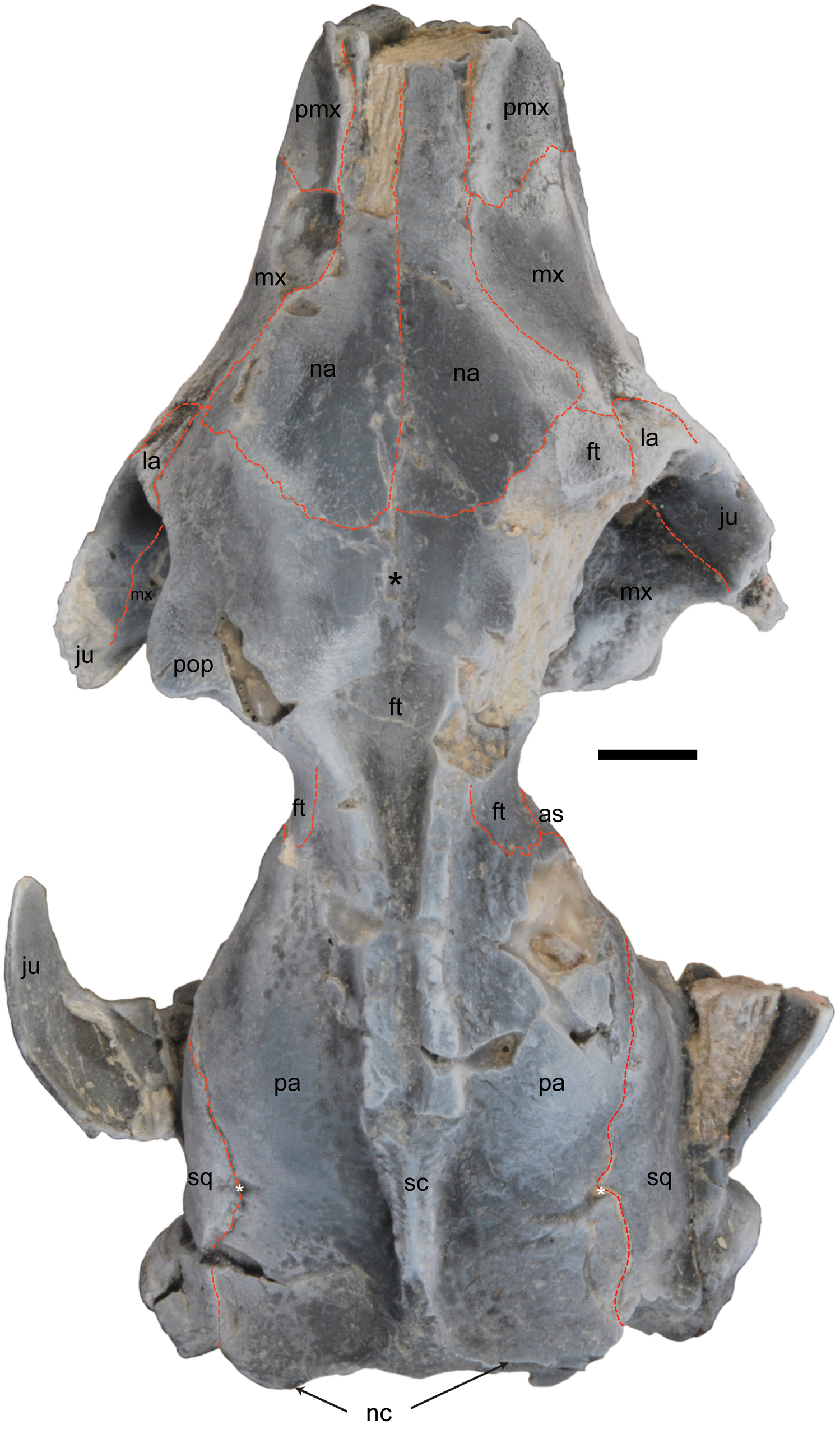
In caudal view the occipital complex (either exoccipital or supraoccipital; “oc” in Fig. 7) contacts the mastoid and squamosal. The caudal surface of the supraoccipital is rugose, likely forming a well-demarcated area of muscle attachments ventral to the well-defined nuchal crest (“nc” in Fig. 7). There is no evidence of any foramina piercing the substance of the supraoccipital on the surface of the bone. In dorsal view (Fig. 11), the location of the suture between the supraoccipital and the parietal is not clearly demarcated. In the absence of a visible suture, the contact between these two bones likely passed along the nuchal crest. This interpretation would be consistent with the anatomy observed in UM 99843, a specimen of Microsyops cf. M. elegans that exhibits rugose, sutural morphology along its nuchal crest (Silcox et al., Reference Silcox, Benham and Bloch2010a, fig. 6A). In UW 12362 the nuchal crest is continuous with the caudal end of the sagittal crest (Fig. 11).
Dorsal view of the cranium of Microsyops annectens (UW 12362), lightly dusted with ammonium chloride. Dashed red lines indicate sutures that can be traced. The white asterisks indicate the foramina for the rami temporales on the parietal-squamosal suture. The black asterisk sits on the remodeled remnants of the metopic suture. Scale bar represents 5 mm. Abbreviations: as = alisphenoid; ft = frontal; ju = jugal; la = lacrimal; mx = maxilla; na = nasal; nc = nuchal crest; pa = parietal; pop = postorbital process; pmx = premaxilla; sc = sagittal crest; sq = squamosal.
Sphenoid complex (=basisphenoid, presphenoid, alisphenoid, orbitosphenoid) and pterygoid
The sphenoid complex forms much of the more rostro-ventral portion of the neurocranium, a small portion of the lateral wall of the neurocranium, and the caudal component of the orbital mosaic. Although sutures separate some elements of the sphenoid complex from one another in UW 12362, in most instances the various parts of this complex are fused seamlessly to one another, which makes it difficult to delineate the boundaries of the various bones.
In ventral view, the basisphenoid contacts the basioccipital, at a roughly horizontal suture near the level of the rostral pole of the promontorium (Figs. 5, 10). The basisphenoid contacts the petrosal at the rostralmost end of the middle ear cavity, and contributes a small, pointed basisphenoid tympanic process (“bstp” in Figs. 5, 9) to the floor of the tympanic cavity. The bstp is horizontal in orientation rather than being vertical—as such it differs in orientation from the other tympanic processes (basioccipital, alisphenoid, and petrosal) and does not contribute to the formation of a ventrally oriented “rim” around the middle ear cavity in this specimen. At the rostromedialmost corner of the middle ear cavity, dorsal to the level of the bstp and rostral to the anterior carotid foramen, there is a tiny (~1.0 mm2) foramen, visible bilaterally in UW 12362 (“vf” in Figs. 5, 9; Fig. 8, slice 319). This opening can be tentatively identified as the vidian foramen (i.e., the caudal opening of the pterygoid canal; “posterior vidian foramen” of Novacek, Reference Novacek1986, p. 6; “posterior opening of the pterygoid canal” of Wible, Reference Wible2008, p. 345) based on its location near the rostromedial extent of the tympanic cavity (e.g., in analogy to Leptictis; Novacek, Reference Novacek1986, fig. 22). A midline crest extends rostrally the length of the basiphenoid and presphenoid (Fig. 10); as noted above, this is continuous with a crest on the basioccipital.
In ventral view (Fig. 10), the presphenoid is continuous with the basiphenoid and is widest along its caudal border. There is a well-defined suture between the presphenoid and the medial aspect of the pterygoid that curves towards the midline from caudal to rostral (Fig. 10). Laterally, the pterygoid is fused seamlessly with the alisphenoid. The rostral border of the pterygoid articulates with the palatine, and the rostral border of presphenoid extends towards the caudal margin of the vomer as a midline ridge (Fig. 10).
In ventral view (Fig. 5, 9), both the alisphenoid and basisphenoid likely contribute to the rostral roof (=epitympanic wing of the sphenoid, sensu MacPhee, Reference MacPhee1981) of the tympanic cavity. The epitympanic wing of the sphenoid (“sw” in Figs. 5, 9) articulates with the petrosal at a well-defined suture and forms the rostral portion of the anterior carotid foramen (“acf”) and foramen for the ramus superior of the stapedial artery (“frs”). A clear indentation for the auditory tube (at) marks the surface of the alisphenoid, extending between the basisphenoid tympanic process and the pterygoid crest (Fig. 9). There is a low, largely rostrocaudally oriented alisphenoid tympanic process (“ap” in Fig. 9), oriented at ~45° to the pterygoid crest, and located caudal to the foramen ovale (“fo” in Fig. 9), that is continuous with the entoglenoid process. A well-defined suture with the squamosal extends onto the process (Fig. 9). On the right side of UW 12362 it is possible to identify the location of the foramina ovale (“fo”; ~2.83 mm2) and rotundum (“fr”; 4.24 mm2) on the ventral surface of the alisphenoid, lateral to the pterygoid crest (“pt” in Fig. 10; Fig. 8, slices 358, 477). On the left side of UW 12362, the foramen ovale is clearly preserved, but the foramen rotundum is damaged, and the pterygoid crest is largely broken away. There is no evidence of an enclosed alisphenoid canal. In lateral views of the neurocranium (Figs. 12–14), the alisphenoid contributes a wing to the rostral aspect of the neurocranium just caudal to the orbitosphenoid that is well demarcated by sutures from the orbitosphenoid, squamosal, parietal, and possibly the frontal (the boundaries of which are not visible in relation to either the orbitosphenoid or alisphenoid).
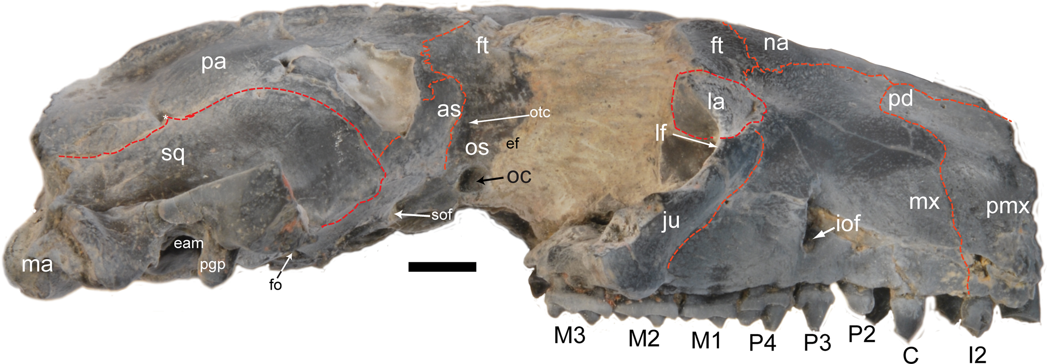
Left lateral view of Microsyops annectens (UW 12362), lightly dusted with ammonium chloride. Dashed red lines indicate sutures between bones. Although most of the bones that make up the orbital mosaic can be clearly delineated, the suture at the front of the orbitosphenoid (os) is not clear. The white asterisk on the suture between the parietal and squamosal indicates a foramen for a ramus temporalis. Scale bar represents 5 mm. Abbreviations: as = alisphenoid; C = canine; eam = external acoustic meatus; ef = ethmoidal foramen; ft = frontal; I = incisor; iof = infraorbital foramen; ju = jugal; la = lacrimal; M = molar; ma = mastoid; mx = maxilla; na = nasal; oc = optic canal; os = orbitosphenoid; otc = orbital opening of the orbitotemporal canal; P = premolar; pa = parietal; pal = palatine; pd = posterodorsal process of the premaxilla; pgp = postglenoid process; pmx = premaxilla; pop = postorbital process; pr? = possible remnant of a process on the jugal; spf = sphenopalatine foramen; sq = squamosal.

Oblique lateral-dorsal view of the left orbital region of Microsyops annectens (UW 12362), lightly dusted with ammonium chloride. Dashed red lines indicate sutures between bones. White asterisk indicates foramen for a ramus temporali. Scale bar represents 5 mm. Abbreviations: as = alisphenoid; ef = ethmoidal foramen; fr = foramen rotundum; ft = frontal; iof = infraorbital foramen; ju = jugal; la = lacrimal; mx = maxilla; na = nasal; of = optic foramen; os = orbitosphenoid; otc = orbital opening of the orbitotemporal canal; pa = parietal; pal = palatine; pop = postorbital process; sof = sphenorbital fissure; spf = sphenopalatine foramen; sq = squamosal.

In ventral view, the orbitosphenoid (“os” in Fig. 10) contacts the alisphenoid caudally at a well-demarcated suture on the left side of the cranium (Fig. 10). It also contacts the palatine rostrally and pterygoid medially in this view, although the relevant sutures are not clearly demarcated. The sphenorbital fissure (“sof”; left ~2.14 mm2) sits just rostral to the foramen rotundum (Fig. 15); on the left side, this foramen is clearly located on the suture between the alisphenoid and orbitosphenoid (Fig. 10). On both sides of the cranium in lateral view (Figs. 12–14) the orbitosphenoid shares a well-defined caudal suture with the alisphenoid caudally. The more rostral part of the orbital region is obliterated on the right side, but on the left there is a short suture between the orbitosphenoid and the palatine at the orbitosphenoid's ventrorostral aspect (Figs. 12, 13). The sutures between the frontal and orbitosphenoid are entirely obliterated on the left side (and missing on the right), so the limits of both bones in the orbital mosaic are not clear. The optic canal (“oc” in Figs. 12–15; average area for right and left sides ~1.6 mm2; Silcox et al., Reference Silcox, Benham and Bloch2010a) pierces the orbitosphenoid at the caudal extent of the orbit. A small (left ~1.07 mm2) foramen located dorsal and caudal to the optic canal can be traced on the endocast (Fig. 15) to the orbitotemporal canal (“otc”; Figs. 12, 15)—as such, this represents the rostral opening of that passageway, which conveys the ramus superior of the stapedial artery and accompanying vein (Wible, Reference Wible1987; this passageway is sometimes referred to as the “sinus canal,” but we follow Wible and Gaudin, Reference Wible and Gaudin2004 and Wible, Reference Wible2008 in using the term “orbitotemporal canal” to reflect the fact that it does not convey a sinus, but rather an artery and vein). Rostral to the opening for the otc on the left side is another small foramen (~2.0 mm2), which can be tentatively identified as an ethmoidal foramen (for the external ethmoidal artery, vein, and/or nerve; “ef” in Figs. 12, 13, 15) based on its location near the likely border between the frontal and the orbitosphenoid and its close relationship to the cribriform plate (Fig. 8, slice 650; Fig. 15). As such, the foramen is in a similar position to the anteroventral ethmoidal foramen in certain mammals (Evans, Reference Evans1993; Wible, Reference Wible2008). The suture with the frontal is not clear and it is not possible to determine whether the ef and/or the otc were located in whole or in part in the orbitosphenoid, or were located instead in the frontal.
Oblique left lateral view of a virtual reconstructions based on high resolution X-ray CT data of Microsyops annectens (UW 12362) (1) with the cranium rendered to be transparent to allow the endocast to be visualized; and (2) a reconstruction of just the endocast (Silcox et al., Reference Silcox, Benham and Bloch2010a) to show the relationship between the external and internal anatomy. Scale bar represents 5 mm. Abbreviations: ef = ethmoidal foramen; fr = foramen rotundum; oc = optic canal; otc = orbitotemporal canal; sof = sphenorbital fissure.

Squamosal
In ventral view the squamosal shares a well-defined suture with the alisphenoid rostrally, and contacts the petrosal more caudally (Fig. 10). The contact between the squamosal and petrosal is marked by a clear suture in the roof of the epitympanic recess (Fig. 5, traced on the right side of the specimen). Lateral to the epitympanic recess, there is a well-excavated fossa incudis that is entirely in the squamosal (Fig. 5). The squamosal aspect of the external acoustic meatus lies ventral to this recess (Fig. 5). More caudally, the squamosal likely forms the lateral aspect of the mastoid process and makes up the posttympanic process of the squamosal, sharing a suture with the petrosal (Figs. 5, 10). Although the precise location of the suture is not clear on UW 12362, it may lie just medial to the strong crest present on the mastoid process (labeled “cr” in Fig. 5), based on a gap between parts of the process visible in the CT reconstruction on the right side (i.e., between “cr” and “ma” in Fig. 5.2). It is likely that damage may have occurred in the plane of articulation between the two bones, a viewpoint supported by the presence of a similarly placed discontinuity on the left side of the specimen (Fig. 5.2)
The groove on the squamosal lateral to the entoglenoid process (“egp”) and medial to the postglenoid process (“pgp”) can be tentatively identified as the glaserian fissure (“gf” on Fig. 5, for the chorda tympani and likely the ramus inferior of the stapedial artery). The pgp is located rostral to the large postglenoid foramen (“pgf”; right ~2.0 mm2; left ~2.1 mm2), which can be traced on the high-resolution CT data into the cranial cavity (Fig. 8, slice 267), ultimately being continuous with the transverse sinus (see Silcox et al., Reference Silcox, Benham and Bloch2010a, fig. 4). Although the pgp is damaged on the left side of UW 12362, on the less-damaged right side, it is clear that this process also extends significantly lateral to the level of this opening (Fig. 5). In ventral view, the glenoid fossa (“glf” in Figs. 5, 10) is present rostral to the postglenoid process. The glenoid fossa is longer (left side of UW 12362 ~6.96 mm) than wide (~5.85 mm) and very flat. It is continuous with the zygomatic process of the squamosal, which articulates with the jugal (Fig. 10). In lateral view, on the preserved left side (Fig. 12), there is no evidence of a subsquamosal foramen on the zygomatic process of the squamosal or on the jugal. The squamosal presumably shared a suture caudally with the supraocciptial, but this is not clearly demarcated (Fig. 7).
In lateral view (Figs. 12, 13), the squamosal shares a long squamous suture with the parietal. A small foramen is present along this suture, which can be traced in the CT data to be continuous with both the endocranial cavity and the postglenoid canal (Fig. 8, slice 267). This opening likely carried blood to and from the temporalis muscle (i.e., from the ramus temporalis of the ramus superior of the stapedial artery; Wible, Reference Wible1987). More dorsally, the squamosal contacts the alisphenoid near the back of the orbit (Fig. 13).
Parietal
In lateral view (Figs. 12, 14), the parietal contacts the squamosal ventrally. In UW 12362 (Microsyops annectens) the suture between the parietal and the supraoccipital is not preserved, but a fossil of M. cf. M. elegans (UM 99843; Silcox et al., Reference Silcox, Benham and Bloch2010a, fig. 6) suggests that the suture runs along the nuchal crest in microsyopids. Rostrally (Fig. 13) the parietal contacts the alisphenoid and the frontal. In dorsal view (Fig. 11), part of the suture with the frontal is visible on both sides of UW 12362, demonstrating that it sat just caudal to the postorbital constriction and had an inter-digitated morphology, although the rostral portion of the suture with the frontal is obliterated at the midline. In UW 12362 (Microsyops annectens), temporal crests extend from the frontal onto the parietals, forming two distinct parasagittal crests for most of the length of the neurocranium. They join to form a single sagittal crest ~14.5 mm from the nuchal crest, just rostral to the level of the postglenoid process, so that the two crests are joined for ~38% of their length. No foramina pass directly through the parietal, although as noted previously, there are bilateral openings on the right and left sutures with the squamosals that presumably housed rami temporales (Fig. 11)
Right lateral view of Microsyops annectens (UW 12362), lightly dusted with ammonium chloride. Dashed red lines indicate sutures between bones. The white asterisk on the suture between the parietal and squamosal indicates a foramen for a ramus temporalis. Scale bar represents 5 mm. Abbreviations: as = alisphenoid; C = canine; eam = external acoustic meatus; ef = ethmoidal foramen; fo = foramen ovale; ft = frontal; I = incisor; iof = infraorbital foramen; ju = jugal; la = lacrimal; lf = lacrimal foramen; M = molar; ma = mastoid; mx = maxilla; na = nasal; oc = optic canal; os = orbitosphenoid; otc = orbital opening of the orbitotemporal canal; P = premolar; pa = parietal; pd = posterodorsal process of the premaxilla; pgp = postglenoid process; pmx = premaxilla; sof = sphenorbital fissure; sq = squamosal.
Frontal
In dorsal view (Fig. 11), the frontal contacts the parietal just caudal to the postorbital constriction. More rostrally the frontal articulates with the lacrimal and maxilla, and forms a very wide U-shaped suture with the nasals (Fig. 11). There is no contact with the premaxilla, which terminates far rostral to the frontomaxillary suture. The frontal has a blunt postorbital process (“pop” in Fig. 11) that is continuous with a crest that sits at the dorsal limit of the orbit, and with the temporal crests caudally. This process is preserved intact on the left side of UW 12362, making it clear that it did not form part of a complete postorbital bar. Remodeling of the bone is evident along the course of the metopic suture, demonstrating that this suture was fused (Fig. 11, black asterisk).
The frontal likely forms most of the medial wall of the orbit, contacting the lacrimal, palatine, and the orbitosphenoid in lateral view (Figs. 12, 13). However, as noted previously, the suture with the orbitosphenoid is totally obliterated, so it is unclear whether the ethmoidal foramen and rostral opening of the orbitotemporal canal were located in whole or in part in the frontal (Figs. 12, 13). The presence or absence of a contact between the frontal and the alisphenoid also cannot be resolved.
Palatine
In ventral view (Fig. 10), the palatine contacts the pterygoid and the vomer, forming the rostral portion of the mesocranial region. This bone exhibits fine, ventrally protruding lateral crests that become continuous with the pterygoid crests distally. In lateral view (Figs. 12, 13, 16), the palatine can be seen to comprise most of the ventral aspect of the medial wall of the orbit, contacting the frontal dorsally, the lacrimal rostrally, the orbitosphenoid distally, and the maxilla ventrally. On the better-preserved left side of UW 12362 (Fig. 11), the orbital exposure of the palatine is pierced by a well-demarcated foramen located dorsal to M3, which can be traced into the substance of the nasal cavity on the high-resolution CT data (Fig. 8, slice 744). This pathway is consistent with identification of the opening as the sphenopalatine foramen that would have conveyed the sphenopalatine artery and vein, and caudal nasal nerve (Wible, Reference Wible2008; “spf” in Fig. 12). The palatine is not well preserved more ventrally on either side in UW 12362, and the position of its suture with the maxilla on the palate is not clear (Fig. 10), so it is not possible to comment on the condition of the caudal aspect of the palatine (e.g., the presence or absence of a posterior palatine torus, or its caudal extent). Visualization of the CT data shows that the palatine overlies the ethmoid, forming a contact in the mesocranial region (Fig. 8, slice 608).
Vomer
In ventral view the vomer contacts the palatine and the most rostral end of the presphenoid (Fig. 10). Visualization of the CT data shows that the vomer overlies the midline of the ethmoid in the mesocranial region (Fig. 8, slice 608).
Ethmoid
The ethmoid is not visible on the external surface of the cranium. In particular, there is no evidence of ethmoid exposure on the medial wall of the orbit, with the ?ethmoidal foramen apparently piercing the frontal and/or orbitosphenoid (Fig. 12). As discussed earlier, this opening can be traced in the CT data into the substance of the ethmoid, to a trough posterolateral to the cribriform plate (Fig. 8, slice 650; Fig. 15). Based on the CT data, the ethmoid extends caudally to approximately the level of the optic foramen (Fig. 8, slice 608). Most of the internal structure of the nasal cavity is poorly preserved, although parts of the cribriform plate are still intact (Fig. 8, slice 650), which is reflected in the complex texture on the ventral aspect of the olfactory bulbs in the endocast (Fig. 15.2).
Lacrimal
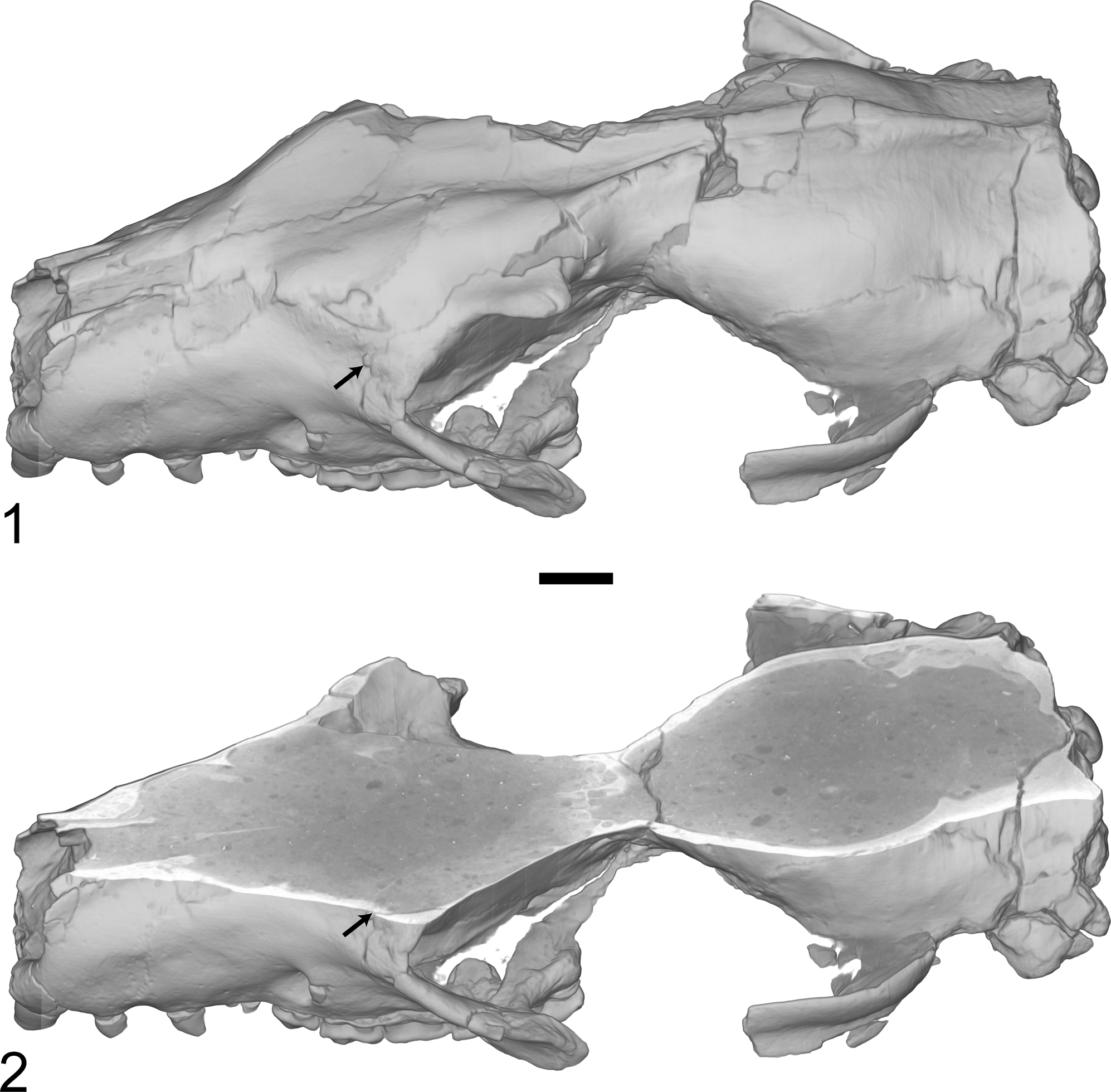
In lateral view the lacrimal is exposed in front of the orbit as a small facial process located between the frontal, maxilla, and jugal, but not contacting the nasals (Figs. 12, 14). In the orbit (Fig. 16), the lacrimal comprises the rostrodorsal corner of the wall and contacts the palatine, frontal, maxilla, and jugal. There is no clearly demarcated lacrimal process. A small pit is visible on the rostral aspect of the left lacrimal of UW 12362 (Fig. 17.1). Visualization of the CT data strongly suggests that this indentation is not perforate (Fig. 17.2), and there is no other evidence of an opening on the facial process of the frontal, demonstrating that this specimen did not have an extraorbital lacrimal foramen.
Oblique caudal-lateral view of the left orbital region of Microsyops annectens (UW 12362), lightly dusted with ammonium chloride. Dashed red lines indicate sutures between bones. Scale bar represents 5 mm. Abbreviations: ef = ethmoidal foramen; ft = frontal; iof = infraorbital foramen; ju = jugal; la = lacrimal; lf = lacrimal foramen; mx = maxilla; na = nasal; pa = parietal; pal = palatine; pmx = premaxilla; pop = postorbital process; sq = squamosal.

Reconstructions of Microsyops annectens (UW 12362) based on the high resolution CT data. The volume rendering in (1) was sliced in the transverse plane to reveal the interior (2). Black arrow (1) illustrates the presence of a pit on the lacrimal bone in the same location as the supposed extraorbital lacrimal foramen in AMNH 55284 (see discussion in text) and (2) demonstrates that the pit is not perforate, and therefore cannot be a lacrimal foramen. Scale bar represents 5 mm.
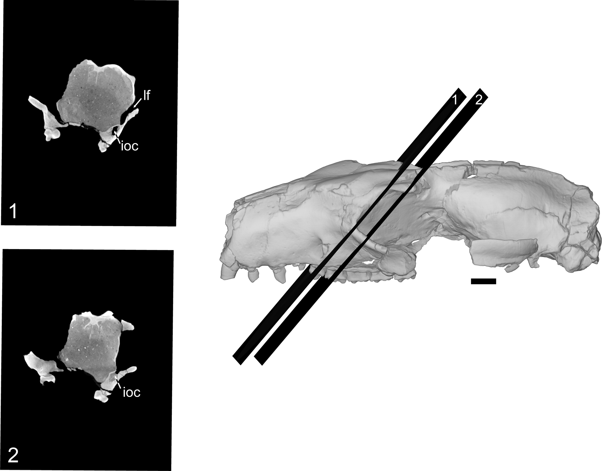
There are two foramina at the rostral end of the orbit in UW 12362 (Fig. 16): one in the maxilla, and the other composed of the lacrimal and jugal (laterally). When traced in the CT data, the more ventral (in the maxilla) of these openings (“iof” on Fig. 16) leads to the infraorbital foramen on the face via the infraorbital canal (Fig. 18.1, 18.2), while the more dorsal opening (between the lacrimal and jugal; “lf” on Fig. 16) extends into the nasal cavity (Fig. 18.1). As such, this second opening can be identified as a large intraorbital lacrimal foramen, which would have conveyed the nasolacrimal duct.
Lateral view of Microsyops annectens (UW 12362) generated as a volume rendering from the high resolution X-ray CT data, with oblique coronal slices showing (1) the opening of the lacrimal foramen (lf) in the orbit and its communication with the nasal cavity, with the infraorbital canal (ioc) running through the maxilla more ventrally; and (2) the opening of the infraorbital canal into the orbit. Scale bar represents 5 mm. Slices are at 75% scale relative to the cranial reconstruction.
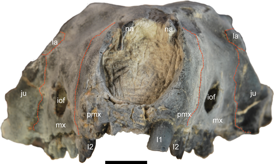
Rostral view of Microsyops annectens (UW 12362), lightly dusted with ammonium chloride. Dashed red lines indicate sutures between bones. Scale bar represents 5 mm. Abbreviations: I = incisor; iof = infraorbital foramen; ju = jugal; la = lacrimal; mx = maxilla; na = nasal; pmx = premaxilla.
Contrast between the internal structure of the mastoid processes in M. annectens (1) and an extant dermopteran (Cynocephalus volans; AMNH 16697) (2). (1) Reconstruction of the caudal portion of UW 12362 in ventral view from high resolution X-ray CT data showing the plane from which the slice given below the reconstruction was taken. Note that the data have been resliced relative to the original dataset to be in a more precisely coronal plane. (2) Reconstruction of the caudal portion of the cranium of modern dermopteran Cynocephalus volans (AMNH 16697) showing the plane of the slice given below the reconstruction. Note the solid mastoid processes and lack of pneumatisation of the lateral walls of the cranium in M. annectens compared to C. volans. Scale bars represent 5 mm.
Jugal
In UW 12362 the rostral and caudal ends of the left jugal are preserved (Fig. 11). In lateral view (Fig. 12), the jugal contacts the maxilla and lacrimal, contributing to the rostral and ventral aspects of the orbit. As noted previously, the jugal also contributes to the lateral wall of the lacrimal foramen (Fig. 16). While the location of the contact is not clearly visible, presumably the caudal end of the jugal contacts the zygomatic process of the squamosal. There is no evidence of a subsquamosal foramen on either the squamosal or the jugal (Fig. 12). The caudal end of the jugal preserved on the left side of UW 12362 preserves what appears to have been the base of a process on the ventral aspect of the bone (“pr?” in Fig. 12), which would have sat lateral to the glenoid fossa. Although it is unclear if the relevant part of the jugal is preserved on either side, in UW 12362 there is no evidence of a frontal process of the jugal that would have contributed to a postorbital bar.
Maxilla
In dorsal view, the maxilla contacts the premaxilla rostrally, the nasal dorsally, and the lacrimal caudally (Fig. 11). There is also evidence of a limited articulation between the frontal and maxilla (Fig. 11). In lateral view, contact with the jugal is also evident (Fig. 12). The maxilla forms the floor of the orbit, contacting the palatine, jugal, and lacrimal. The orbital process of the palatine and its contact with the lacrimal excludes the maxilla from contacting the frontal in the orbit (Fig. 16), which means that the maxilla does not contribute to the medial wall of the orbit (as previously observed for this specimen by Wible and Covert, Reference Wible and Covert1987). The large infraorbital canal extends from the orbit at the level of the caudal edge of M1 (Figs. 16, 18) to the infraorbital foramen on the snout (left ~ 7.3 mm2 at its opening; Fig. 19), which is located above the mesial edge of P4. Damage to the palate has made the ventral contacts of the maxilla difficult to ascertain, apart from the suture with the premaxilla (Fig. 10). Other than the incisive foramina, which sit on the premaxillary-maxillary suture, there are no other clearly demarcated foramina piercing the maxilla on its ventral aspect (Fig. 10).
Nasals
As is evident in dorsal view (Fig. 11), the nasals are broken rostrally in UW 12362, so it is not possible to ascertain whether or not they would have overhung the nasal cavity. They are relatively narrow rostrally at their contacts with the premaxillae in dorsal view, but begin to broaden distally at approximately the level of the facial opening of the infraorbital canal, ultimately forming a very broad suture with the frontals, and comprising a significant proportion of the snout (i.e., that part of the cranium that extends rostral to the zygomatic arch, to the end of the premaxilla; Fig. 11). The nasals are excluded dorsally from contact with the lacrimal by the contact between the maxilla and frontal (Fig. 11). In rostral view (Fig. 19), the nasals sit internal to both the premaxilla and the maxilla, curving over to meet at the midline, forming a barrel vault shape. As such, these bones are not as narrow as they appear in the dorsal view of the cranium, even at their rostral ends, because they are overlain by adjacent bones.
Premaxillae
In lateral view, the premaxillae are substantial bones, comprising approximately 1/3rd of the length of the snout (Figs. 12, 14). Their only contacts are with the nasals and the maxillae—although they do exhibit posterodorsal processes that extend distally along their contact with the nasals, these extend only to approximately the level of the distal margin of C1 (Figs. 12, 14), terminating far from the rostral end of the frontal. On the ventral aspect of the cranium the premaxillary-maxillary suture can be traced directly behind the I2 near the tip of the snout (Fig. 10)—as such, the premaxillae only contribute a small part of the palate (<20%). Large, separate incisive foramina (left ~8.4 mm2) occur on this suture ventrally.
Preservation
UW 12362 is substantively complete. The only parts of the cranium that are missing bilaterally are the ectotympanics, ear ossicles, lateral sections of the jugals, and the rostral portions of the nasals. Otherwise, all cranial bones are represented on at least one side. Elements missing unilaterally include the palatal process of the left maxilla, the caudal part of the left palatine, and the right, lateral orbital wall, including portions of the frontal, palatine, and orbitosphenoid. There is also more minor damage to features, such as the pterygoid plates, the ventral portions of which are missing. The specimen is largely undistorted, although the left side is deflected upwards slightly relative to the right side.
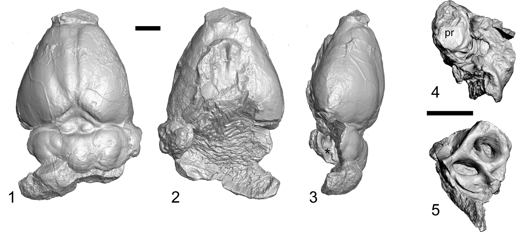
UW 14559 consists of two pieces (Fig. 4): a natural endocast preserved with fragments of the right auditory region, and a portion of the left petrosal (and likely some fused elements of the squamosal) that preserves the promontorium, much of the roof of the tympanic cavity, and aspects of the internal surface including the subarcuate fossa and foramina for cranial nerves VII and VIII.
Natural endocast and cranial fragments of Microsyops annectens (UW 14559), from UW locality V-78001 in the Blue Point Marker horizon, Carter Mountain, northwestern Wyoming, reconstructed from high resolution X-ray CT data. Natural endocast in (1) dorsal; (2) ventral; and (3) left lateral views. Asterisk in (3) is on the bone of the endocranial surface of the right petrosal, for orientation. Left petrosal/squamosal fragment in (4) ventral; and (5) endocranial views; “pr” in (4) indicates the promontorium. Scale bars represent 5 mm.
Comparisons
Comparisons were made to fossil specimens of Labidolemur kayi Simpson, Reference Simpson1929 (USNM 530208, 530221; UM 41869), Dryomomys szalayi Bloch et al., Reference Bloch, Silcox, Boyer and Sargis2007 (UM 41870), Tinimomys graybulliensis Szalay, Reference Szalay1974 (USNM 461201, USNM 461202), Ignacius graybullianus Bown and Rose, Reference Bown and Rose1976 (USNM 421608), Phenacolemur jepseni Simpson, Reference Simpson1955 (AMNH 48005), Carpolestes simpsoni Bloch and Gingerich, Reference Bloch and Gingerich1998 (UM 82670, 82688, 85177, 86273, 101923, 101963; USNM 482354), Plesiadapis tricuspidens Gervais, Reference Gervais1877 (MNHN CR-125), Plesiadapis cookei Jepsen, Reference Jepsen1930 (UM 87990), Microsyops knightensis (AMNH 55286), Megadelphus lundeliusi (AMNH 55284), and Palaechthon nacimienti Wilson and Szalay, Reference Wilson and Szalay1972 (UKMNH 9557). Extant taxa examined include Ptilocercus lowii Gray, Reference Gray1848 (USNM[MA] 291272, 488061; YPM 10179), Galeopterus variegatus (Audebert, Reference Audebert1799) (AMNH 106628; USNM[MA] 83276, 84421, and 307553; Silcox collection), and Cynocephalus volans (Linnaeus, Reference Linnaeus1758) (AMNH 203255; FMNH 56442; UF-M 3290; UMMZ 117122; AMNH 16697).
Remarks
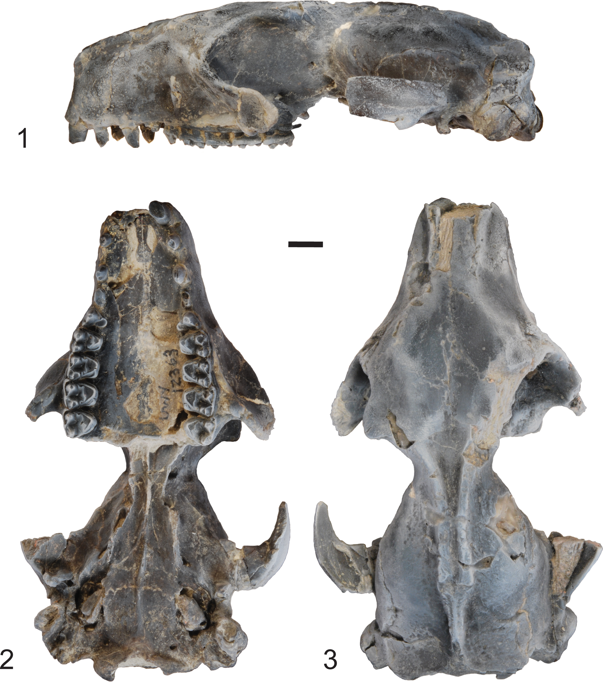
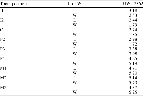
UW 12362 (Fig. 3) can be attributed to Microsyops annectens based on size (Table 1), and on the presence of upper molars with relatively bunodont cusps and well-defined, V-shaped centrocristae, following diagnoses from Szalay (Reference Szalay1969b) and Gunnell (Reference Gunnell1989). UW 14559 is attributed to M. annectens based on similarity in size and morphology to that of UW 12362. In particular, both specimens exhibit a strong mastoid process with a distinctive rounded crest (“cr” on Figs. 5, 6), as well as a promontorium that is somewhat deflated in appearance and has well-demarcated, posteromedially oriented (with respect to the basicranium) grooves for the internal carotid artery and for both stapedial and promontorial branches (Figs. 5, 6).
Cranium of Microsyops annectens (UW 12362) from UW locality V-78001 in the Blue Point Marker horizon, Carter Mountain, northwestern Wyoming in (1) left lateral; (2) ventral; and (3) dorsal views, lightly dusted with ammonium chloride. Scale bar represents 5 mm. Note that the specimen number transcribed on the specimen is incorrect.
Dental measurements of UW 12362 (Microsyops annectens). Values represent the average of measurements from the right and left sides when both are preserved. I = Incisor; C = Canine; P = Premolar; M = Molar; L = Length; W = Width.
Results
Microsyopidae.—
Microsyops knightensis AMNH 55286
This specimen was originally discussed by McKenna (Reference McKenna1966), and subsequently described and figured by Szalay (Reference Szalay1969b), MacPhee et al. (Reference MacPhee, Novacek and Storch1988), and Gunnell (Reference Gunnell1989). Although compressed dorsoventrally, the specimen includes much of the cranium caudal to the level of M1, and previous to the current account, represented the best-preserved microsyopid specimen with respect to the basicranium. In most respects, M. annectens is similar in its preserved anatomy to that of AMNH 55286. In dorsal view, the most obvious contrast is that AMNH 55286 bears only a single sagittal crest, which originates near the caudal end of the zygomatic arch (see McKenna, Reference McKenna1966, fig. 4), whereas the sagittal crest in UW 12362 extends farther rostrally, melding with the temporal lines, and is doubled for most of its length (Fig. 11). The two specimens also differ in the position of the foramina on the dorsal aspect of the braincase, which are located on the suture between the squamosal and parietal in M. annectens (Fig. 11), but pass directly through the parietal in AMNH 55286. Szalay (Reference Szalay1969b, p. 288) considered these foramina to be related to the venous system. Because they would have underlain the temporal muscle, they are interpreted here instead as having carried the rami temporales of the ramus superior of the stapedial artery following Wible (Reference Wible2008).
In ventral view, most aspects of the basicranial anatomy are similar, including the absence of a bulla or significant tympanic processes of the petrosal. As discussed previously, the rostral tympanic process of the petrosal sometimes (McKenna, Reference McKenna1966; Szalay, Reference Szalay1969b) has been interpreted as an attachment point for another bone, possibly forming a bulla in AMNH 55286, but as in UW 12362, there is no positive evidence in support of this interpretation. One apparent difference is the presence of a small basisphenoid tympanic process in UW 12362, which is not present bilaterally in AMNH 55286. Although the relevant area is broken on the right side in AMNH 55286, it is well preserved on the left, making it clear that this is a real contrast (as reported by MacPhee et al., Reference MacPhee, Novacek and Storch1988). Apart from this difference, our interpretation of the tympanic roof for UW 12362 is consistent with that portrayed for AMNH 55286 by MacPhee et al. (Reference MacPhee, Novacek and Storch1988, fig. 13).
In both specimens, there are grooves that run from medial to the lateral across the promontorium of the petrosal that are interpreted to mark the course of the promontorial and stapedial branches of the internal carotid artery. There is a contrast in the interpretation of AMNH 55286 between Szalay (Reference Szalay1969b) and MacPhee et al. (Reference MacPhee, Novacek and Storch1988) with respect to the presence or absence of a foramen for the ramus superior of the stapedial artery on the suture between the epitympanic wing of the sphenoid and the petrosal. This foramen is labelled by Szalay (Reference Szalay1969b, fig. 17; his “?fsst”), but is not indicated by MacPhee et al. (Reference MacPhee, Novacek and Storch1988, fig. 13). In AMNH 55286, this area is damaged on the left side of the skull, and missing entirely on the right. However, the identification of a similarly positioned foramen in UW 12362 (“frs” in Fig. 9) suggests that that Szalay (Reference Szalay1969b) may have been correct about the presence of a foramen in that area. However, Szalay (Reference Szalay1969b, p. 290) also reported the presence of a possible groove for the ramus posterior of the stapedial artery, absent in UW 12362, and not identified by MacPhee et al. (Reference MacPhee, Novacek and Storch1988). The presence of such a groove would be surprising, because when present that vessel generally does not leave a trace on the bone (Wible, Reference Wible1987). The location of the groove indicated by Szalay (Reference Szalay1969b, fig. 17; not labeled, but indicated with shading caudal to the fenestra cochleae [labeled “fer”]) is adjacent to the fenestra cochleae rather than the fenestra vestibuli (Szalay, Reference Szalay1969b, fig. 17), which is not consistent with an interpretation that it is associated with a branch of the stapedial artery. Our re-examination of AMNH 55286 found no evidence of a well-defined groove, making M. knightensis similar to UW 12362 in this respect, contra Szalay (Reference Szalay1969b). While Szalay (Reference Szalay1969b) also identified a foramen for a medial entocarotid artery, it has since been inferred that eutherians primitively lacked that vessel (Presley, Reference Presley1979; see also Cartmill and MacPhee, Reference Cartmill, MacPhee and Luckett1980; MacPhee, Reference MacPhee1981; Wible, Reference Wible1983; MacPhee and Cartmill, Reference MacPhee, Cartmill, Swisher and Erwin1986; Gunnell, Reference Gunnell1989), and we follow MacPhee and Cartmill (Reference MacPhee, Cartmill, Swisher and Erwin1986) and MacPhee et al. (Reference MacPhee, Novacek and Storch1988) in re-identifying the relevant opening as the cochlear canaliculus. In sum, to the extent that it can be deduced from bony structures, it appears as though the pattern of the arterial circulation that passed through the middle ear was identical in M. knightensis and M. annectens.
Oblique view of the left side of the basicranium of Microsyops annectens (UW 12362), lightly dusted with ammonium chloride. The specimen has been tipped rostromedially to allow for better visualization of the roof of the tympanic cavity and the cochlear canaliculus. Red dashed lines indicate sutures between bones. White dashed arrows indicate auditory tube (at) and glassierian fissure (gf). White dotted lines on the promontorium indicate the inferred pathway of the internal carotid artery and promontorial and stapedial branches. The white asterisk indicates the very small caudal tympanic process of the petrosal. Scale bar represents 5 mm. Abbreviations: acf = anterior carotid foramen; ap = tympanic process of the alisphenoid; as = alisphenoid; at = auditory tube; bo = basioccipital; bs = basisphenoid; bstp = basisphenoid tympanic process; cc = cochlear canaliculus; eg = entoglenoid process; er = epitympanic recess; ex = exoccipital; fc = fenestra cochleae; fi = fossa incudis; fm = foramen magnum; fo = foramen ovale; frs = foramen for the ramus superior of the stapedial artery; fv = fenestra vestibuli; gf = glaserian fissure; glf = glenoid fossa; hf = hypoglossal foramen; ips = inferior petrosal sinus; ma = mastoid; pa = parietal; pet = petrosal; pgf = postglenoid foramen; pgp = postglenoid process; plf = posterior lacerate (jugular) foramen; pr = promontorium; pt = pterygoid; rtpp = rostral tympanic process of the promontorium; sq = squamosal; sw = epitympanic wing of the sphenoid; vf = vidian foramen.

While Gunnell (Reference Gunnell1989) suggested that the tympanohyal was absent in M. knightensis (AMNH 55286) we note that the relevant area is broken away on the right side of this specimen, and the base of the process is present on the left, with only its tip broken away. In support of this interpretation, the new specimen of M. annectens (UW 12362) has a well-preserved tympanohyal (Fig. 10). This suggests that microsyopids had a facial nerve that would have exited the ear cavity via a stylomastoid foramen primitivum. Both M. annectens and M. knightensis have a shallow facial sulcus located lateral to the fenestrae cochleae and vestibuli supporting the inference that microsyopids had a facial nerve that passed through the middle ear (consistent with interpretation by MacPhee et al.1988; contra Szalay, Reference Szalay1969b).
Megadelphus lundeliusi AMNH 55284
This specimen was first discussed by McKenna (Reference McKenna1966), and subsequently described and figured by Szalay (Reference Szalay1969b) and Gunnell (Reference Gunnell1989). The cranium is notably larger than that of UW 12362 (estimated cranial length for AMNH 55284 = 105 mm [Szalay, Reference Szalay1969b] compared to cranial length as preserved in UW 12362 = 78.60). Although missing the ventral part of the braincase, including the ear region, AMNH 55284 is nearly completely preserved rostrally. In dorsal view, M. annectens (UW 12362; Fig. 11) has a sagittal crest that is lower and originates farther caudally than that of M. lundeliusi (McKenna, Reference McKenna1966, fig. 8). In both dorsal and lateral view the relationships among the bones making up the rostral aspect of the cranium are similar in M. lundeliusi and M. annectens. In particular, both have long snouts, with very large premaxillae that extend caudally by way of processes that pass beyond the level of the canine but do not reach the frontal (Fig. 14; Szalay, Reference Szalay1969b, fig. 18; note that the premaxilla is erroneously portrayed as missing this process in Szalay, Reference Szalay1969b, fig. 23). Both have a similar orbital mosaic, with a large orbital palatine process that contacts the lacrimal, excluding the frontal from contact with the maxilla (Fig. 12; Szalay, Reference Szalay1969b, fig. 18).
Szalay (Reference Szalay1969b) interpreted the lacrimal foramen of M. lundeliusi as being extraorbital likely based on the presence of a small pit (indicated, but not labelled in Szalay, Reference Szalay1969b, fig. 18) on the facial exposure of the lacrimal. A similarly located pit is visible on the left lacrimal of M. annectens (UW 12362; Fig. 17.1). However, visualization of CT data for M. annectens does not provide support for the interpretation that this indentation is perforate (Fig. 17.2). Although, in the absence of CT data for M. lundeliusi (AMNH 55284), it is unclear whether or not the relevant pit is more extensive in that specimen, other aspects of the anatomy suggest that M. lundeliusi was similar to M. annectens in having an intraorbital lacrimal foramen. Specifically, the lacrimal foramen is one of two foramina located at the rostral extent of the orbit in UW 12362. Two similar foramina, labeled as infraorbital foramina, were previously described in M. lundeliusi as occurring in the maxilla (Szalay, Reference Szalay1969b, fig. 19). Re-study of AMNH 55284 demonstrates that the more dorsal foramen is actually located closer to the dorsal aspect of the orbit than originally thought, and is located in the lacrimal rather than the maxilla, making it a likely candidate for the lacrimal foramen. As such, the morphology of AMNH 55284 is entirely consistent with that of UW 12362, suggesting that they both have an intraorbital lacrimal foramen.
Non-microsyopid plesiadapiforms
Of the 10 families of non-microsyopid plesiadapiforms (Silcox et al., Reference Silcox, Bloch, Boyer, Chester and López-Torres2017), four are known from reasonably complete crania: Plesiadapidae Trouessart, Reference Trouessart1897 (Russell, Reference Russell1959, Reference Russell1964; Gingerich, Reference Gingerich1976; Boyer, Reference Boyer2009; Boyer et al., Reference Boyer, Scott and Fox2012), Paromomyidae Simpson, Reference Simpson1940 (Szalay, Reference Szalay1972; Kay et al., Reference Kay, Thorington and Houde1990, Reference Kay, Thewissen and Yoder1992; Bloch and Silcox, Reference Bloch and Silcox2001; Silcox, Reference Silcox2003), Carpolestidae Simpson, Reference Simpson1935 (Bloch and Silcox, Reference Bloch and Silcox2006), and Micromomyidae Szalay, Reference Szalay1974 (Bloch et al., Reference Bloch, Chester and Silcox2016). More fragmentary remains are known for Palaechthonidae (Szalay, Reference Szalay1969b) (Kay and Cartmill, Reference Kay and Cartmill1977; Chester et al., Reference Chester, Williamson, Bloch, Silcox and Sargis2017, Reference Chester, Williamson, Bloch, Silcox and Sargis2019) and Picrodontidae Simpson, Reference Simpson1937 (Matthew, Reference Matthew1917), while there are no substantive cranial remains, apart from dentaries and some fragmentary maxillae, known for Saxonellidae (Russell, Reference Russell1964), Toliapinidae Hooker et al., Reference Hooker, Russell and Phélizon1999, Picromomyidae Rose and Bown, Reference Rose and Bown1996, or Purgatoriidae (Van Valen and Sloan, Reference Van Valen and Sloan1965).
Several basic similarities in the overall construction of the cranium of microsyopids are shared with other plesiadapiforms. In general, plesiadapiforms have skulls that show little cranial flexion and have relatively long snouts, particularly compared with those of euprimates, with micromomyids being the most extreme in inferred relative snout length (Bloch et al., Reference Bloch, Chester and Silcox2016, fig. 20). This configuration is associated with having relatively large premaxillae that house enlarged upper central incisors. Among plesiadapiforms, there is some variation in the mosaic of bones on the snout. Although plesiadapiforms generally have a posterodorsal process of the premaxilla that extends caudally (Figs. 12, 14), that process is relatively much longer in plesiadapids than in other taxa, extending caudally to contact the frontal (Gingerich, Reference Gingerich1976, fig. 33; Boyer et al., Reference Boyer, Scott and Fox2012). Plesiadapids and caropolestids differ from other plesiadapiforms (including M. annectens; Fig. 11) in having a narrow contact between the nasals and the frontals (Bloch et al., Reference Bloch, Chester and Silcox2016). Although M. annectens is similar to plesiadapids (and different from other plesiadapiforms) in lacking a lacrimal tubercle, it differs from all other non-microsyopid plesiadapiforms in having a fully intraorbital lacrimal foramen. As in Plesiadapis Gervais, Reference Gervais1877, though, the lacrimal of M. annectens has a substantial facial process. In other plesiadapiforms, this bone is confined to the orbital rim (e.g., Kay et al., Reference Kay, Thewissen and Yoder1992, fig. 5A; Bloch and Silcox, Reference Bloch and Silcox2006, fig. 3).
Unlike that of euprimates, all known plesiadapiforms had strong postorbital constrictions and relatively laterally oriented orbits, lacking complete postorbital bars. Microsyopids are distinct from other plesiadapiforms (with the possible exception of P. nacimienti; Kay and Cartmill, Reference Kay and Cartmill1977), however, in possessing postorbital processes of the frontal. In the absence of cranial specimens for more primitive microsyopids (but see White et al., Reference White, Bloch and Silcox2016), it remains unclear whether this trait evolved within the family, or is plesiomorphic (i.e., based on its shared presence in all living euarchontan groups). The well-preserved orbital mosaic of UW 12362 (Fig. 16) confirms a prior inference (based on Megadelphus lundeliusi; Szalay, Reference Szalay1969b, fig. 18) that microsyopids differ from other plesiadapiforms (i.e., Plesiadapis, Dryomomys Bloch et al., Reference Bloch, Silcox, Boyer and Sargis2007, Carpolestes Simpson, Reference Simpson1928, Ignacius Matthew and Granger, Reference Matthew and Granger1921) in lacking a significant maxillary contribution to the medial orbital wall, excluded by a large orbital process of the palatine that contacts the lacrimal (Wible and Covert, Reference Wible and Covert1987; Bloch et al., Reference Bloch, Chester and Silcox2016). This difference may be significant because euprimates are typically like non-microsyopid plesiadapiforms in having a maxillary contribution to the orbital wall (Wible and Covert, Reference Wible and Covert1987).
All known plesiadapiforms have sagittal crests, but there is variation in the point at which the temporal lines converge. For example, in the paromomyid Ignacius graybullianus, the temporal lines unite much more rostrally than that of M. annectens (e.g., compare Kay et al., Reference Kay, Thorington and Houde1990, fig. 1a to Fig. 1), although this configuration varies even among microsyopids. Where known (e.g., Carpolestes, Plesiadapis, Ignacius), plesiadapiforms also generally had nuchal crests, as in M. annectens, and foramina magnum that open caudally (Fig. 7), a configuration that is presumably associated with their relatively unflexed crania.
Like other plesiadapiforms (Kay and Cartmill, Reference Kay and Cartmill1977; Kay et al., Reference Kay, Thewissen and Yoder1992; Bloch and Silcox, Reference Bloch and Silcox2006; Boyer et al., Reference Boyer, Scott and Fox2012; Bloch et al., Reference Bloch, Chester and Silcox2016), M. anncectens had large infraorbital foramina. Microsyops annectens more closely resembles Plesiadapis than other plesiadapiforms in its relatively longer infraorbital canal. Those two taxa are also more similar to one another than either is to other plesiadapiforms in having a relatively long mesocranial region (Bloch and Silcox, Reference Bloch and Silcox2006; Bloch et al., Reference Bloch, Chester and Silcox2016), although the proportions of this region remain speculative in micromomyids since the relevant specimens are damaged.
There is considerable variation in the configuration of the basicranial region among plesiadapiforms. Perhaps the most marked contrast between microsyopids and other plesiadapiforms is the apparent absence of a bony auditory bulla in the former. Among non-microsyopid plesiadapiforms there are at least two different patterns in terms of the relationship between the bulla and the other bones of the basicranium. In paromomyids there is a well-demarcated suture on the tympanic roof between the petrosal and the bone making up the bulla (inferred to be the entotympanic; Kay et al., Reference Kay, Thorington and Houde1990, Reference Kay, Thewissen and Yoder1992; Bloch et al., Reference Bloch and Silcox2001; Silcox, Reference Silcox2003). This suture is absent in all other families known from adequate material, with apparent continuity between the bulla and petrosal in micromomyids, carpolestids, and plesiadapids (Russell, Reference Russell1959; Szalay, Reference Szalay1972; Szalay et al., Reference Szalay, Rosenberger and Dagosto1987; Bloch and Silcox, Reference Bloch and Silcox2006; Boyer, Reference Boyer2009; Bloch et al., Reference Bloch, Chester and Silcox2016). Leaving aside the complex question of whether or not this means that the bulla was partly or entirely petrosal in origin in those taxa (e.g., as discussed by MacPhee et al., Reference MacPhee, Cartmill and Gingerich1983; MacPhee and Cartmill, Reference MacPhee, Cartmill, Swisher and Erwin1986; Bloch and Silcox, Reference Bloch and Silcox2006; Boyer et al., Reference Boyer, Scott and Fox2012; Bloch et al., Reference Bloch, Chester and Silcox2016), this conformation does suggest that the caudal and rostral tympanic processes of the petrosal were enlarged over the condition in M. annectens, in which they are quite diminutive (Figs. 5, 9). Also lacking in M. annectens (and M. knightensis) are bony septae associated with the promontorium; this contrasts with the situation in all other plesiadapiforms, although there is some variation in how these septae are arranged (Szalay, Reference Szalay1972; Kay et al., Reference Kay, Thewissen and Yoder1992; Bloch and Silcox, Reference Bloch and Silcox2006; Boyer, Reference Boyer2009; Boyer et al., Reference Boyer, Scott and Fox2012; Bloch et al., Reference Bloch, Chester and Silcox2016).
The configuration of the well-demarcated grooves on the promontorium for the internal carotid artery and its promontorial and stapedial branches of M. annectens are similar to what would be expected in a primitive eutherian (see Wible, Reference Wible1987, fig. 1), with the internal carotid artery entering the ear posteromedially, and with both branches being substantial. Most other plesiadapiforms (paromomyids, micromomyids, and plesiadapids; Kay et al., Reference Kay, Thewissen and Yoder1992; Bloch et al., Reference Bloch, Chester and Silcox2016; but see Boyer et al., Reference Boyer, Scott and Fox2012) show evidence of reductions to the internal carotid circulation, based on the small size of the posterior carotid foramen and/or the narrowness of grooves or bony tubes associated with the intratympanic route of the internal carotid plexus and any remnants of the related arteries. The one exception to this generalization is Carpolestes simpsoni, which has grooves that are more substantial on the promontorium, consistent with the internal carotid artery being functional (Bloch and Silcox, Reference Bloch and Silcox2006). In spite of this one area of similarity, the ear regions of Carpolestes and M. annectens are profoundly different, with the former exhibiting a narrow central stem (also seen in paromomyids and plesiadapids; micromomyids are too damaged to assess adequately; Kay et al., Reference Kay, Thewissen and Yoder1992; Bloch and Silcox, Reference Bloch and Silcox2006; Bloch et al., Reference Bloch, Chester and Silcox2016) and (uniquely among plesiadapiforms) an apparent division in the tympanic cavity proper into two chambers (Bloch and Silcox, Reference Bloch and Silcox2006). Microsyops annectens also lacks the distinctive lateral entrance of the internal carotid nerves to the tympanic cavity observed in paromomyids (Kay et al., Reference Kay, Thewissen and Yoder1992) and plesiadapids (Boyer et al., Reference Boyer, Scott and Fox2012). While M. annectens has a large mastoid process, it lacks the marked inflation of the squamosal observed in the micromomyid Dryomomys szalayi (Bloch et al., Reference Bloch, Chester and Silcox2016). Microsyops annectens is similar to micromomyids in the inferred pathway of the facial nerve through the middle ear in an open sulcus, although differing in that its exit would have been through a stylomastoid foramen primitivum rather than a stylomastoid foramen definitivum (sensu MacPhee, Reference MacPhee1981), based on the inferred absence of a bony bulla. By contrast, paromomyids, plesiadapids, and carpolestids (along with euprimates) have been interpreted as having the facial nerve enclosed to at least some degree in a bony canal (Kay et al., Reference Kay, Thewissen and Yoder1992; Bloch and Silcox, Reference Bloch and Silcox2006; Boyer et al., Reference Boyer, Scott and Fox2012).
In summary, the anatomy of the basicranium of Microsyops annectens shows that it lacked essentially all of the distinctive, presumably derived features observed in other plesiadapiform taxa. As such, this region sheds little light on the relationships of microsyopids to other plesiadapiform families.
Apatemyidae
In order to assess the polarity of cranial traits in microsyopids, it is necessary to seek out one or more taxa that could serve as relevant outgroups. One obvious choice might be leptictids, a group known from extensive and comprehensively described (Novacek, Reference Novacek1986) cranial fossils. However, recent phylogenetic analyses (e.g., O'Leary et al., Reference O'Leary, Bloch, Flynn, Gaudin, Giallombardo, Giannini, Goldberg, Kraatz, Luo, Meng, Ni, Novaceck, Perini, Randall, Rougier, Sargis, Silcox, Simmons, Spaulding, Velazco, Weksler, Wible and Cirranello2013) have found that leptictids are very distantly related to Euarchonta, suggesting that they may not be the best option. Also, the best-preserved fossils (Novacek, Reference Novacek1986) for the family belong to the Oligocene aged Leptictis dakotensis Leidy, Reference Leidy1868, and as such are notably more recent than UW 12362, separated in time from their last common ancestor by tens of millions of years.
An alternative is Apatemyidae Matthew, Reference Matthew1909, a group with a long history of tentative associations with Primates specifically or euarchontans more generally (reviewed by McKenna, Reference McKenna1963). Silcox et al. (Reference Silcox, Bloch, Boyer and Houde2010b) suggested that they were euarchontoglirans with weak support for a closer relationship to Glires Linnaeus, Reference Linnaeus1758 than to Euarchonta Waddell et al., Reference Waddell, Okada and Hasegawa1999. As possible primitive euarchontoglirans, with well-preserved cranial fossils known from the Paleocene and Eocene (Matthew, Reference Matthew1921; Jepsen, Reference Jepsen1934; Koenigswald, Reference Koenigswald1990; Koenigswald et al., Reference Koenigswald, Ruf and Gingerich2009; Silcox et al., Reference Silcox, Bloch, Boyer and Houde2010b), apatemyids are potentially valuable for outgroup comparisons.
The discussion below focusses on Labidolemur kayi, the oldest and most primitive member of the Apatemyidae for which cranial fossils are known. It is known from cranial fossils from the late Paleocene and early Eocene of Wyoming (Silcox et al., Reference Silcox, Bloch, Boyer and Houde2010b). Like plesiadapiforms, L. kayi has a long, relatively unflexed cranium with large premaxillae that house enlarged incisors, and large infraorbital foramina. The conformation of bones on the snout is different in L. kayi than in M. annectens, with the caudal end of the nasals being narrow, and the presence of a contact between the premaxilla and the frontals. Like M. annectens (but in contrast with that of other plesiadapiform families) the lacrimal foramen is intraorbital. The orbital mosaic is not well preserved in any specimen of L. kayi, but the position of the inferred fronto-maxillary suture suggests that the palatine was not expanded in the orbit (Silcox et al., Reference Silcox, Bloch, Boyer and Houde2010b, fig. 16), although it also suggests that there was not a substantial contribution from the maxilla to the medial orbital wall, implying that it was somewhat different from M. annectens, but also from other plesiadapiforms.
In the ear region, there are numerous general anatomical similarities between L. kayi and M. annectens, including the presence of a broad central stem, the likely absence of a bony auditory bulla, the presence of transpromontorial grooves for the promotontorial and stapedial branches coming from an internal carotid artery that entered the ear posteromedially, and a foramen faciale that opened into the ear cavity, implying a passage for the facial nerve that was not contained in a bony tube. These similarities support the inference that the similar traits in M. annectens were primitive for Primates or Euarchonta (or possibly even Euarchontoglires). Interestingly, the rostral and caudal tympanic processes of the petrosal are actually better developed in L. kayi than in M. annectens. They form part of a “rim” around the tympanic cavity, to which a tympanic process of the basisphenoid also contributes, extending more ventrally than the small basisphenoid tympanic process in M. annectens. This “rim” provides a clearer location for the support of a covering of the tympanic cavity (whether it be bony or cartilaginous) than anything preserved in M. annectens, highlighting how little evidence there is for any articulation with a bony bulla in the latter.
There are some differences between the basicranial morphology of L. kayi and M. annectens. In particular, the route of the ramus inferior of the stapedial artery is inferred to be different, with it passing medial to the glaserian fissure in L. kayi (rather than in that fissure), and with the ramus infraorbitalis of the ramus superior of the stapedial artery passing through an alisphenoid canal, a feature that is absent from M. annectens. The foramen for the ramus superior of the stapedial artery is also somewhat differently positioned, passing through the tegmen tympani in L. kayi, rather than through a remnant of the piriform fenestra. Lastly, L. kayi lacks a distinct foramen rotundum, making M. annectens more like plesiadapoid plesiadapiforms (Russell, Reference Russell1964; Bloch and Silcox, Reference Bloch and Silcox2006) than apatemyids in this feature. These differences highlight the lack of clearly derived similarities between apatemyids and microsyopids (Silcox et al., Reference Silcox, Bloch, Boyer and Houde2010b)
Dermoptera
Szalay et al. (Reference Szalay, Rosenberger and Dagosto1987) suggested that Microsyopidae might have a special relationship to Dermoptera, citing three proposed similarities in the cranium. The first was based on inferred similarities in the composition of the tympanic floor, with these authors suggesting that, not only was a bulla present in microsyopids, but that it was partially composed of a rostral entotympanic as in dermopterans. As discussed earlier, the record of microsyopid crania known to date fails to add any positive evidence that members of this family possessed a bony bulla, and the supposed articulation for the bulla may be better interpreted as a low rostral tympanic process of the petrosal, to which nothing bony need have attached. As MacPhee et al. (Reference MacPhee, Cartmill and Rose1989, p. 344) noted, in the “absence of any decisive evidence about the nature of the tympanic floor in microsyopids” it is not clear how one would “infer that a specific bone (rostral entotympanic) was present.” Further, there is a common developmental pattern for the entotympanic in dermopterans and scandentians (Wible and Martin, Reference Wible, Martin and MacPhee1993), so similarities to microsyopids with regards to a possible entotympanic would not necessarily signal a special relationship to Dermoptera alone.
The two other features listed by Szalay et al. (Reference Szalay, Rosenberger and Dagosto1987, p. 88) include “a flat and circular expansion of the petrosal, and evidence of the squamosal air spaces.” These two features are related in dermopterans, and are both associated with the very highly pneumatized nature of the dermopteran cranium, with the large mastoid process containing air spaces that are continuous with the highly pneumatized lateral walls of the caudal part of the braincase (Fig. 20.2; MacPhee et al., Reference MacPhee, Cartmill and Rose1989, fig. 8). Indeed, MacPhee et al. (Reference MacPhee, Cartmill and Rose1989) argued that the “mastoid” of dermopterans is developed as a posterior expansion of the epitympanic-paratympanic cavity, making it likely non-homologous with other mastoid processes, which develop from a separate locus of pneumatization centered around the posterior semicircular canal. Visualization of CT data reveals that Microsyops annectens completely lacks any pneumatization of the caudal cranium, with the mastoid itself appearing solid, and there being no air spaces extending from it to the lateral walls of the neurocranium (Fig. 20.1; the absence of such air spaces was also inferred from this specimen by Wible and Covert, Reference Wible and Covert1987). Because there is no evidence for an epitympanic-paratympanic cavity in microsyopids, it seems likely that the mastoid process, although phenetically similar, derived from a different developmental origin. Similarly, it is not clear on what basis Szalay et al. (Reference Szalay, Rosenberger and Dagosto1987) inferred the presence of “squamosal air spaces” in microsyopids. There is no evidence for such air spaces in the CT data for M. annectens (Fig. 20.1).
Dermopteran crania are highly specialized in several other respects, including the presence of a tubular external acoustic meatus formed by the ectotympanic (Hunt and Korth, Reference Hunt and Korth1980), the complete involution of the internal carotid artery during ontogeny (Wible, Reference Wible1993), the absence of the postglenoid vein (Wible, Reference Wible1993), and the absence of the tegmen tympani, so that the petrosal makes little contribution to the tympanic roof (Wible and Covert, Reference Wible and Covert1987). In the absence of any known ectotympanics for microsyopids, it is unclear whether or not they possessed an ectotympanic external acoustic meatus. However, the presence of grooves on the promontorium for the stapedial and promontorial arteries (Figs. 5, 6, 9), the presence of a well-developed postglenoid foramen that is continuous with the endocranial space (Fig. 8, slice 267), and the presence of an expansive petrosal contribution to the tympanic roof (Figs. 5, 9; as also observed by Wible and Covert, Reference Wible and Covert1987) suggest that the derived traits of the dermopteran cranial circulation and tympanic roof are not shared by Microsyops annectens (or any other member of the family known from relevant material).
Microsyopids are similar to dermopterans in having a blunt postorbital process of the frontal (Fig. 11; Hunt and Korth, Reference Hunt and Korth1980, fig. 3). In dermopterans, there is also a frontal process of the jugal, extending towards the postorbital process of the frontal. The relevant region is not preserved in UW 12362 (Fig. 11), nor in most other partial microsyopid crania (e.g., Megadelphus lundeliusi AMNH 55284 [McKenna, Reference McKenna1966, fig. 8]). However, the relevant region is preserved in Microsyops knightensis (AMNH 55286; McKenna, Reference McKenna1966, fig. 4), and although damaged, the jugal orbital process appears to be absent (also as portrayed by Szalay, Reference Szalay1969b, fig. 16). This suggests that the orbit would not have been as extensively ringed by bone in microsyopids as it is in dermopterans. There are other superficial differences in the shape of the cranium, including the presence, in dermopterans, of two distinct crests extending from the postorbital processes that generally remain separate, although the degree to which they approach one another varies between the two genera of dermopterans (Stafford and Szalay, Reference Stafford and Szalay2000, fig. 4). In both cases, however, there is a clear contrast with the distinct sagittal crest of microsyopids. Both genera of dermopterans also have relatively wide, evenly rounded rostral palates (Stafford and Szalay, Reference Stafford and Szalay2000, fig. 4), which contrast with the narrower rostral portion of the palate in M. annectens (Fig. 10), a feature that is likely associated with the loss of I1 in dermopterans. Also likely associated with the loss of I1, the premaxilla forms a smaller portion of the facial skeleton in dermopterans, and lacks the posterodorsal process of the premaxilla that is evident in M. annectens (Fig. 12; also seen in Megadelphus lundelusi [Szalay, Reference Szalay1969b, fig. 18]). Therefore, although similar in the presence of the postorbital process, the overall gestalt of the cranium is quite different.
It is worth noting, however, that with the possible exception of the shape and proportions of the premaxilla, most of the cranial features that differ between dermopterans and microsyopids represent autapomorphous specializations on the part of Dermoptera. As such, their absence in Microsyopidae would not rule this family out of a position as a stem member of Dermoptera. The only clear candidate for a character supporting such a relationship, however, is the postorbital process of the frontal, which is a trait that appears many times in mammalian evolution (e.g., megachiropterans, artiodactyls), and one that is present as part of the postorbital bar in both other living groups of euarchontans.
Scandentia
Scandentians possess a well-developed entotympanic bulla that is continuous with the largely entotympanic roof of the tympanic cavity (Wible, Reference Wible2011). They possess bony tubes for the stapedial and promontorial arteries, which are formed from a combination of the entotympanic and petrosal, with Tupaia Raffles, Reference Raffles1821 and Ptilocercus differing in the degree to which the two bones contribute (Wible, Reference Wible2011). The facial nerve is enclosed in a bony tube formed from the petrosal, and there is an expanded caudal process of this bone, which forms an expanded tegmen tympani that rooves the entire ossicular chain (Wible and Covert, Reference Wible and Covert1987). In all these ways, scandentians differ from Microsyops annectens. It is worth considering, however, to what degree the differences between the two groups rest on the presence in scandentians of a well-developed entotympanic—that is, if this bone were missing, how similar would the two groups appear? Interestingly, the main contrasts that remain (i.e., partial bony tubes for the branches of the internal carotid artery, petrosal enclosure of the facial nerve, expanded tegmen tympani and caudal tympanic process of the petrosal) are precisely those features that have been identified as candidate synapomorphies for a euprimate-scandentian clade (Wible and Covert, Reference Wible and Covert1987). As such, if microsyopids are primates, then the absence of these traits in the family implies that these features must have developed in parallel in Scandentia and Euprimates Hoffstetter, Reference Hoffstetter1977. Although this may seem unlikely, the support found in large scale analyses (e.g., Nie et al., Reference Nie, Fu, O'Brien, Wang, Su, Tanomtong, Volobouev, Ferguson-Smith and Yang2008; O'Leary et al., Reference O'Leary, Bloch, Flynn, Gaudin, Giallombardo, Giannini, Goldberg, Kraatz, Luo, Meng, Ni, Novaceck, Perini, Randall, Rougier, Sargis, Silcox, Simmons, Spaulding, Velazco, Weksler, Wible and Cirranello2013) for a monophyletic Sundatheria Olson et al., Reference Olson, Sargis and Martin2005 (Scandentia + Dermoptera) that excludes euprimates, or for a closer relationship of Dermoptera to Primates than Scandentia (e.g., Janečka et al., Reference Janečka, Miller, Pringle, Wiens, Zitzmann, Helgen, Springer and Murphy2007; Li and Ni, Reference Li and Ni2016; Mason et al., Reference Mason, Li, Minx, Schmitz, Churakov, Doronina, Melin, Dominy, Lim, Springer and Wilson2016; Asher et al., Reference Asher, Smith, Rankin and Emry2019) would also imply independent evolution of these features in Scandentia and Euprimates, as they are absent in dermopterans.
There are few candidate features for shared derived traits between microsyopids and scandentians. Both groups have postorbital processes of the frontal (Fig. 11), but unlike the short, blunt, and incomplete process of M. annectens, the process is narrower and joins an orbital process of the jugal in scandentians to form a complete postorbital bar (Wible, Reference Wible2011, fig. 2). One similarity shared by M. annectens and Scandentia is the presence of the posterodorsal process of the premaxilla (Fig. 12; Wible, Reference Wible2011, fig. 3)—as a result, the configuration of the premaxilla in UW 12362 is actually more similar to that in Scandentia than Dermoptera. With the possible exception of this trait, however, there is no compelling cranial evidence to link microsyopids to scandentians.
Assessment of cranial characters in a phylogenetic context
As noted above, UW 12362 was included in the coding of Microsyopidae by Silcox et al. (Reference Silcox, Bloch, Boyer and Houde2010b), producing a data matrix that was subsequently modified by Bloch et al. (Reference Bloch, Chester and Silcox2016). In order to consider the relevance of the cranial partition to the results of that analysis, the cranial characters from the Bloch et al. (Reference Bloch, Chester and Silcox2016; numbers 69–113) matrix were assessed in Mesquite version 3.6 (build 917; Maddison and Maddison, Reference Maddison and Maddison2018) using the “trace character history option” under “Parsimony Ancestral States.” Microsyopids have 20 character states that are reconstructed as being primitive for all the included taxa, and four character states that are reconstructed as possible synapomorphies for Microsyopidae, although all four traits exhibit extensive homoplasy throughout the phylogeny. The point at which the microsyopid state evolved is ambiguous for 17 characters; of these, microsyopids have a state that may be a primate synapomorphy for five (characters 78, 79, 88, 91, and 93), although all these states also appear in other, non-primate groups. There are two traits for which the microsyopid state is reconstructed as primitive for Euarchontoglires: character 69 [1: moderate snout length] and character 95 [1: pathway for the ramus inferior of the stapedial artery location within Glaserian fissure with chorda tympani nerve].
In sum, there are no unambiguous synapomorphies in the cranial anatomy of microsyopids for Primates or Euarchonta. Of the two candidate euarchontogliran synapomorphies, for character 69 the relevant trait appears in various other taxa (i.e., leptictids and some hedgehogs), and for character 95, the state for microsyopids is being tentatively inferred from indirect data. This is not to say that the inclusion of Microsyopidae is unimportant to the Bloch et al. (Reference Bloch, Chester and Silcox2016) analysis—indeed when they are excluded, the inferred relationships among Euarchontoglires changes (with apatemyids and Glires being closer to Primates than are either Scandentia or Dermoptera). But the cranial partition is not the source of Microsyopidae's influence over the results of the analysis.
Discussion and conclusions
The current paper provides the first comprehensive description of UW 12362, a specimen of M. annectens discovered in 1978. It is certainly the best-preserved cranium known for a microsyopid, and arguably the best cranium available for any plesiadapiform. Some isolated details of the external cranial anatomy of UW 12362 have been mentioned in previous works (Wible and Covert, Reference Wible and Covert1987; MacPhee et al., Reference MacPhee, Novacek and Storch1988, Reference MacPhee, Cartmill and Rose1989), but prior to this paper, only the endocranial anatomy had been described in detail (Silcox et al., Reference Silcox, Benham and Bloch2010a). The new fossil is significant in a number of ways, providing a nearly complete picture of the sutural contacts between bones of the cranium, and new perspective on some issues of anatomy that had previously been unclear (e.g., the position of the lacrimal foramen).
The cranial details of UW 12362 fail to provide any clearer resolution to the question of the broader relationships of Microsyopidae within Euarchonta, or even Mammalia. There are no clear cranial synapomorphies shared by microsyopids, euprimates, and other plesiadapiforms, so if all of these groups are members of a monophyletic Primates (as several analyses have suggested; Bloch et al., Reference Bloch, Silcox, Boyer and Sargis2007; Silcox et al., Reference Silcox, Bloch, Boyer and Houde2010b; Chester et al., Reference Chester, Williamson, Bloch, Silcox and Sargis2017; Fig. 2), then the implication is that there are no cranial synapomorphies for Primates sensu lato. This same conclusion has been reached by previous authors who referenced UW 12362. For example, MacPhee et al. (Reference MacPhee, Novacek and Storch1988, p. 37) wrote that “If the dental traits apparently shared by Microsyops and (other) primates are truly synapomorphies…then the primate basicranial morphotype is…not distinguishable from that of the basal eutherian.” Wible and Covert (Reference Wible and Covert1987, p. 18) also likened microsyopids to “the pattern reconstructed for primitive eutherians…” The one proposed alternative based on cranial data, linking microsyopids to dermopterans to the exclusion of Primates (Szalay et al., Reference Szalay, Rosenberger and Dagosto1987), is supported by little beyond the presence of a blunt postorbital process of the frontal, a trait that is of questionable value in light of the presence of postorbital processes (albeit modified into postorbital bars) in Scandentia and Euprimates, and in other mammals (e.g., Carnivora Bowditch, Reference Bowditch1821, Artiodactyla Owen, Reference Owen1848). The lack of support for a sister taxon relationship between Scandentia and Euprimates to the exclusion of Dermoptera in large-scale analyses that include molecular data (e.g., Janečka et al., Reference Janečka, Miller, Pringle, Wiens, Zitzmann, Helgen, Springer and Murphy2007; O'Leary et al., Reference O'Leary, Bloch, Flynn, Gaudin, Giallombardo, Giannini, Goldberg, Kraatz, Luo, Meng, Ni, Novaceck, Perini, Randall, Rougier, Sargis, Silcox, Simmons, Spaulding, Velazco, Weksler, Wible and Cirranello2013; Mason et al., Reference Mason, Li, Minx, Schmitz, Churakov, Doronina, Melin, Dominy, Lim, Springer and Wilson2016; Asher et al., Reference Asher, Smith, Rankin and Emry2019) would imply that the complete postorbital bar observed in Euprimates and Scandentia likely arose independently, as did other proposed (e.g., by Wible and Covert, Reference Wible and Covert1987) primate-scandentian cranial synapomorphies, such as petrosal contributions to the tubes for the facial nerve and stapedial artery.
Prior to 1990, conceptions of cranial anatomy in plesiadapiforms were mostly limited to Plesiadapis (Russell, Reference Russell1959, Reference Russell1964; Gingerich, Reference Gingerich1976) and microsyopids (McKenna, Reference McKenna1966; Szalay, Reference Szalay1969b), with other specimens that were described (e.g., Szalay, Reference Szalay1972; MacPhee et al., Reference MacPhee, Cartmill and Gingerich1983) being incomplete in ways that made them difficult to interpret correctly (see discussions in Kay et al., Reference Kay, Thewissen and Yoder1992; Silcox, Reference Silcox2001). However, since the publication of a well-preserved cranium of Ignacius graybullianus in 1990 (Kay et al., Reference Kay, Thorington and Houde1990, Reference Kay, Thewissen and Yoder1992), there has been considerable improvement in our knowledge of the cranial anatomy of plesiadapiforms (Bloch and Silcox, Reference Bloch and Silcox2006; Boyer, Reference Boyer2009; Boyer et al., Reference Boyer, Silcox, Bloch, Coleman and Dobrota2011, Reference Boyer, Scott and Fox2012; Bloch et al., Reference Bloch, Chester and Silcox2016; White et al., Reference White, Bloch and Silcox2016). The over-riding story of this expanded sample is one of variety rather than uniformity. Although there are some traits that may link some plesiadapiforms to primates, such as the contribution of the maxilla to the orbital wall (Wible and Covert, Reference Wible and Covert1987) and the possible petrosal contributions to the bulla in non-microsyopid plesiadapiforms (Russell, Reference Russell1959; Bloch and Silcox, Reference Bloch and Silcox2006; Bloch et al., Reference Bloch, Chester and Silcox2016), plesiadapiform families seem to have developed unique, presumably autapomorphous features of their own, such as the highly pneumatized mastoid and squamosal of micromomyids (Bloch et al., Reference Bloch, Chester and Silcox2016) and the apparently doubled tympanic cavity of carpolestids (Bloch et al., Reference Bloch and Silcox2006).
What does all this mean for understanding the relationships of microsyopids? Simply put, cranial anatomy generally, and basicranial anatomy specifically, should not be treated as the touchstone upon which such understanding must be sought. This is not to suggest that cranial anatomy is not important to an understanding of the phylogenetic relationships of these groups. However, as with any anatomical region, the pace of evolutionary change needs to be considered in how data are used to understand questions of inter-relationships among groups. If microsyopids are euarchontans, then their generally primitive basicranial anatomy means that little evolutionary change occurred in this region in the stem lineage of Euarchonta (or of Euarchontoglires or Boreoeutheria). In contrast, within Euarchonta, the high degree of variation and homoplasy revealed by our growing understanding of plesiadapiform cranial anatomy, and our revised perspectives on the cranial traits shared by Scandentia and Euprimates, suggest that within the group there was a considerable amount of homoplastic and autapomorphous change shaping cranial evolution.
These perspectives highlight the importance of an integrated approach to phylogenetic inference that includes cranial anatomy along with dental and postcranial traits (and molecular data when available). In particular, for microsyopids, tentatively associated postcranial materials (Szalay and Drawhorn, Reference Szalay, Drawhorn and Luckett1980; Bloch et al., Reference Bloch, Chester and Holroyd2015) have suggested possible dermopteran affinities for the group. More concrete conclusions on microsyopid affinities may need to await discovery (or publication) of clearly dentally associated postcranial remains.
Acknowledgments
UW 12362 was scanned by T. Ryan. Photographs in Figures 3, 5, 7, 9–14, 16, and 19 were taken by Z. Randall. The endocast of UW 12362 (pictured in Fig. 15) was segmented by A.E. Benham. AMNH 16697 (pictured in Fig. 20) was CT scanned by K. Selig. Thanks to A. Walker, P. Halleck, A. Grader, and T. Ryan (Penn State, Center for Quantitative Imaging) and J. Gladman and D. Boyer (Duke University) for help with CT scanning; and M. Novacek, J. Meng (American Museum of Natural History), and M. Cassiliano (University of Wyoming) for access to material. Thanks to J. Wible and an anonymous reviewer for comments that substantively improved this paper. This research was supported by NSF research grants BCS-0003920 to A. Walker, EF-0629836 to JIB, MTS, and E. Sargis, BCS-1440558 to JIB, and NSERC discovery grants to MTS.
This work is dedicated to the memory of our coauthor G. F. Gunnell, a great paleontologist, and the kindest and most generous of colleagues. Without a doubt, in his lifetime, Gregg forgot more about microsyopids than we will ever know.



