INTRODUCTION
Toxoplasma gondii infections are widely prevalent in human beings and other animals worldwide (Dubey and Beattie, Reference Dubey and Beattie1988). Humans become infected post-natally by ingesting tissue cysts from undercooked meat, consuming food or drink contaminated with oocysts, or by accidentally ingesting oocysts from the environment. However, only a small percentage of exposed adult humans develop clinical signs.
Most T. gondii isolates from human and animal sources in Northern America and Europe have been grouped into 1 of 3 clonal lineages by multi-locus enzymes electrophoresis, PCR-RFLP and microsatellite typing (Dardé et al. Reference Dardé, Bouteille and Perstreal1992; Howe and Sibley, Reference Howe and Sibley1995; Ajzenberg et al. Reference Ajzenberg, Bañuls, Tibayrenc and Dardé2002a, Reference Ajzenberg, Cogné, Paris, Bessières, Thulliez, Filisetti, Pelloux, Marty and Dardéb). Even though the difference among these 3 genotypes at the genome sequence level is less than 1%, they have strikingly different virulence phenotypes in mice, with type I strains uniformly lethal in outbred mice. In contrast, type II and III strains are significantly less virulent (Sibley and Boothroyd, Reference Sibley and Boothroyd1992). It is of great interest to investigate whether the genotypes are related to the disease manifestations in human toxoplasmosis (Boothroyd and Grigg, Reference Boothroyd and Grigg2002). Some studies reported that type I strains were more frequently associated in congenital and ocular toxoplasmosis in human patients based on one marker at the SAG2 locus or a very limited number of genetic loci (Howe et al. Reference Howe, Honoré, Derouin and Sibley1997; Fuentes et al. Reference Fuentes, Rubio, Ramírez and Alvar2001; Grigg et al. Reference Grigg, Ganatra, Boothrooyd and Margolis2001; Aspinall et al. Reference Aspinall, Guy, Roberts, Joynson, Hyde and Sims2003; Kahn et al. Reference Khan, Su, German, Storch, Clifford and Sibley2005; Vallochi et al. Reference Vallochi, Muccioli, Martins, Silveira, Belfort and Rizzo2005). Severe toxoplasmosis was reported in patients from French Guiana in association with atypical isolates of T. gondii (Dardé et al. Reference Dardé, Villena, Pinon and Beguinot1998; Carme et al. Reference Carme, Bissuel, Ajzenberg, Bouyne, Aznar, Demar, Bichat, Louvel, Bourbigot, Peneau, Neron and Dardé2002). However, we must be cautious about the interpretation of these studies, as the genetic markers used have limited power to distinguish parasite isolates (Su et al. Reference Su, Zhang and Dubey2006). For those T. gondii isolates that do belong to the same clonal lineage, biological information obtained from one representative may successfully predict the others because of their identical genetic background. The same may not be true for strains with different genetic makeup, particularly when frequent genetic crosses occur in the population. In such cases, there is low linkage disequilibrium and the association of genotypes with phenotypes is diminished (Su et al. Reference Su, Zhang and Dubey2006). One also needs to consider that genetic characterization of T. gondii isolates from humans is limited to isolates from patients ill with toxoplasmosis. To our knowledge there is no report of the genetic diversity of T. gondii isolates circulating in the general human population.
Recent studies on T. gondii populations in the animal populations started to reveal the diversity of the parasite. We have recently found that the isolates of T. gondii from Brazil are biologically and genetically different from those in North America and Europe (Dubey et al. Reference Dubey, Graham, Blackston, Lehmann, Gennari, Ragozo, Nishi, Shen, Kwok, Hill and Thulliez2002, Reference Dubey, Cortés Vecino, Vargas-Duarte, Sundar, Velmurugan, Bandini, Polo, Zambrano, Mora, Kwok, Smith and Su2007a; Lehmann et al. Reference Lehmann, Marcet, Graham, Dahl and Dubey2006). Toxoplasma gondii isolates from asymptomatic chickens from Brazil were more pathogenic to mice than isolates from Europe or North America, irrespective of the genotype (Dubey et al. Reference Dubey, Gennari, Labruna, Camargo, Vianna, Marcet and Lehmann2006a). Additionally, most isolates from chickens from Brazil were not clonal (Dubey et al. Reference Dubey, Cortés Vecino, Vargas-Duarte, Sundar, Velmurugan, Bandini, Polo, Zambrano, Mora, Kwok, Smith and Su2007a). These worldwide studies in free-range chickens are important since chickens become infected mostly by feeding from ground contaminated with oocysts, and thus prevalence of T. gondii in chickens is a good indicator of the strains prevalent in their environment (Ruiz and Frenkel, Reference Ruiz and Frenkel1980).
In the present study, we attempted to isolate and genotype T. gondii from chickens from Guyana, South America and compare the results with those obtained from other countries in South America.
MATERIALS AND METHODS
Naturally-infected chickens
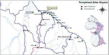
During June, July, and August 2006 a total of 76 free-range chickens were obtained in 6 batches from 17 properties from various locations on coastal Guyana, with 2 collections being made 32 km (Soesdyke) and 92 km (Wismar) south of Georgetown, the latter being out of the coastal zone. Collection sites were separated from each other by a minimum of 6 km (Fig. 1).
Map of Guyana showing the collection sites.

Chickens were purchased, killed by cervical dislocation, and samples of brain, whole heart, and blood were collected from each chicken and kept at 4°C until sent with cold packs by air to Beltsville, Maryland. Two to five days elapsed between killing of chickens and receipt of samples at Beltsville.
Serological examination
Sera of chickens were tested for T. gondii antibodies using 8 dilutions, from 1:5 to 1:640 with the modified agglutination test (MAT) as described by Dubey and Desmonts (Reference Dubey and Desmonts1987).
Bioassay of chickens for T. gondii infection
Tissues of all chickens were bioassayed for T. gondii infection. Brains, and hearts of 50 chickens with titres of 1:5 or higher were bioassayed individually in outbred female Swiss Webster mice obtained from Taconic Farms, Germantown, New York, as described (Dubey et al. Reference Dubey, Graham, Blackston, Lehmann, Gennari, Ragozo, Nishi, Shen, Kwok, Hill and Thulliez2002). Brains and hearts from 26 seronegative (MAT <1:5) chickens were pooled for specimens in each batch (5, 8, 5, 6, 2 chickens in batches 1–5; all chickens were seropositive in the sixth batch and bioassayed in mice. Tissues were homogenized, digested in acidic pepsin, washed, and aliquots of homogenates inoculated subcutaneously into 4 mice (Dubey, Reference Dubey1998).
Mice that received chicken tissues were examined for T. gondii infection. Tissue imprints of lungs and brains of mice that died were examined for T. gondii tachyzoites or tissue cysts. Survivors were bled on day 41 post-inoculation (p.i.) and a 1:25 dilution of serum from each mouse was tested for T. gondii antibodies with the MAT. Mice were killed 43 days p.i. and brains of all mice were examined for tissue cysts as described (Dubey and Beattie, Reference Dubey and Beattie1988). The inoculated mice were considered infected with T. gondii when tachyzoites or tissue cysts were found in tissues.
Genetic characterization for T. gondii
Toxoplasma gondii DNA was extracted from the tissues of all infected mice from each group and strain typing was initially performed using the PCR-RFLP markers SAG1, SAG2, SAG3, BTUB and GRA6. These 5 markers allow us to quickly characterize all samples and to identify potential mixed infection in chickens. One or two representative DNA extracts from mice infected with the same chicken sample were genotyped with 6 additional genetic markers including c22-8, c29-2, L358, PK1, a new SAG2 and Apico to further identify isolates with high resolution (Dubey et al. Reference Dubey, Patitucci, Su, Sundar, Kwok and Shen2006b; Su et al. Reference Su, Zhang and Dubey2006).
RESULTS
Antibodies to T. gondii were found in 50 (65·8%) of 76 chickens with titres of 1:5 in four, 1:10 in one, 1:20 in five, 1:40 in seven, 1:80 in six, 1:160 in eight, 1:320 in four, 1:640 or higher in 15.
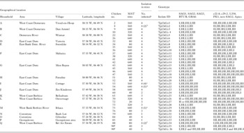
A total of 36 T. gondii isolates were obtained from tissues of 35 chickens with titres of 1:20 or higher (Table 1). These isolates were designated TgCkGy 1–36. Two isolates were obtained from chicken no. 90, from which 1 of the 4 mice infected with tissues of the chicken had mixed allele types at loci SAG3 and PK1, indicating potential mixed infection, and this isolate is designated as TgCkGy36 (Tables 1 and 2). Five of the 36 T. gondii isolates (TgCkGy 2, 21, 22, 29, 34) were mildly pathogenic for mice (Table 1). In 4 of these isolates only 6 of 16 infected mice died of toxoplasmosis. In only 1 of these isolates (TgCkGy 21) all 4 inoculated mice died of acute toxoplasmosis on day 23 after receiving chicken tissues (Table 1).
Isolation of Toxoplasma gondii from free-range chickens from Guyana, South America

a Four mice were inoculated with tissues of each chicken.
b Two mice died on day 12 p.i.
c All mice died on day 23 p.i.
d One mouse died on day 23 p.i.
e One mouse died on day 17 p.i.
f Two mice died on days 19 and 24 p.i.
g Mixed infection seen in this chicken.
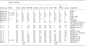
Genotyping of these 36 isolates with 11 markers revealed 9 genotype groups with isolate TgCkGy36 showing mixed infection (Tables 1 and 2). One third (12/36) of the isolates belong to genotype no. 1, which was widespread in the areas sampled. Genotype no. 9 has type III alleles at all loci and likely belongs to the clonal type III lineage. A few pairs of genotypes differ only at 1 locus, including genotype nos 2 and 3 at locus Apico, genotype nos 4 and 5 at locus PK1, and genotype nos 7 and 8 at locus Apico, indicating each pair of genotypes are closely related.
Summary of genotyping of Toxoplasma gondii from chicken from Guyana

a Cannot distinguish II or III.
DISCUSSION
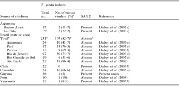
The present study found that T. gondii isolates from Guyana were biologically and genetically different from Brazilian chicken isolates, irrespective of the location in Brazil. Table 3 summarizes information on mouse pathogenicity of isolates from chickens from South American countries. Differences in murine pathogenicity of isolates from asymptomatic chickens from countries in South America are evident; isolates from Brazil and Colombia were more pathogenic than from neighbouring countries. Among these countries, extensive data are available only from Brazil. Mouse pathogenicity was not affected by region of Brazil; T. gondii strains from the very northern state (Pará) were similar phenotypically to strains from the southern state, Rio Grande do Sul.
Mouse mortality patterns of Toxoplasma gondii isolates from chickens from South America

a 100% mortality in Swiss Webster mice inoculated with chicken tissues.
b Data applicable to Brazil.
Recent studies using 11 PCR-RFLP markers indicated that virtually all isolates from 2 widely separated regions (Pará and Rio Grande do Sul) of Brazil were genetically distinct and this diversity was also evident among isolates from Guyana in the present study. At present, no correlation between mouse pathogeniciy and T. gondii genotype is established, and this may due to the great plasticity of gene expression in the parasite. Furthermore, there is no clear evidence that mouse pathogenicity of T. gondii isolates has any correlation with pathogencity in domestic animals or humans. Therefore, the concept of virulence in T. gondii is limited to the mouse model.
A comparison with previously published genotyping data from South and Central American isolates including dogs (Dubey et al. Reference Dubey, Cortés Vecino, Vargas-Duarte, Sundar, Velmurugan, Bandini, Polo, Zambrano, Mora, Kwok, Smith and Su2007b, Reference Dubey, Su, Cortés, Sundar, Gomez-Marin, Polo, Zambrano, Mora, Lora, Jimenez, Kwok, Shen, Zhang, Nieto and Thulliezc), cats (Dubey et al. Reference Dubey, Su, Cortés, Sundar, Gomez-Marin, Polo, Zambrano, Mora, Lora, Jimenez, Kwok, Shen, Zhang, Nieto and Thulliez2006c; Su et al. Reference Su, Zhang and Dubey2006) and chickens (Dubey et al. Reference Dubey, Patitucci, Su, Sundar, Kwok and Shen2006b, Reference Dubey, Su, Cortés, Sundar, Gomez-Marin, Polo, Zambrano, Mora, Lora, Jimenez, Kwok, Shen, Zhang, Nieto and Thulliezc, Reference Dubey, Sundar, Pineda, Kyvsgaard, Luna, Rimbaud, Oliveira, Kwok, Qi and Sue, Reference Dubey, Sundar, Gennari, Minervino, Farias, Ruas, dos Santos, Cavalcante, Kwok and Su2007a) showed that genotype nos 1, 4, 5, 6 and 7 are unique in chickens from Guyana, whereas genotype nos 2, 3, 8 and 9 were more widely spread. Both genotype nos 2 and 3 were found in chickens from Pará in Brazil, a location close to Guyana (Dubey et al. Reference Dubey, Cortés Vecino, Vargas-Duarte, Sundar, Velmurugan, Bandini, Polo, Zambrano, Mora, Kwok, Smith and Su2007a). Genotype no. 8 was found in chickens from Costa Rica (Dubey et al. Reference Dubey, Patitucci, Su, Sundar, Kwok and Shen2006b), Nicaragua (Dubey et al. Reference Dubey, Sundar, Pineda, Kyvsgaard, Luna, Rimbaud, Oliveira, Kwok, Qi and Su2006e) and Pará in Brazil (Dubey et al. Reference Dubey, Cortés Vecino, Vargas-Duarte, Sundar, Velmurugan, Bandini, Polo, Zambrano, Mora, Kwok, Smith and Su2007a), indicating that it is widespread in South and Central America. Genotype no. 9 (type III lineage), one of the 3 predominant clonal lineages found in North America and Europe (Dardé et al. Reference Dardé, Bouteille and Perstreal1992; Howe and Sibley, Reference Howe and Sibley1995; Ajzenberg et al. Reference Ajzenberg, Bañuls, Tibayrenc and Dardé2002a), was also found in chickens from Brazil (Dubey et al. Reference Dubey, Cortés Vecino, Vargas-Duarte, Sundar, Velmurugan, Bandini, Polo, Zambrano, Mora, Kwok, Smith and Su2007a), Chile (Dubey et al. Reference Dubey, Su, Cortés, Sundar, Gomez-Marin, Polo, Zambrano, Mora, Lora, Jimenez, Kwok, Shen, Zhang, Nieto and Thulliez2006c), Costa Rica (Dubey et al. Reference Dubey, Patitucci, Su, Sundar, Kwok and Shen2006b) and Nicaragua (Dubey et al. Reference Dubey, Sundar, Pineda, Kyvsgaard, Luna, Rimbaud, Oliveira, Kwok, Qi and Su2006e), and dogs from Brazil (Dubey et al. Reference Dubey, Cortés Vecino, Vargas-Duarte, Sundar, Velmurugan, Bandini, Polo, Zambrano, Mora, Kwok, Smith and Su2007c), confirming its global presence. In summary, T. gondii isolates in chickens from Guyana are highly diverse, with a variety of genotypes existing both locally and globally.