Introduction
Chronic rhinosinusitis is a common medical disorder affecting a significant number of the general population each year, with annual US costs related to medical and surgical care estimated at six billion dollars.Reference Pleis and Coles1 Frontal sinus disease is often caused by inflammation of the mucosa, creating secondary obstruction within the frontal sinus drainage pathway, specifically at the level of the frontal recess. Symptoms of recurrent frontal sinusitis may include frontal headache, a sensation of frontal pressure, nasal discharge and nasal obstruction.Reference DelGaudio, Wise and Wise2 The goal of functional endoscopic sinus surgery is to provide surgical access to the obstructed area, in order to resect impinging anatomical structures and to allow drainage and ventilation to resume.Reference Jacobs3
A full understanding of the complex and highly variable sinonasal anatomy is critical in the pre-operative evaluation and treatment of frontal sinus disease, both to enhance treatment efficacy and to minimise the risk of surgical complications.Reference Bolger, Butzin and Parsons4 In addition to endoscopy, multi-detector, triplanar computed tomography (CT) is used to provide a pre-operative anatomical survey. Although CT imaging is considered a standard means of documenting general anatomy as well as the presence of paranasal sinus mucosal disease, there are limitations to the utility of conventional, two-dimensional images. In any one orthogonal plane, such images are unable to fully depict the complex three-dimensional structure and variable relationships of the frontal sinus, its outflow tract and the associated pattern of impinging ethmoid air cells. Reliance on two-dimensional CT imaging may limit the surgeon's ability to conceptualise these relationships during pre-operative planning. Many studies have established the relationship between the presence and size of various anatomical structures, using triplanar, two-dimensional images. However, to the best of our knowledge, none have made use of three-dimensional metrics to evaluate these structures, enabling spatial analysis of the relationships of the surrounding anatomy.Reference Lee, Kuhn and Citardi5–Reference Zhang, Han, Ge, Xian, Zhou and Fan7
In this study, we attempted to use three-dimensional imaging to better characterise the three-dimensional relationships between the frontal sinus drainage pathway and the surrounding air cells, including the agger nasi cells, frontal cells and ethmoid cells. The goal of this study was to determine whether this novel method of visualising the anatomy of the frontal sinus drainage pathway might provide additional valuable radiological information which could affect clinical decision-making and guide medical or surgical treatment.
Materials and methods
This study was conducted with the approval of our institutional review board (approval number 06-54). Multislice CT scans of the paranasal sinuses acquired from 29 adult patients who had undergone paranasal sinus CT, based on appropriate clinical indications, were evaluated retrospectively at a tertiary care centre. Patients ranged in age from 19 to 83 years. Patients with a history of prior sinonasal surgery were excluded.
Patients were imaged on a four-slice CT scanner (Sensation; Siemens, Erlangen, Germany) with the following parameters: slice thickness 0.75 mm, field of view 14 cm, 140 mA, 120 kV and a pitch of 0.9. An attending neuroradiologist evaluated the two-dimensional images for sinonasal anatomical variations and mucosal disease. Images with complete opacification of the frontal sinus or an absence of agger nasi cells were excluded. The imaging data, stored in a Digital Imaging and Communication in Medicine file, were imported into ITK-SNAP visualisation version 1.4 software (Cognitica, Chapel Hill, North Carolina, USA) running on a laptop computer (Dell Latitude D600, Round Rock, Texas, USA).
Using standard craniocaudal dimensions to reduce the influence of height on volume, several structures were manually segmented for three-dimensional reconstruction (Figure 1). These included the upper frontal sinus drainage pathway volume, reflecting the relative volume of the frontal recess (including part of the frontal sinus, and extending to the floor of the frontal recess). More specifically, the superior margin of this volume was set at the most anterior skull base insertion point of the middle turbinate, and the volume was segmented caudally for a craniocaudal distance of 3.75 mm. This point of measurement was used instead of the frontal sinus ostium plane, to reduce potential influences on frontal recess volume based on frontal sinus volume or the volume of the nasofrontal beak. A lower frontal sinus drainage pathway volume (divided by the uncinate process into a lateral infundibular segment and a medial meatal segment) was segmented for a craniocaudal distance of 4 mm. Additionally, agger nasi cells and any adjacent ethmoid cells (i.e. frontal, supraorbital ethmoid, suprabullar and ethmoid bulla cells) were segmented.
Full-screen view of three-dimensional reconstruction of segmented volumes, showing posterior view with triplanar two-dimensional computed tomography views. The frontal recess (light blue) demonstrates a meatal drainage pattern, medial to the uncinate process (green and light purple portions). In addition, the agger nasi cell is depicted in dark purple; in this case, the agger nasi cell does not appear to significantly obstruct the frontal sinus drainage pathway.

Subsequently, three-dimensional volume renderings were reviewed in an interactive free rotation mode, as well as using standardised anterior, lateral and bilateral anterior oblique projections. An example of image output is provided in Figure 1. When reviewing these images, we focused on the morphological impact that the agger nasi cells and surrounding air cells had on the frontal sinus drainage pathway. In selected cases, we were also able to evaluate the ethmoid bulla and the suprabullar cells and recesses. Altogether, this entire process required approximately 20 minutes per study in experienced hands.
Results and analysis
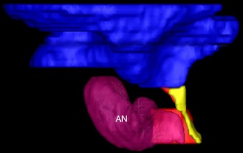
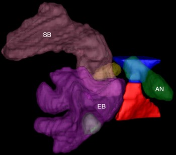
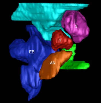
Two sides were excluded on the basis of absent agger nasi cells, for a total of 56 sinuses evaluated for 29 patients. Three-dimensional reconstruction based on paranasal sinus CT images was successfully performed using segmentation on all patients. Of these, 30 sinus cavities had evidence of frontal sinus drainage pathway disease. Selected examples are shown as Figures 2 to 7, demonstrating the three-dimensional reconstruction of the frontal sinus drainage pathway, which enhances the clinical usefulness of the images.
Three-dimensional reconstruction of segmented volumes, lateral view, indicating the frontal recess (blue), meatal drainage pattern (medial to uncinate process; yellow) and agger nasi cell (AN; purple). This image demonstrates a relatively unobstructed pattern of flow.

Three-dimensional reconstruction of segmented volumes, oblique view, indicating the frontal recess (blue), infundibular drainage pattern (lateral to uncinate process; red) and agger nasi cell (AN; purple). This image demonstrates a relatively large agger nasi cell impinging on the drainage pathway. Post = posterior; med = medial; lat = lateral; ant = anterior

Three-dimensional reconstruction of segmented volumes, lateral view, indicating the frontal recess (blue), drainage pathway (yellow), agger nasi cell (AN; purple) and ethmoid bulla (EB; green). The agger nasi cell impinges on the frontal recess, creating a concavity in the blue portion of the image. The ethmoid bulla impinges on the drainage pathway, as evidenced by the depression created in the yellow portion of the image. Note that these impinging air cells create a tortuous frontal sinus drainage pathway.

Three-dimensional reconstruction of segmented volumes, lateral view, indicating the frontal recess (blue), drainage pathway (red), agger nasi cell (AN; green), ethmoid bulla (EB; purple) and suprabullar cell (SB; brown). The agger nasi cell appears to impinge on the frontal recess, while the suprabullar cell and ethmoid bulla appear to affect the width of the drainage pathway (the blue and red portions of the image).

Three-dimensional reconstruction of segmented volumes, anterior view, indicating the frontal recess (light blue), ethmoid bulla (EB; dark blue), drainage pathway (green), agger nasi cell (AN; orange), and type one (1) and type three (3) frontal cells. In this image, the frontal cells impinge on the frontal recess, as evidenced by the depression created in the light blue portion of the image.

Three-dimensional reconstruction of segmented volumes: (a) lateral view and (b) medial view. The image indicates the frontal sinus (pink), suprabullar cell (SB; pink), agger nasi cell (AN; blue) and an additional drainage pathway (red). The frontal sinus drains into the suprabullar cell, and an accessory pathway (better shown on the medial view) is seen originating just inferior to the frontal sinus and draining posteromedially. Ant = anterior; med = medial; lat = lateral; post = posterior

Three-dimensional reformations allowed excellent depiction of anatomical relationships, often in a more intuitive fashion on the four images provided, compared with the 200–300 axial source images and additional post-processed coronal and sagittal images which are typically evaluated. While triplanar two-dimensional imaging, reviewed in real-time, can help improve understanding of the regional anatomy, three-dimensional imaging was able to provide the same, if not greater, degree of understanding without the need for meticulous (and sometimes time-consuming) scrolling through the imaging in multiple planes.
In many cases, we were able to demonstrate the influence of the agger nasi cells and frontal cells, when present, on the frontal sinus drainage pathway. The three-dimensional images enabled an anatomical survey of the region, confirming the presence or absence of anatomical stenoses suspected on the two-dimensional imaging. Additionally, by varying alpha transparency values, it was possible to demonstrate the combined impact of the anterior ethmoid labyrinth, as opposed to only visualising a cell or group of cells fortuitously set in the same orthogonal plane.
Most commonly, an increased degree of tortuosity and arcing of the lower frontal sinus drainage pathway segment was noted, with focal defects of the upper pathway segments (Figure 4). Notably, this degree of tortuosity did not always correlate with the size of the agger nasi cells; the actual morphology of these cells appeared to play a more important role in determining whether deformity was created. It is notable that this tortuosity was not anticipated on two-dimensional images in all cases. Additionally, in some cases, the ethmoid bulla and suprabullar cells were noted to have an impact on the course of the frontal sinus outflow tract. Three-dimensional imaging allowed ready conceptualisation of more complicated cases, including a case with apparent suprabullar drainage of the frontal sinus.
Discussion
The anatomical variation of the frontal sinus region has been well documented in cadaveric studies.Reference van Alyea8–Reference van Alyea10 The frontal sinus may be continuous with the infundibulum, communicate with the frontal recess and middle meatal region, or drain directly to the suprabullar region. Anterior ethmoid cells (such as frontal cells, supraorbital cells, agger nasi cells, intersinus septal cells and frontal recess cells) can impinge on the frontal sinus drainage pathway, as well as cause anatomical variation of the ostiomeatal unit.Reference Jacobs3, Reference van Alyea8–Reference Wormald12 These cells must be exposed, identified and adequately resected if identified as the cause of frontal sinus drainage pathway obstruction. Inadequate treatment of obstructing air cells limits the efficacy of frontal sinus surgery. Furthermore, restenosis and obstruction of the frontal sinus drainage pathway can occur if cells are only partially removed.Reference Wormald13 In addition, this region is adjacent to several important structures potentially at risk during surgery, including the cribriform plate, orbit and anterior ethmoidal artery. All these factors necessitate a full understanding of the patient's regional anatomy prior to undertaking any frontal sinus surgery.
Computed tomography is essential in the pre-operative assessment of anatomical variation, mucosal disease, anatomical relationships and osseous integrity.Reference Jacobs3 Coronal slices enable visualisation of frontal sinus pneumatisation and location of the supraorbital plate. Anterior ethmoid air cells that may impinge on frontal sinus drainage may also be seen on this view. Sagittal imaging can contribute to the assessment of the frontal sinus, nasofrontal duct and patterns of impingement.Reference Jacobs, Lebowitz, Sorin, Hariri and Holliday14–Reference Kew, Rees, Close, Sdralis, Sebben and Wormald16 In the typical circumstance, the surgeon must be able to mentally integrate the combined two-dimensional images into a three-dimensional representation of the frontal sinus and its outflow tract. In addition, focal anatomical constriction or abnormality seen on any single two-dimensional image may be shown to be less significant on three-dimensional imaging (Figure 1). Three-dimensional imaging may create more representative and intuitive images, which may serve as an adjunct to cross-sectional, two-dimensional imaging.
Wormald suggested that surgeons use two-dimensional CT scanning to identify ‘building blocks’, in order to develop a three-dimensional impression of the frontal recess and its outflow tract.Reference Wormald13 Volume rendering of segmented volumes takes this methodology a step further, by presenting an intuitive three-dimensional model.Reference Jun, Song, Cho, Park, Lee and Chang17 In addition to providing an understanding of these complex anatomical relationships, three-dimensional reconstruction can identify the spatial relationship between pathological areas and common surgical landmarks. Three-dimensional rendering can serve as a teaching tool for medical trainees, with which to improve their understanding of the variable anatomical configurations of the frontal sinus drainage pathway. In addition, such images can provide a blueprint for surgical planning.
Other applications of three-dimensional segmentation include the quantitative measurement of such parameters as length, area and volume for different regions, when object boundaries are defined. In a concurrent analysis, Wang et al. have used three-dimensional segmentation to calculate the volume of cells in the frontal sinus drainage pathway. They found that frontal sinus drainage pathway volume was significantly smaller in patients with radiological evidence of frontal sinus and outflow tract disease (E Y Wang, unpublished data).
The limitations of this study are related to the methodology of acquiring three-dimensional imaging. Current automatic segmentation protocols can be of decreased accuracy in delineating paranasal sinus volumes. This may be related to increased image noise from the high resolution, thin-slice data required to delineate the complex regional anatomy. In addition, the septations between air cells are of minimal thickness, which reduces their CT attenuation and renders an attenuation-based threshold of segmentation difficult to achieve; this is further exacerbated by the deossification and presence of mucosal thickening that is often present in rhinosinusitis. Consequently, all three-dimensional images were rendered manually in this study. This required time-intensive post-processing, introducing the potential for researcher subjectivity.
• Frontal sinus drainage is affected by anterior ethmoid pneumatisation patterns, which may contribute to the development of frontal sinus disease
• Two-dimensional computed tomography images can be limited in their depiction of such anatomical variations
• In this study, three-dimensional, reformatted images of segmented volumes aided delineation of the spatial relationships of the frontal sinus, frontal sinus drainage pathway, infundibular and meatal direction of drainage, agger nasi cells, ethmoid bulla cells, supraorbital cells, and suprabullar cells
• Three-dimensional, reformatted images of frontonasal anatomy enable improved understanding of the frontal sinus drainage pathway anatomy and the spatial relationships between ethmoid air cells
• Such images may provide a useful adjunct to surgical planning and education
Future studies will include a prospective analysis of patients with and without frontal sinus disease, in order to determine if our three-dimensional images are useful and comparable to CT scans for disease identification and pre-operative surgical planning. We are also actively investigating more sophisticated automatic segmentation protocols, in order to decrease the time and effort involved in segmenting these volumes to such an extent that ready clinical application would be feasible. Three-dimensional reconstructions improve anatomical understanding and possibly may alter the surgical plan; therefore, we feel that pre-operative analysis of patients with frontal sinus disease, using this technique, is valuable and provides a useful adjunct to standard two-dimensional imaging.
Conclusion
Three-dimensional depictions of segmented volumes of interest are useful adjuncts to the radiological evaluation of the frontal sinus drainage pathway, and enable improved understanding of regional anatomy for surgical and educational purposes. Further study is required to fully detail the clinical and anatomical significance of three-dimensional morphological parameters.