Introduction
Heat stress (HS), defined as exposure to environmental hot temperatures, results in hyperthermia, which accounts for one of the most important causes of infertility in cattle during the summer. The effects of heat stress on female fertility reduction are pronounced during the oestrus phase of the cycle when oocytes within graffian follicles resume their maturation to be prepared for fertilization (Gwazdauskas, Reference Gwazdauskas1985; Hansen, Reference Hansen2019). Heat stress at this time induces a reduction in de novo synthesis of intracellular proteins, level of mRNA and marked changes in the cytoskeleton, impairing nuclear and cytoplasmic maturation; however, the consequences go beyond and are manifested in the evident reduction in the development of the embryo (Miętkiewska et al., Reference Miętkiewska, Kordowitzki and Pareek2022; Payton et al., Reference Payton, Rispoli, Saxton and Edwards2011; Roth and Hansen, Reference Roth and Hansen2005).
HS is well demonstrated to induce the overproduction of ROS (Reactive Oxygen Species), which is unable to be neutralized through non-enzymatic and enzymatic antioxidant defense mechanisms in the cells, leading to oxidative stress (Ayemele et al., Reference Ayemele, Tilahun, Lingling, Elsaadawy, Guo, Zhao, Xu and Bu2021). The antioxidant enzymes include superoxide dismutase (SOD), catalase (CAT) and glutathione peroxidase (GPX) that counteract the generation of ROS sequentially at the first line of protection, in which superoxide anions are dismutated to H2O2 by SOD and then neutralized to H2O by CAT or GPX (Silva and Silva, Reference Silva and Silva2023). Moreover, the intracellular pathways promoted by the antioxidant system maintain the steady state of cell redox status. Recently, the master regulator of redox status and detoxification response in cells has been introduced as a nuclear factor (erythroid-derived 2)-like 2 (Nrf2) transcription factor, which induces the transcription of several genes related to the elimination of ROS (Rungratanawanich et al., Reference Rungratanawanich, Abate, Serafini, Guarienti, Catanzaro, Marziano, Memo, Lanni and Uberti2018). In recent years, to resolve the problem of heat stress-induced oxidative stress, numerous studies have considered the supplementation with antioxidants in vitro and in vivo (Hussain, Reference Hussain2023; Torres-Osorio et al., Reference Torres-Osorio, Urrego, Echeverri-Zuluaga and López-Herrera2019).
Gamma-oryzanol is a natural nutraceutical derived from rice bran oil and consists of a mixture of ferulic acid esters, sterols and triterpene alcohols and has been recently introduced as an antioxidant characterized by the ability to scavenge free radicals (Islam et al., Reference Islam, Nagasaka, Ohara, Hosoya, Ozaki, Ushio and Hori2011; Minatel et al., Reference Minatel, Francisqueti, Corrêa and Lima2016). Different in vivo experiments have demonstrated the antioxidant potential of gamma-oryzanol (ORY) in boar (Chanapiwat and Kaeoket, Reference Chanapiwat and Kaeoket2015), ram (Escobar et al., Reference Escobar, Lopes, Malavolta, Ramalho, Missio, Pinto, Soares, Leivas, dos Santos Brum and Cibin2019), stallion (Arlas et al., Reference Arlas, Pederzolli, Terraciano, Trein, Bustamante-Filho, Castro and Mattos2008), rat (Ghatak and Panchal, Reference Ghatak and Panchal2012) and mice (Son et al., Reference Son, Rico, Nam and Kang2011; Spiazzi et al., Reference Spiazzi, Manfredini, da Silva, Flores, Izaguirry, Vargas, Soares and Santos2013). Moreover, some in vitro experiments have evaluated the mechanism of ORY action in human embryonic kidney cells (Rungratanawanich et al., Reference Rungratanawanich, Abate, Serafini, Guarienti, Catanzaro, Marziano, Memo, Lanni and Uberti2018), hepatic (Huang et al., Reference Huang, Jiang, Zhu, Ma, Ou, Luo, Wu, Wen, Tan and Yi2020) and HepG2 cells (Zhu et al., Reference Zhu, Xu, Guo, Liu, Chang and Wang2024), human prostate cancer cells (Klongpityapong et al., Reference Klongpityapong, Supabphol and Supabphol2013), rooster semen (Najafi et al., Reference Najafi, Daghigh-Kia, Mehdipour, Mohammadi and Hamishehkar2022) and buffalo sperm (Inyawilert et al., Reference Inyawilert, Rungruangsak, Liao, Wirojwutthikul, Phinyo, Tang, Wanangkarn and Tiantong2022).
Heat stress (HS) has a detrimental impact on oocyte development, disrupts nuclear organelles and the cytoskeleton, reduces polar body (PB) extrusion, impairs endoplasmic reticulum and mitochondrial function, induces cell apoptosis and causes oxidative stress in both oocytes and cumulus cells (Wang et al., Reference Wang, Sui, Miao, Liu, Zhou, Ge and Tan2009); to study its effects and evaluate potential treatments, in vitro models are commonly used to simulate HS by exposing COCs to a controlled temperature of 41 °C throughout the maturation period (Residiwati et al., Reference Residiwati, Azari-Dolatabad, Tuska, Sidi, van Damme, Benedetti, Montoro, Luceno, Pavani, Opsomer and van Soom2021; Toosinia et al., Reference Toosinia, Davoodian, Arabi and Kadivar2024); this controlled environment allows researchers to investigate strategies for mitigating the adverse effects of HS.
It was hypothesized in the present study that bovine oocytes undergoing maturation in vitro under hyperthermia might benefit from supplementation with ORY. To the best of our knowledge, there is not any study yet to evaluate the ameliorating effect of ORY during the maturation of heat-shocked bovine oocytes. Therefore, designing an in vitro model to mimic the heat-shock condition, we supplemented the maturation media with ORY to culture heat-shocked bovine oocytes and evaluated their maturation and consequent development. The matured oocytes were subjected to analysis for their nuclear maturation, fertilizability, development to the blastocyst and changes in abundance of transcripts related to antioxidative status.
Materials and methods
ORY in the form of powder was obtained from Sigma-Aldrich (CDS021604). ORY powder, using ethanol as a vehicle, was tested at 0–100 μM; 0 μM (ethanol only) served as the control. The concentration of 5 μM ORY was selected as optimal for bovine oocyte development. The final ethanol concentration in the culture media was <1% (v/v). All the other chemicals used in this research, except for those stated, were purchased from Sigma Chemicals Co. (St. 85 Louis, MO, USA) as well.
Recovery and maturation of oocytes
Bovine ovaries (predominantly of the Holstein breed) were collected from a local slaughterhouse, embedded in a warmed normal saline solution and transported to the laboratory within 2 h, where evaluated morphologically to determine normal 3–8 diameter follicles for aspiration of their cumulus–oocyte complexes (COCs). Microscopic evaluation was performed to separate all the morphologically normal COCs based on the presence of at least three layers of cumulus cells without any signs of degeneration or expansion around an oocyte with evenly granulated cytoplasm. COCs were washed repeatedly in HEPES-buffered medium 199 (9.5 mg M199, 0.42 mg NaHCO3, 0.02 mg Na Pyruvate, 2.38 mg HEPES(Free acid), 2.6 mg HEPES(Na Salt), 0.06 mg Penicillin and 0.05 mg Streptomycin per ml) supplemented with 10% FBS and then into droplets of preincubated b-TCM (9.5 mg M199, 2.1 mg NaHCO3, 0.04 mg Na Pyruvate, 0.06 mg Penicillin and 0.05 mg Streptomycin per ml) supplemented with 0.05 IU/ml FSH and 12% FBS as maturation media. The experimental groups were defined as Treatment (addition of 5 µM ORY to IVM (In Vitro Maturation) media) and PC or NC (positive and negative controls without ORY). COCs were randomly cultured in media, 10 each 50 µl droplet, under mineral oil 5% CO2 in a humidified atmosphere for 22 h at 38.5°C for NC and 41°C for PC and Treatment groups.
Assessment of nuclear maturation
Matured COCs, at the end of IVM, were treated with 0.2 mM Hyaluronidase in HEPES-TCM (serum-free) for a minute, then washed and subjected to gentle pipetting to remove the cumulus cover. After washing in H-TCM, denuded oocytes were stained with Hoechst 33342 and evaluated for nuclear maturation status with an Epifluorescent microscope. The presence of a PB in perivitelline space or two shiny spots in ooplasm was determined as metaphase II (MII) stage, while metaphase plate or irregular networks of condensed chromatin were recorded as metaphase I (MI). Clumped or slightly condensed chromatin was recorded as germinal vesicle breakdown (GVBD). Oocytes with condensed or non-condensed fluorescent foci different from other groups were categorized as fragmented (Figure 1).
Photomicrograph illustrating various nuclear stages of bovine oocytes matured in vitro, labelled with Hoechst 33342. (A) Germinal Vesicle (GV) stage; (B) Germinal Vesicle Breakdown (GVBD); (C) Metaphase I (MI) stage; (D) Metaphase II (MII) stage; (E) Fragmented oocyte.

The production of embryo
Mature COCs were subjected to fertilization in vitro at the end of IVM. All the expanded COCs were washed three times with HEPES-SOF (synthetic oviductal fluid) and transferred to drops of TALP (Tyrode’s albumin lactate pyruvate) supplemented with heparin and 0.6% BSA as IVF media, 10 COCs each 50µL droplet, incubated at 39°C and 5% CO2 in a humidified atmosphere.
Bovine fresh sperm were collected from the epididymis of slaughtered bulls (predominantly Holstein) and incubated in HEPES-TCM supplemented with BSA for 1 h at room temperature and then swam up by pouring into the bottom of a microtube containing SOF media supplemented with BSA for 20 min at 39°C and centrifuging the upper layer at 200×g for 2 min. The pellet is composed of highly motile spermatozoa, which co-incubated with COCs at a concentration of 1 × 10⁶ motile sperm/ml. The next day, the presumptive zygotes were manipulated to be denuded from cumulus cover, washed with HEPES-SOF supplemented with 2% BSA, evaluated morphologically under a stereomicroscope, and normal ones without any signs of degeneration or atresia were selected and transferred to 20 µl drops of IVC-SOF media composed of SOF (0.33 mM Na pyruvate, 3.3 mM Na Lactate, 1.5 mM glucose, 25 mM NaHCO3, 1.19 mM KH2PO4, 1.78 mM CaCl2.2H2O, 0.5 mM MgCl2.6H2O, 107.7 mM NaCl, 7.16 mM KCl, plus amino acids (2% (v/v) BME amino acid solution, 1% (v/v) MEM amino acid solution) and antibiotics (0.06 mg/ml Penicillin and 0.05 mg/ml Streptomycin), 8% BSA (fatty acid-free BSA) and 10% charcoal-stripped FBS, six zygotes each, and incubated for 8 days at 39°C in a humidified atmosphere with 5% CO2 and 7% O2. The day of fertilization was considered as day 0; at day 2nd, cleaved embryos were determined, and then at the 8th day, the number of blastocysts was recorded.
Gene expression analysis
Extraction of RNA, qualification and reverse transcription
Five replicates of pooled denuded mature oocytes and the respective CCs (Cumulus Cells) of 25 matured COCs were subjected to total RNA extraction. FavorPrepTM Blood/Cultured Cell Total RNA (Favorgen, Ping-Tung, Taiwan) was employed for this purpose. Upon the user manual, binding (FRAB) buffer (350 μl) and β-Mercaptoethanol reagent (3.5 μl) were mixed with samples and centrifuged through a mini-column filter (2 min at full speed) to remove supernatant and transfer to a new tube, mix with ethanol (70%) and repeat the centrifugation. Then, two different washing buffers were employed to wash the filters. After drying the filters, elution in RNase-free water was performed.
Spectrophotometry (LSPR spectrophotometer MPANM96, Nanomabna, Iran) was used to determine the quality and quantity of extracted RNA. Absorbance at 260 and 280 nm was determined to calculate the ratio of A260/A280. The ratio greater than 1.8 was ensured to go beyond the next step; A260 was used to determine the concentration of total RNA to ensure 1µg total RNA was incorporated into the synthesis of cDNA.
Reverse transcription for synthesis of cDNA was performed using M-MLV reverse transcriptase (YektaTajhizAzma, Iran). Two mixtures were prepared. Mixture A (13.5 µl) is composed of extracted RNA (1 µl), random hexamer (0.5 µl) and oligo dT (0.5 µl). Mixture B (6.5 µl) is composed of 5× first strand buffer (4 µl), dNTPs (1 µl), RNsin (0.5 µl) and M-MLV (1 µl). MixA was incubated at 70°C for 5 min, chilled on ice and utilized with MixB to incubate at 37°C for 60 min and finally stopped by heating at 70°C for 5 min.
Relative quantification
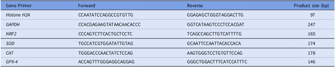
For this purpose, Rotor-Gene Q 6000 (Qiagen, Hilden, Germany) was employed to perform Real-time PCR in a triplicate manner. A reaction mixture (20 µl) was prepared for each sample composed of 10 µl of SYBR master mix (RelQ Plus 2× Master Mix Green; Ampliqon, Denmark), 0.5 µM of each specific primer, DNase-free distilled water and 1 µl cDNA. The following thermocycling conditions were performed: preheating at 95°C for 15 min, 40 cycles of amplification at 95°C for 15 sec, 30 sec at 60°C and 30 sec at 72°C. The primer sequences are listed in Table 1.
The list of primers used for RT-PCR

The relative expression of each target gene was normalized to Histone H2a and GAPDH as housekeeping genes, which were validated for their stabilities by BestKeeper software (Technical University of Munich, Munich, Germany). The reaction efficiency was analyzed by LinRegPCR software version 2012.0 (Amsterdam, Netherlands), and the relative expression was calculated using comparative Ct (2-∆∆Ct 170) methods (Pfaffl et al., Reference Pfaffl, Tichopad, Prgomet and Neuvians2004; Ruijter et al., Reference Ruijter, Ramakers, Hoogaars, Karlen, Bakker, van den Hoff and Moorman2009).
Statistics
Data, presented as the mean ± SEM, were analyzed using one-way ANOVA followed by post-hoc Tukey with SPSS statistical software package, version 20.0 (SPSS Inc., Chicago, Illinois, USA). Arcsin transformation of proportional data was performed before analysis. The differences at the level of P < 0.05 were considered significant.
Results
ORY improves the nuclear maturation of bovine heat-shocked oocytes
The results of the comparison among treatment groups, PC and NC are presented in Table 2. Comparison between groups NC and PC shows that heat shock decreased and increased the proportions of MII (0.42 ± 0.03 vs 0.79 ± 0.03) and MI (0.38 ± 0.03 vs 0.12 ± 0.02) oocytes, respectively (P < 0.05), while the proportion of GV/Frag remained unchanged.
The results of the maturation of COCs of groups NC, PC and Treatment

COCs = cumulus–oocyte complexes; MII = metaphase II; MI = metaphase I; GVBD = germinal vesicle breakdown; GV = germinal vesicle; Frag = fragmented oocyte. NC: maturation in no supplemented media at 38.5˚C; PC: maturation in no supplemented media at 41˚C; Treatment: maturation in ORY supplemented media at 41˚C; the statistical differences between groups are indicated by different superscripts in each column (P < 0.05); results from five replicates.
In the comparison of groups that matured under hyperthermia, treatment of maturing oocytes with ORY improved the proportion of MII oocytes (0.78 ± 0.03 vs 0.42 ± 0.03) while decreased MI oocytes (0.10 ± 0.02 vs 0.38 ± 0.03) (P < 0.05) compared with PC group. The proportion of GV/Frag remained unchanged too.
There was no significant difference between groups NC and Treatment with ORY in the parameters.
ORY improves the development of bovine heat-shocked oocytes to embryos
Embryonic development of matured oocytes of three groups Treatment, PC and NC was assessed on days 2nd and 8th for their cleavage and blastocyst rates, respectively (Table 3). Results of comparison between groups NC and PC show that heat shock decreased the proportions of cleaved embryos (0.33 ± 0.03 vs 0.64 ± 0.03) and total blastocysts (0.14 ± 0.02 vs 0.29 ± 0.03) (P < 0.05).
The results of the subsequent development of matured COCs of groups NC, PC and Treatment to the blastocyst stage

COCs = cumulus–oocyte complexes. NC: maturation in no supplemented media at 38.5˚C; PC: maturation in no supplemented media at 41˚C; Treatment: maturation in ORY supplemented media at 41˚C; the statistical differences between groups are indicated by different superscripts in each column (P < 0.05); results from five replicates.
In the comparison of groups derived from heat-shocked oocytes, treatment with ORY increased the proportions of cleavage (0.52 ± 0.04 vs 0.33 ± 0.03) and total blastocyst (0.30 ± 0.03 vs 0.14 ± 0.02) rates (P < 0.05).
There was no significant difference between groups NC and Treatment with ORY in these parameters.
ORY induces the transcription of genes related to antioxidative capacity
To investigate the effect of heat shock on the endogenous enzymatic antioxidant defense, the transcription levels of NRF2, SOD, CAT and GPX-4 genes were evaluated.
Comparison of transcripts in oocytes related to groups NC and PC revealed that heat shock elevated the transcription of NRF2, and treatment of maturing oocytes with ORY made upregulation of NRF2, SOD, CAT and GPX-4 transcriptions (Figure 2).
Relative expression of NRF-2, SOD, CAT and GPX-4 genes in oocytes of NC, PC and T groups. Different letters show a significant difference (results from five replicates).

Comparison of transcripts in CCs related to groups NC, PC and treatment revealed that ORY treatment made upregulation of GPX-4 transcripts. No significant changes in transcription of other genes were detected (Figure 3).
Relative expression of NRF-2, SOD, CAT and GPX-4 genes in CCs of NC, PC and T groups. Different letters show a significant difference (results from five replicates).

Discussion
Upon our results, heat-shocked oocytes achieved more proportions in the MII stage, cleavage and total blastocysts when treated with ORY during maturation, so nuclear maturation and the developmental competence of oocytes to the embryo were improved. It’s well known that a series of nuclear and cytoplasmic events during the process of maturation makes the oocytes acquire the capacity for fertilization and subsequent development, but so many confounding factors have been introduced to disturb the intracellular balance and attenuate the outcomes, one of them is the high level of ROS responsible for damage to the quality of oocytes leading to a decrease in fertilization rate (Agarwal et al., Reference Agarwal, Said, Bedaiwy, Banerjee and Alvarez2006). The overproduction of ROS induces the cascade of apoptosis, fragmentation of DNA and errors in the formation of the spindle, so impairs the progression to the second stage of meiosis (Pandey et al., Reference Pandey, Yadav, Premkumar, Tiwari, Pandey and Chaube2024; Roth and Hansen, Reference Roth and Hansen2005). Moreover, lipid peroxidation exerts damage to the membranes of gametes compromising their ability for binding and fusion (Tarin, Reference Tarin1996). On the other hand, making a disturbance in viability, ROS keeps cumulus cells from carrying out their role in the enrichment of oocytes (Russell et al., Reference Russell, Gilchrist, Brown and Thompson2016). The adverse impact of heat stress on the cellular function of gonads, hormonal axis, follicular development, oocyte growth and ageing of oocytes has made it one of the most noticeable causes of reduced reproduction efficiency of dairy and beef cattle (Becker et al., Reference Becker, Collier and Stone2020). HS is well introduced to disturb intracellular balance and impairs cytoskeletal modification and oocyte development (Roth and Hansen, Reference Roth and Hansen2004, Reference Roth and Hansen2005).
Several experiments have addressed pronounced properties of ORY in the regulation of redox homeostasis and oxidative stress in vivo and in vitro, e.g. antihyperlipidemic, antidiabetic, anti-inflammatory and antioxidant effects (Chalermpong et al., Reference Chalermpong, Chaiyavat, Sunee, Maitree and Busaban2012; Islam et al., Reference Islam, Nagasaka, Ohara, Hosoya, Ozaki, Ushio and Hori2011; Minatel et al., Reference Minatel, Francisqueti, Corrêa and Lima2016; Parrado et al., Reference Parrado, Miramontes, Jover, Márquez, Angeles Mejias, Collantes de Teran, Absi and Bautista2003; Sakai et al., Reference Sakai, Murata, Tsubosaka, Ushio, Hori and Ozaki2012). ORY has been demonstrated to trigger the nuclear factor erythroid 2-related factor 2 (NRF2) pathway for scavenging free radicals (Rungratanawanich et al., Reference Rungratanawanich, Abate, Serafini, Guarienti, Catanzaro, Marziano, Memo, Lanni and Uberti2018). This pathway is mentioned a lot in the area of antioxidant function.
The present study compared the expression of NRF2 transcript during the maturation of bovine oocytes between normal and heat-shock conditions and showed enhanced expression in ORY and heat-treated oocytes along with higher maturation and development as well; the upregulation exhibits the triggering for compensating the higher stress during heat-shock condition. Meanwhile, three downstream transcripts, SOD, CAT and GPX-4, were considered to evaluate the effect of treatment as another possible mechanism related to the ORY effect. NRF2 is demonstrated to induce the transcription of SOD and CAT genes as downstream antioxidant targets (Shaw and Chattopadhyay, Reference Shaw and Chattopadhyay2020) to convert tandemly superoxide radicals into water and oxygen. This was evident in the present study. Moreover, being detected in several cytosolic organelles including mitochondria, ER (Endoplasmic Reticulum) and nucleus, GPX-4 participates in the cell defense through the conversion of harmful peroxides into less detrimental byproducts such as water and corresponding alcohols, scavenging the accumulating ROS, suppressing apoptosis and protecting the cell membranes (Herbette et al., Reference Herbette, Roeckel-Drevet and Drevet2007; Ighodaro and Akinloye, Reference Ighodaro and Akinloye2018). The balance among the upregulation of mRNA that codes for these antioxidant enzymes was observed in our results in which upregulation of these enzymes coincided with greater development of treated heat-shocked oocytes. On the other hand, during oxidative stress, NRF2 translocates to the nucleus and binds to the antioxidant response element to induce the expression of downstream antioxidant genes and initiate the transcription of various proteins that mitigate stress-induced cellular damage (Kensler et al., Reference Kensler, Wakabayashi and Biswal2007); the balance among these key antioxidant enzymes, SOD, CAT and GPX, has proven to be more critical than any single enzyme in achieving optimal antioxidant activity against oxidative stress. Upon our results in the PC group, oocyte stress levels were sufficiently high to activate the NRF2 signalling pathway, leading to NRF2 upregulation as a protective response. However, an imbalance in antioxidant enzymes likely limited their protective effect, and the NRF2 pathway was unable to fully compensate for the stress. In contrast, antioxidant supplementation in the treatment group augmented the antioxidant defense system and helped mitigate stress.
Nuclear maturation involves coordinated reorganization of the cytoskeleton and chromatin. HS impairs the resumption of meiosis and blocks the progression from MI to MII, therefore attenuating the ability of oocytes to be fertilized (Roth and Hansen, Reference Roth and Hansen2005). Also making alterations in the structure of intracellular filaments and tubules, HS compromises the rearrangement of the cytoskeleton, which nuclear maturation is dependent on (Gallicano, Reference Gallicano2001; Tseng et al., Reference Tseng, Chen, Chou, Yeh and Ju2004). HS results in chromatin aggregation and reduction of meiotic spindle size of bovine oocytes, which might alleviate separation of the chromosomes during embryonic cleavage as well (Ju et al., Reference Ju, Jiang, Tseng, Parks and Yang2005). In line with these properties, the nuclear maturation outcome of the PC group was affected by HS in terms of lower MII and more GVBD and MI stages, which were reversed in the treated group significantly. Altogether, these findings suggest that ORY supplementation of maturation media provides heat-shocked bovine oocytes with a suitable environment for maturation and subsequent development in vitro.
On the other hand, the results of transcription analysis in CCs of treated groups indicated the role of ORY in protecting CCs against oxidative imbalance. It’s well demonstrated that cumulus cells surrounding oocytes have an essential role in appropriate oocyte maturation through the enrichment of oocytes with requirements for subsequent development to EGA (Embryonic Genome Activation) that could not be provided on its own (Hashimoto et al., Reference Hashimoto, Saeki, Nagao, Minami, Yamada and Utsumi1998; Salhab et al., Reference Salhab, Tosca, Cabau, Papillier, Perreau, Dupont, Mermillod and Uzbekova2011). Another constituent of ORY is considered to be phytosterols. These substances play the role of cholesterol in living organisms, for the synthesis of essential biomolecules such as steroids (Kritchevsky and Bonfield, Reference Kritchevsky and Bonfield2012). Therefore, the synthesis of steroids in the cumulus cover during maturation might be improved by ORY supplementation to assist in the better development of oocytes. We assume that the improvement in the outcome of IVM and IVP (In Vitro Production) might be partly related to this effect of ORY, which demands a more detailed analysis relevant to the underlying mechanisms in the next experiments.
Conclusions
Our results demonstrated that ORY in maturation media improves the nuclear maturation of heat-shocked bovine oocytes and upregulates the expression of antioxidant enzyme genes Nrf2, CAT, SOD and GPX; these oocytes demonstrated higher potential for development translated in increased cleavage and blastocyst rates. These findings indicate that ORY supplementation during maturation protects bovine oocytes from heat shock and enhances their developmental capacity.
Data availability
The datasets generated and/or analyzed during this study are available from the corresponding author upon reasonable request.
Acknowledgements
The authors would like to acknowledge the Research Institute of Animal Embryo Technology for technical support and Shahrekord University for financial support.
Author contributions
Ali Kadivar: Formal analysis, Investigation, Writing – review & editing. Nahid Davoodian: Formal analysis, Investigation, Writing – review & editing. Najmeh Davoodian: Conceptualization, Design, Methodology, Investigation, Writing – original draft, Writing – review & editing.
Funding
This work was supported by Shahrekord University, Shahrekord, Iran (grant numbers: 0GRD34M40481).
Competing interests
The authors have no conflicts of interest concerning the manuscript to declare.
Ethical standards
No approval of research ethics committees was required as the study used slaughterhouse materials.