Introduction
Diabetes mellitus is a common metabolic disease which causes variable impairment of various body systems. A relationship between diabetes mellitus and hearing loss has been found in many clinical investigations.Reference Ravecca, Berrettini, Bruschini, Segnini and Sellari-Franceschini1–Reference Cullen and Cinnamond4 The literature reports rates of diabetes-related hearing loss of up to 93 per cent.Reference Parving, Elberling, Balle, Parbo, Dejgaard and Parving5 The hearing loss related to diabetes mellitus has been described as bilateral, progressive, sensorineural and present at high frequencies.Reference Parving, Elberling, Balle, Parbo, Dejgaard and Parving5 The sensorineural hearing loss (SNHL) related to diabetes mellitus has been attributed to localised inner-ear microangiopathy, neuronal degeneration and diabetic encephalopathy, but the observed relationship is inconsistent.Reference Park, Park and Choi6–Reference Sasso, Salvatore, Tranchino, Cozzolino, Caruso and Persico9 Hearing loss associated with diabetes mellitus may be the result of derangement in the metabolism of glucose and the related hyperactivity of free oxygen radicals and increased oxidative stress. Excess synthesis of oxygen free radicals, with resultant toxic effects on the inner ear, may result in the early auditory dysfunction seen in diabetes mellitus, which progresses without microvascular or neural alterations.Reference Lisowska, Namysłowski, Morawski and Strojek8
Oxidative stress is thought to be increased in the presence of increased free radical production and/or impaired antioxidant mechanisms. In diabetes mellitus, increased free radical formation has been attributed to non-enzymatic protein glycation, glucose auto-oxidation and activation of the polyol pathway, due to a hyperglycaemic environment.Reference Hunt, Smith and Wolff10 Free oxygen radicals are molecules that have one or more unpaired electrons in their outer shell. They are produced during various aerobic metabolic reactions within the body; endogenous sources comprise mitochondrial leakages, the immune system, and enzymatic and auto-oxidation reactions. They are extremely reactive and can bind various molecules, leading to changes in deoxyribonucleic acid (DNA) nucleic acid bases, injury to various proteins and peroxidation of membrane lipids. Tissue injury results. Under normal physiological conditions, such free radical induced tissue injury is controlled by the antioxidant defence system. A variety of natural antioxidants exist to scavenge free radicals and to prevent oxidative damage. The enzymatic (or intracellular) group of antioxidants includes catalase, superoxide dismutase and glutathione peroxidase. In addition to enzymatic antioxidants, there also exist natural antioxidants which are derived from natural sources via dietary intake, such as vitamins A, C and E and the carotenoids. Numerous small molecules with antioxidant properties are also synthesised or produce within the body (e.g. glutathione and uric acid).Reference Ramakrishna and Jailkhani11
A number of studies have evaluated the role of oxidative stress in diabetes and its complications.Reference Bhatia, Shukla, Venkata Madhu, Kaur Gambhir and Madhava Prabhu12–Reference Lehmann and Schleicher15 However, the effect of oxidative stress on auditory impairment in diabetic patients has not previously been reported. This study was undertaken to investigate the status of oxidative stress in type two diabetic patients with and without SNHL, and to compare these patients with non-diabetic subjects.
Materials and methods
Sixty-three patients with type two diabetes mellitus but with no signs of microangiopathy or peripheral neuropathy were selected from amongst diabetic patients previously diagnosed according to the World Health Organization criteria of 1998. Diabetic complications due to microangiopathy or peripheral neuropathy had been excluded by clinical examination (including neurological assessment, using the Michigan diabetic neuropathy score, and ophthalmological assessment) and laboratory investigation (including assessments of nerve conduction velocity, auditory brainstem responses and 24 hour urinary albumin excretion rate). All the diabetic patients received oral anti-diabetic therapy.
The control group was composed of 37 sex-, age- and body weight matched, healthy adults. None of the diabetic patients or control subjects had a history of otological or central nervous system disease, had received ototoxic medication or antioxidant supplementation, or had been exposed to unsafe noise levels. All subjects gave their informed consent to participate in the study, according to the guidelines of the local ethics committee. The clinical details of all the study and control subjects are presented in Table I.
Clinical characteristics of DM and control subjects

Data (apart from subject number and sex) expressed as mean ± standard deviation. DM = diabetes mellitus; M = male; F = female; yrs = years; BMI = body mass index
Following clinical history-taking and examination, subjects' auditory function was evaluated using pure tone audiometry (using an Interacoustics AC 5 clinical audiometer; Assens, Denmark) and tympanometry (using a Zodiac 901 middle ear analyser; Madsen, Denmark). A normal otoscopic appearance, an A-type tympanogram and the absence of an air–bone gap were inclusion criteria for all subjects. Pure tone averages were calculated over the entire frequency range (i.e. 0.25, 0.5, 1, 2, 3, 4, 6 and 8 kHz) for each ear. Bilateral pure tone averages worse than 30 dB HL were observed in 28 of the 63 study group subjects and in 18 of the 37 control group subjects; all these patients were confirmed as suffering SNHL. Subjects with bilateral pure tone averages between 0 and 20 dB HL were confirmed as having normal hearing. Both the study and the control groups were divided into two subgroups according to hearing levels (i.e. either ≤20 dB HL or ≥30 dB HL).
Fasting venous blood samples from all subjects were drawn into heparin-free tubes during routine blood sampling, for biochemical analysis. After immediate centrifugation (2000 g for 15 minutes at +4°C), serum samples were stored frozen at −70°C. The analyses undertaken are described below. All analyses used commercial chemicals supplied by Sigma (St Louis, Missouri, USA). Protein levels were measured according to the method of Lowry et al. Reference Lowry, Rosebrough, Farr and Randall16
Serum antioxidant enzyme
The total activity of (Cu–Zn and Mn) superoxide dismutase (EC 1.15.1.1) was determined according to the method of Sun et al. Reference Sun, Oberley and Li17 The principle of the method is based on inhibition of nitroblue tetrazolium reduction by the xanthine–xanthine oxidase system as a superoxide generator. Activity was assessed in the ethanol phase of the supernatant after 1.0 ml of ethanol–chloroform mixture (5:3, volume per volume) was added to the same volume of sample and centrifuged. One unit of superoxide dismutase was defined as the amount causing 50 per cent inhibition in the nitroblue tetrazolium reduction rate. The superoxide dismutase activity was expressed as U/ml. Glutathione peroxidase activity was measured by the method of Paglia and Valentine.Reference Paglia and Valentine18 The enzymatic reaction in the tube (containing Nicotinamide adenine dinucleotide phosphate, reduced glutathione, sodium azide and glutathione reductase) was initiated by the addition of H2O2, and the change in absorbance at 340 nm was monitored by a spectrophotometer. Activity was expressed as U/l.
Thiobarbituric acid reactive substance
The tissue level of thiobarbituric acid reactive substance was determined by the method of Esterbaur and Cheeseman,Reference Esterbaur and Cheeseman19 based on reaction with thiobarbituric acid at 90–100°C. In this reaction, malondialdehyde or malondialdehyde-like substances and thiobarbituric acid react to produce a pink pigment with an absorption maximum at 532 nm. The reaction was performed at pH 2–3 and 90°C for 15 minutes. The sample was mixed with two volumes of cold 10 per cent (weight per volume) trichloroacetic acid to precipitate the protein. The precipitate was pelleted by centrifugation, and an aliquot of the supernatant was reacted with an equal volume of 0.67 per cent (weight per volume) thiobarbituric acid in a boiling water bath for 10 minutes. After cooling, the absorbance was read at 532 nm. Results were expressed as µmol/l, according to the standard graphic prepared from measurements with a standard solution (1,1,3,3-tetramethoxypropane).
Tissue protein carbonyl content
The tissue protein carbonyl content was determined spectrophotometrically, based on reaction of carbonyl groups with 2,4-dinitrophenylhydrazine to form 2,4-dinitrophenylhydrazone (according to the method of Levine et al. Reference Levine, Garland, Oliver, Amici, Climent, Lenz, Ahn, Shaltiel, Stantman and Packer L Glazer20). The reagent originally used for proteins subjected to metal-catalysed oxidation was 2,4-dinitrophenylhydrazine. Results were given as nmol/ml.
Nitric oxide
Nitric oxide measurement is very difficult in biological specimens. Therefore, tissue nitrite (NO2−) and nitrate (NO3−) were estimated as indices of NO production. Samples were initially deproteinised with Somogyi reagent. After conversion of nitrate to nitrite by copperised cadmium granules, total nitrite (i.e. nitrite + nitrate) was measured by a spectrophotometer at 545 nm. A standard curve was established with a set of serial dilutions (10−8–10−3 mol/l) of sodium nitrite. The resulting equation was then used to calculate the unknown sample concentrations. Results were expressed as µmol/l.Reference Cortas and Wakid21
Vitamins
Serum levels of vitamins A, C and E were measured using a high performance liquid chromatography system with fluorescence detection (Chromsystems GmbH, München, Germany).
Statistical analysis
The Kolmogorov–Smirnov test was used to evaluate whether the distribution of variables was normal. Therefore, a two independent sample test was used to compare the biochemistry parameters of the study and control groups. Pearson's correlation analysis was used to test correlations between all variables, and was considered significant when the p value was less than 0.05. Analyses were performed using the Statistical Package for the Social Sciences (version 15.0 demo) software.
Results
Some of the basic clinical characteristics of the diabetic patients and controls are summarised in Table I. All subjects were matched for age, sex and body mass index.
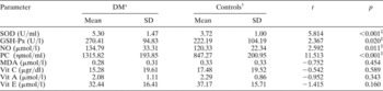
Serum superoxide dismutase activity and serum levels of glutathione, total nitrite and protein oxidation products (PC) were significantly higher in diabetes patients, compared with controls (p < 0.001, p = 0.020, p = 0.011 and p < 0.001, respectively). Serum levels of vitamins E, C and A were lower in diabetic patients compared with controls, but this difference was not statistically significant (Table II).
Oxidant and antioxidant status of diabetic and control subjects

* n = 63; †n = 37. ‡p < 0.05, compared with control values. DM = diabetes mellitus; SD = standard deviation; SOD = superoxide dismutase; GSH-Px = glutathione peroxidase; NO = nitric oxide; PC = protein oxidation products; MDA = malondialdehyde; Vit = vitamin
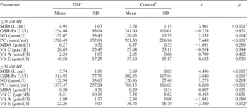
Both the study and control groups were divided into two subgroups based on hearing status, and the results for this analysis are presented in Table III. In subjects with normal hearing, serum NO levels were found to be significantly higher in diabetic patients compared with controls (p = 0.014); serum superoxide dismutase activity and serum protein oxidation product levels were also elevated in diabetic patients compared with controls. In subjects with hearing impairment (both diabetic patients and controls), there was a significant increase in glutathione peroxidase activity and higher levels of superoxide dismutase activity and protein oxidation products, compared with subjects with normal hearing.
Oxidant and antioxidant status of DM and control subjects divided by hearing impairment: ≤20 vs ≥30 dB HL

* For diabetic (DM) subjects, n = 35 for hearing ≤20 dB HL, n = 28 for hearing ≥30 dB HL. †For control subjects, n = 19 for hearing ≤20 dB HL, n = 18 for hearing ≥30 dB HL. ‡p < 0.05, compared with control values. SD = standard deviation; SOD = superoxide dismutase; GSH-Px = glutathione peroxidase; NO = nitric oxide; PC = protein oxidation products; MDA = malondialdehyde; Vit = vitamin
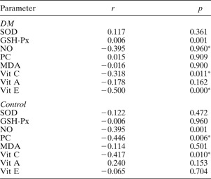
In the diabetic group, clear, negative correlations were observed between serum levels of NO and vitamins C and E and hearing impairment (r = −0.395, r = −0.318 and r = −0.500, respectively). In the control group, a negative correlation was observed between serum vitamin C levels and hearing impairment (r = 0.417).
Correlation between biochemical parameters and hearing impairment in DM and control subjects

* p < 0.05. DM = diabetes mellitus; SOD = superoxide dismutase; GSH-Px = glutathione peroxidase; NO = nitric oxide; PC = protein oxidation products; MDA = malondialdehyde; Vit = vitamin
Discussion
In recent years, substantial research work has been carried out on the involvement of oxidant and antioxidant factors in hearing impairment. It has been claimed that free radicals may play a central role in certain inner-ear disorders, such as acoustic trauma, labyrinthitis, some drug ototoxicities, Ménière's disease and presbyacusis.Reference Chisolm, Willott and Lister22–Reference Takumida, Anniko, Popa and Zhang24 Some studies have noted that reactive oxygen species (this term includes both free oxygen radicals and nonradical derivatives of oxygen) are generated in the cochlea and are involved in the pathogenesis of acoustic injury of the cochlea.Reference Yamane, Nakai, Takayama, Konishi, Iguchi and Nakagawa25, Reference Ohlemiller, Wright and Dugan26 Recently, Hu et al. showed increased reactive oxygen species (ROS) activity at the outer hair cells after high noise exposure in chinchillas.Reference Hu, Henderson and Nicotera27 Several studies have also focused on oxidative stress as a common factor in diabetes mellitus and its complications.Reference Bhatia, Shukla, Venkata Madhu, Kaur Gambhir and Madhava Prabhu12–Reference Lehmann and Schleicher15 In the current study, we evaluated oxidative stress as a potential cause of diabetic hearing impairment. The results suggest that increased oxidative stress may play an important role in this process.
Proteins and lipids are among the prime targets of oxidative stress; the action of free radicals results in the formation of carbonyl groups in proteins and lipid peroxides in lipids. Serum malondialdehyde levels (as a marker of lipid peroxidation) and serum protein oxidation product (PC) levels (as a marker of protein oxidation) have been frequently used to determine the oxidant/antioxidant balance in diabetic patients.Reference Gallou, Ruelland, Legras, Maugendre, Allannic and Cloarec28, Reference Altomare, Vendemiale, Chicco, Procacci and Cirelli29 Protein oxidation does not have the features of a chain reaction, in contrast to lipid peroxidation. When plasma proteins are destroyed by peroxidation, the resulting compounds have a comparatively long half-life. Therefore, the evaluation of protein oxidation products (PC) in plasma is an accepted marker of free radical intensity. Few studies have investigated protein oxidation as a pathogenetic factor in disease progression, including diabetes mellitus.Reference Heistad30 In our study, we found significantly elevated protein oxidation product (PC) levels in diabetic patients, but no difference in malondialdehyde levels, compared with controls. In the literature, several authors have reported increased levels of lipid peroxides in diabetic patients,Reference Mooradian31–Reference Griesmacher, Kindhauser, Andert, Schreiner, Toma and Knoebl33 while others could find no significant increase.Reference Velázquez, Winocour, Kesteven, Alberti and Laker34 In diabetes mellitus, which is associated with increased oxidative stress, low levels of lipid peroxidation may be the consequence of increased NO levels.Reference Miles, Bohle, Glassbrenner, Hansert, Wink and Grisham35, Reference Lüscher and Noll36 However, in the light of our results, it may also be the case that protein oxidation is more significant than lipid peroxidation in the progression of diabetic hearing impairment.
Ischaemic damage has been considered as a promotional factor in the development of acute and chronic, structural and functional disturbances of the cochlea. Tissue damage occurring during ischaemia, and especially during prolonged hypoperfusion and reperfusion, has increasingly been ascribed to free radicals.Reference Evans and Halliwell37 Free radicals have also been implicated in cochlear damage associated with ischaemia.Reference Seidman, Shivapuja and Quirk38 Nitric oxide has emerged as one of the most important molecules released from the endothelium and from a variety of other tissues. Nitric oxide is a free radical which can act as a neurotransmitter, and also in a paracrine or autocrine manner, to produce diverse cellular responses, both beneficial and detrimental. Superoxide neutralises NO, and the peroxynitrite formed is a source of hydroxyl radicals that may cause endothelial damage.Reference Elliott, Cockcroft, Groop, Viberti and Ritter39 Furthermore, reaction between superoxide and NO may hypothetically inhibit lipid peroxidation.Reference Miles, Bohle, Glassbrenner, Hansert, Wink and Grisham35, Reference Lüscher and Noll36 However, another well known function of NO is to increase blood flow to target organs via vasodilation and inhibition of platelet adhesion. It has been demonstrated that cochlear blood flow is regulated by production of NO from cochlear vascular walls.Reference Brechtelsbauer, Nuttall and Miller40, Reference Ren, Nuttall and Miller41 In diabetes mellitus, as in other cardiovascular risk factors, impaired endothelium-dependent vasodilation is a consequence of impaired NO synthesis by oxidants.Reference Cohen42 However, a number of studies have observed impairment of NO production in diabetic animalsReference Durante, Sen and Sunahara43, Reference Mayhan44 and in diabetic humans.Reference Vanizor, Orem, Karahan, Kiran, Erem and Aliyazicioğlu45 Conflicting results have also been published.Reference Aydin, Orhan, Sayal, Ozata, Sahin and Işimer46, Reference Chiarelli, Cipollone, Romano, Tumini, Costantini and di Ricco47 However, none of these studies investigated the association between NO synthesis and diabetic complications. In the current study, we observed significantly higher serum levels of NO end-products in diabetic patients without hearing loss, but not in diabetic patients with hearing loss, compared with controls. Additionally, a clear, negative correlation was also observed between serum NO levels and hearing impairment in diabetic patients. These results suggest that NO may have a protective effect on diabetic hearing loss.
In a healthy population, oxidative stress is countered by various antioxidants. However, some studies of antioxidant enzyme activities in diabetes mellitus have found, controversially, that levels of reactive free oxygen radicals are greatly increased.Reference Sharma, Kharb, Chugh, Kakkar and Singh48 A few authorsReference Matkovics, Varga, Szabó and Witas49, Reference Rema, Mohan, Bhaskar and Shanmugasundaram50 have reported increased glutathione peroxidase activity in type two diabetic patients, but othersReference Gallou, Ruelland, Legras, Maugendre, Allannic and Cloarec28, Reference Matkovics, Kotormán, Varga, Hai, Salgó and Novák51 have reported decreased glutathione peroxidase activity. The present study found a significant increase in glutathione peroxidase activity in diabetic patients, compared with controls. Analogously, there are reports of both increasedReference Matkovics, Kotormán, Varga, Hai, Salgó and Novák51 and decreasedReference Sundaram, Bhaskar, Vijayalingam, Viswanathan, Mohan and Shanmugasundaram52 superoxide dismutase activity in diabetic patients, compared with controls. In our study, we observed increased levels of superoxide dismutase activity in diabetic patients, compared with controls.
• Diabetes mellitus is associated with progressive, bilateral, high-frequency sensorineural deafness
• The mechanisms of hearing loss in diabetics have traditionally been explained in terms of microangiopathy, neuropathy and encephalopathy
• This study suggests that oxidative imbalance may cause or contribute to hearing loss in diabetic patients
In addition to antioxidant enzyme activity, the present study evaluated the capacity of the antioxidant system to neutralise or scavenge free radicals generated under normal or pathological conditions, by measuring the total antioxidant status. The body also produces numerous small molecules which reflect the status of vitamins A, C and E.Reference Martín-Gallán, Carrascosa, Gussinye and Domínguez53–Reference Ford, Mokdad, Giles and Brown55 Vitamins A, C and E are extracellular, non-enzymatic antioxidants which are derived from natural sources by dietary intake and which delay or inhibit the oxidative process. People with diabetes may have greater antioxidant requirements because of increased production of free radicals.Reference Sharma, Kharb, Chugh, Kakkar and Singh48 Furthermore, some studies have demonstrated the benefits of using antioxidants, such as vitamins E and C, in reducing oxidative tissue damage and preventing dysfunction in several pathological conditions,Reference Nakamura, Ozaki, Fuchinoue, Teraoka and Ota56–Reference Shimizu, Araujo, Borges, de Tolosa and Seguro58 including diabetes.Reference Bursell, Clermont, Aiello, Aiello, Schlossman and Feener59 In the current study, we detected a significant negative correlation between hearing impairment and levels of vitamin E and vitamin C in the diabetic group. We also found a significant negative correlation between hearing impairment and vitamin C levels in the control group. These results suggest that vitamins C and E may have a protective effect against the development of hearing impairment in diabetic patients. In the geographical region in which our research was based, the staple diet is based mainly on fruit and vegetables, which contain a variety of antioxidants. This may be the reason for our subjects' altered levels of these nonenzymatic antioxidants. Recently, Le and Keithley showed that dietary intervention with a mixture of fruits, vegetables and some antioxidants reduced the magnitude of age-related cochlear degeneration in dogs.Reference Le and Keithley60 They also noted that the combination of multiple antioxidants in the diet was more effective than specific molecular compounds.
Conclusion
Oxidative imbalance may cause or contribute to hearing loss in diabetic patients. Our results suggest that, in this process, increased protein oxidation may be more important than lipid peroxidation. Nitric oxide may have a protective effect against the development of hearing impairment. Some nonenzymatic antioxidants, such as vitamins C and E, may also help prevent such impairment. These antioxidants appear to act cooperatively in vivo to provide greater protection against free radical or reactive oxygen species (ROS) damage than that provided by any single antioxidant acting alone. Further studies are needed to explore the mechanism of hearing deterioration in diabetic patients, and the role of oxidants and antioxidants in this process.