Image case
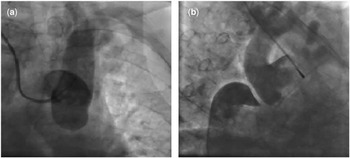
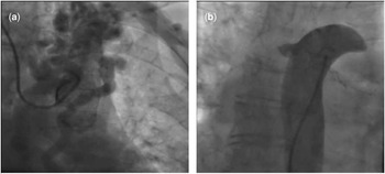
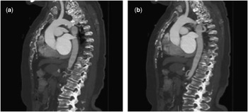
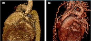
A 75-year-old woman who had been treated for hypertension for 35 years presented at an outpatient clinic complaining of dyspnea and chest pain. She had a transradial coronary angiogram displaying insignificant coronary artery disease 3 years ago. No history of prior cardiac surgery was observed. Physical examination revealed severe hypertension and significant pressure difference between the upper limbs. The blood pressure of the right and left arm were 200/125 and 170/90 mmHg, respectively. Transthoracic echocardiogram showed an ejection fraction of 50%, moderate left ventricular hypertrophy, mild mitral and tricuspid regurgitation. An aortic angiography demonstrated no flow into the descending aorta and showed the giant internal thoracic artery draining into the descending aorta, which functions as a collateral (Figs 1a and 2b). Simultaneous injection of both the aortic stumps showed a complete interruption of the aortic arch at the isthmus with a very large collateral vessel off the ascending aorta that drains to the descending aorta (Figs 1b and 2a). Contrast-enhanced CT confirmed interruption of the aorta exposing latter filling of the descending aorta with collaterals (Fig 3). Volume-rendered images depict the absence of continuity of the arcus aorta distal to the left subclavian artery and also giant collaterals (Fig 4). She was closely followed up with anti-hypertensive therapy for 2 years. Aortic interruption is an extremely rare congenital malformation. Only few case reports exist reporting late adulthood survivors. There are few sexagenerians and less septuagenerians in the literature.Reference Teskin, Gullu, Senay, Okten and Alhan 1 The presented case is one of the oldest survivors reported. This case shows that simultaneous stump aortography is an adequate technique for diagnosis of aortic interruption.
( a ) On aortic angiography, the contrast did not pass into the descending aorta. ( b ) Simultaneous aortic angiography depicted interrupted segment distal to the left subclavian artery.

( a ) Late phase of ascending aortic angiography displayed the giant thoracic artery. ( b ) Descending aortic angiography displayed giant collateral draining into the aorta.

Contrast-enhanced tomography displayed ( a ) discontinuity of the arcus aorta. ( b ) Latter filling of the descending aorta via posterior collaterals were clearly displayed.

Volume-rendering 64-multidetecter CT explicitly depicted ( a ) interrupted aortic segment distal to the left subclavian artery and ( b ) giant collaterals.

Acknowledgement
None.
Conflicts of Interest
None.