


The World Health Organization classification of ovarian tumours includes dozens of diagnostic categories, categorising tumours that can arise from a range of cell types, including germ cells, sex-cord cells, stromal cells and surface epithelial cells (Ref. Reference Tavasolli and Devilee1). Tumours within the ‘surface epithelial stromal’ category account for the largest group of ovarian neoplasms; they can be further classified as benign, borderline or malignant. Malignant tumours within this group (ovarian carcinomas) are responsible for most of the morbidity and mortality due to ovarian cancer. The WHO designation ‘surface epithelial stromal’ reflects the historical view that the epithelial component of these tumours arises from the single cell layer that covers the ovary or forms invagination cysts within the ovary (Ref. Reference Auersperg2). It should be noted that this belief has recently been called into question for the high-grade serous subtype, and there is compelling evidence that many of these tumours arise from the fallopian tube epithelium (Ref. Reference Crum3).
Clinicians treat ovarian carcinomas as a single entity, although pathologists classify them into serous, clear cell, endometrioid, mucinous and other rare subtypes. The serous group can be divided further into high-grade and low-grade types. The frequency of histological subtypes differs dramatically between those diagnosed in low-stage, where the majority are clear cell, endometrioid and mucinous carcinoma (Ref. Reference Gilks4), whereas high-stage carcinomas are predominantly of high-grade serous subtype (Refs Reference Seidman5, Reference Soslow6). Oncologists have noted that the dismal response rate of clear cell and mucinous carcinomas to chemotherapy (15%) contrasts sharply with that of high-grade serous carcinoma (80%) (Refs Reference Takano7, Reference duBois8, 9). Recently, there have been significant advances in the understanding of underlying molecular abnormalities in different subtypes of ovarian carcinoma (Ref. Reference Shih and Kurman10), resulting in improved reproducibility of subtype diagnosis (through the use of immunohistochemical markers). This progress in histopathological subtype assignment has led to more accurate separation of high-grade serous carcinoma from endometrioid, clear cell and undifferentiated carcinomas; tumours formerly diagnosed as high-grade endometrioid, mixed serous and clear cell, or mixed serous and undifferentiated are, in most cases, best considered to be high-grade serous carcinoma (Refs Reference Soslow6, Reference Han11). This approach, which is supported by the use of immunohistochemical biomarkers such as the Wilms' tumour gene WT1 (Refs Reference Gilks4, Reference Köbel12, Reference Waldstrom and Grove13), is being widely adopted by diagnostic pathologists (Refs Reference Soslow6, Reference Al Hussaini14). At the same time, criteria for separation of high-grade serous carcinoma from low-grade serous carcinoma have emerged and have been shown to be reproducible (Refs Reference Malpica15, Reference Malpica16). Low-grade serous carcinomas differ from high-grade serous carcinomas with respect to associated precursor lesions (Ref. Reference Shih and Kurman10), molecular events during oncogenesis (Refs Reference Singer17, Reference Singer18) and natural history, including response to chemotherapy (Refs Reference Crispens19, Reference Gershenson20). The evidence supporting routine separation of these groups of tumours is now compelling (Ref. Reference Kurman and Shih21). The ability to reliably identify high-grade serous carcinomas has opened the way for subtype-specific studies on molecular events during oncogenesis. Here, we review the pathogenesis of high-grade serous carcinoma, which is responsible for most ovarian cancer deaths, leaving aside clear cell, endometrioid, mucinous and low-grade serous subtypes. An overview of the clinical and pathological differences between these subtypes is provided in Table 1.
Ovarian carcinoma subtypes
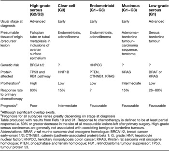
aAlthough significant overlap exists.
bPrognosis for all subtypes varies greatly depending on stage at diagnosis
Table produced with results from Refs Reference Shih and Kurman10 and Reference Bell91. Response to chemotherapy is defined to be at least partial response i.e. 50% or greater decrease in the size of all measurable lesions left after primary surgery. High-grade serous carcinomas are generally not associated with coexisting benign or borderline tumours.
Abbreviations: BRAF, v-raf murine sarcoma viral oncogene homologue; BRCA1/2, breast cancer early-onset 1/2; CTNNB1, catenin (cadherin-associated protein) beta 1; G, grade; HNF, hepatocyte nuclear factor; HNPCC, heriditary nonpolyposis colon cancer; KRAS, Kirsten rat sarcoma viral oncogene homologue; PTEN, phosphatase and tensin homologue; RB1, retinoblastoma tumour suppressor; TP53, tumour protein 53.
High-grade serous carcinomas: pathological and clinical features
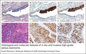
High-grade serous carcinomas are the most common subtype of ovarian carcinoma, accounting for approximately 70% of all ovarian carcinomas and for almost 90% of stage III or IV tumours (Refs Reference Seidman5, Reference Gilks22). They are only rarely diagnosed while confined to the ovaries (Ref. Reference Soslow6). High-grade serous carcinomas are characterised microscopically by high-grade nuclei with remarkable nuclear pleomorphism. High-grade serous carcinomas are highly proliferative tumours that are typically sensitive to chemotherapy, with response rates of 80% (Ref. Reference duBois8). However, tumour recurrence and progression to chemoresistance is usual and high-grade serous carcinomas account for a disproportionately high percentage of deaths due to ovarian cancer. The early dissemination can be explained by recent data suggesting that these tumours arise from the epithelium of the fimbriated end of the fallopian tube, with early intraperitoneal spread. This is based mainly on studies of early lesions in women at high risk of ovarian carcinoma development because of germ-line mutations in the early-onset breast cancer genes BRCA1/2 (Ref. Reference Medeiros23). There is also evidence of tubal mucosal involvement in cases of sporadic high-grade serous carcinoma (Ref. Reference Kindelberger24), implicating the fallopian tube as the primary site of high-grade serous carcinomas. These fallopian tube precursor lesions characteristically show overexpression of the tumour protein TP53 by immunohistochemistry, indicative of an underlying TP53 missense mutation, and increased expression of the proliferation marker Ki-67 (Fig. 1). They also show chromosomal instability (Ref. Reference Salvador25). Thus, even the earliest recognisable in-situ phase of high-grade serous carcinoma shows evidence of genomic instability, TP53 mutation and dramatically increased proliferation. These changes underlie the characteristic natural history of high-grade serous carcinoma (rapid tumour growth with a short latent phase, favourable initial response to DNA damaging chemotherapeutic regimens such as platinum), and the molecular events underlying these early, common (i.e. present in most high-grade serous carcinomas) changes are the focus of this review.
Histological and molecular features of in situ and invasive high-grade serous carcinoma. (a) Tubal intraepithelial carcinoma (TIC, arrow) and adjacent normal fallopian tube epithelium (haemotoxylin and eosin stain). (b) Diffuse expression of TP53 in nuclei of TIC and abrupt negativity of adjacent normal epithelium. (c) High proliferation rate of TIC with more than 50% of nuclei labelled with the proliferation marker Ki-67. Normal epithelium shows occasional proliferating cells in the basal layer (arrow). (d) High-grade serous carcinoma with remarkable nuclear pleomorphism and hyperchromatic enlarged nuclei (short arrow), high mitotic activity (long arrow) and high apoptotic activity (framed) (haemotoxylin and eosin stain). (e) Diffuse expression of TP53 in more than 80% of nuclei in a high-grade serous carcinoma, indicating an underlying TP53 missense mutation. (f) High proliferation rate with >50% of nuclei labelled with the proliferation marker Ki-67 in a high-grade serous carcinoma (magnification in all figures, x200).
The term ‘pelvic serous carcinoma’ has been proposed for tumours currently designated, somewhat arbitrarily, as being primary peritoneal, tubal or ovarian high-grade serous carcinoma (Ref. Reference Salvador26), as there is nothing that can distinguish between these tumours at the molecular level. Although the following discussion refers to high-grade serous carcinoma of the ovary, it applies equally to high-grade serous carcinomas of peritoneal or tubal origin.
BRCA1/2
Patients with hereditary breast/ovarian cancer syndrome carry germline mutations in one allele of either BRCA1 or BRCA2; the proteins encoded by these genes repair double-stranded DNA breaks via homologous recombination, among other functions. In contrast to single-strand DNA damage, which is repaired by base excision repair (BER), nucleotide excision repair (NER) or mismatch repair (MMR), double-stranded DNA breaks are particularly hazardous to the cell because they can lead to genomic rearrangements. Double-stranded DNA breaks are repaired by homologous recombination using a sister chromatid (available during S and G2 phases) as a template. BRCA1 appears to have a relatively early regulatory role in homologous recombination. Phosphorylated in response to double-stranded DNA breaks, it becomes a component of the BRCA1-associated genome surveillance complex (BASC) where it activates an initial DNA processing machinery. Unlike BRCA1, BRCA2 plays a more direct role in binding the recombinase Rad51 and localising to the site of DNA damage (Ref. Reference Tutt27). A deficiency in BRCA function leads to a switch to a non-homologous mechanism of DNA repair, e.g. nonhomologous end joining (NHEJ), which is also required to generate the diversity of B cells and T-cell receptors. This mechanism of repair is error prone, resulting in a ‘mutator phenotype’ (Ref. Reference Tutt28).
BRCA1/2 germline mutation
A significant proportion of ovarian carcinomas arise on a hereditary basis. The prevalence of BRCA mutations in Jewish women diagnosed with ovarian carcinoma is 29–41% in Israel (Refs Reference Modan29, Reference Moslehi30). A population-based study of 341 patients with high-grade serous carcinoma from the Canadian province of Ontario showed a 16.4% prevalence of germ-line BRCA1/2 mutations (10.9% BRCA1 and 5.5% BRCA2) (Ref. Reference Risch31). By contrast, in ovarian carcinomas other than high-grade serous carcinomas, germline BRCA mutations were identified in only 4% of cases. Using a different methodology, another study described a rate of germline BRCA1/2 mutations of 19% in a cohort of 92 ovarian carcinomas of all histological types (14% BRCA1 and 5% BRCA2) (Ref. Reference Hilton32). Although BRCA1/2 mutations are occasionally reported in tumours of endometrioid or clear cell histology and even borderline tumours, after contemporary gynaecopathological review, we and others have found BRCA1/2 germline mutations exclusively in high-grade serous carcinomas (Refs Reference Press33, Reference Shaw34). In this series of 49 ovarian carcinoma cases, 10 out of 38 (26%) high-grade serous carcinomas were from patients with germline BRCA1/2 mutations (21% BRCA1; 5% BRCA2). Recent studies on prophylactic salpingo-oophorectomy specimens from patients with BRCA1/2 mutations showed exclusively serous histology in precursor lesions of the fallopian tube (Ref. Reference Medeiros23). Thus, germline mutations are associated with high-grade serous subtype and, although there are differences depending on the population studied, they are present in a significant minority of cases of high-grade serous carcinoma (16–29%).
BRCA1/2 loss by somatic events
Somatic mutations in BRCA1 and BRCA2 are uncommon (Ref. Reference Foster35) but BRCA1 dysfunction as a result of epigenetic silencing through promoter hypermethylation is a common event in sporadic high-grade serous carcinomas, and is present in 33% of all high-grade serous carcinomas in our centre (Ref. Reference Press33). Epigenetic silencing of BRCA2, by contrast, occurs rarely, if ever (Ref. Reference Collins36). BRCA2 function might be abrogated by amplification and overexpression of the BRCA2-interacting gene EMSY. The EMSY gene located on chromosome 11q is amplified in 18% of high-grade serous carcinomas and overexpression of EMSY leads to a chromosomal instability phenoptype identical to that observed with loss of BRCA2 (Refs Reference Brown37, Reference Raouf38). Most high-grade serous carcinomas have some genetic or epigenetic changes that result in BRCA dysfunction (but this is encountered rarely, if ever, in other subtypes of ovarian carcinoma) (Ref. Reference Press33).
BRCA1/2: clinical implications and future directions
BRCA-associated hereditary ovarian carcinomas showed a longer recurrence-free and overall survival after adjuvant therapy than nonhereditary cases (Refs Reference Boyd39, Reference Ben David40). Similarly, sporadic ovarian carcinomas with low BRCA1 mRNA levels showed longer overall survival (Ref. Reference Quinn41). It is not yet clear whether the improved survival rates are due to a difference in the natural history of ovarian cancer in the two subgroups – for example, association with a different immunological response (indicated by intratumoural CD8-positive lymphocytes) – or are the result of a greater sensitivity of BRCA-associated tumours to DNA-crosslinking agents, such as platinum; the latter notion is supported by in vitro data (Refs Reference Bhattacharyya42, Reference Narod and Boyd43). There is compelling evidence that cells harbouring BRCA defects are more sensitive to agents that target other DNA repair pathways, for example, inhibition of BER is selectively lethal in BRCA-deficient cells. Poly(ADP-ribose) polymerase family, member 1 (PARP1) is involved in BER by protecting the single-strand breaks that are produced by DNA-crosslinking agents, such as platinum, or endonuclease action during the BER process. PARP1 inhibition leads to persistent single-strand breaks, and if these are encountered by the replication fork during mitosis, arrest occurs with generation of double-stranded DNA breaks that cannot be repaired by homologous recombination in a BRCA-deficient state, leading to cellular crisis and cell death (Ref. Reference Farmer44). Although it appears that BRCA1/2-deficient high-grade serous carcinomas are more sensitive to platinum-based chemotherapy and are highly sensitive to PARP inhibitors, these changes can be reversible. Secondary mutations in the BRCA1/2 genes that restore the wild-type reading frame and functional protein can give rise to acquired chemoresistance (Refs Reference Sakai45, Reference Edwards46, Reference Swisher47). It remains to be seen how common this phenomenon is.
Where are we now in terms of assessing BRCA status in ovarian carcinoma samples? Although there is considerable variation based on local differences in the frequency of founder mutations, we estimate that 15–20% of high-grade serous carcinomas are hereditary in our population, justifying referral of all women presenting with high-grade serous carcinoma for genetic counselling. This in turn could lead to screening for BRCA1/2 germ line mutations and risk-reducing salpingo-oophoroectomy for affected family members (Ref. Reference Narod and Boyd43). Genetic testing takes many weeks and currently can not be used to guide therapy in patients presenting with high-grade serous carcinoma, where there is an urgent need to initiate chemotherapy. As BRCA status is a critical predictor of responsiveness to specific therapies, the development of assays that could be used to provide routine rapid assessment of BRCA function in ovarian cancers to guide adjuvant therapy choices would be of great value. Immunohistochemistry approaches for detection of BRCA1 or BRCA2 protein are not currently applicable in practice because of technical failings (low level protein expression, available antibodies, gene expression only during S and G2 phase). It would theoretically be possible to use a combination of quantitative mRNA analysis and methylation-specific PCR to assess BRCA status. This would identify cases with loss of BRCA1 or BRCA2 expression, and further enable subcategorisation of those cases with loss of BRCA1 as being due to either epigenetic (promoter methylation) or genetic mechanisms. There are formidable challenges to overcome before this could be used routinely. However, such an approach to routine classification of BRCA status could address the question of whether different mechanisms of BRCA dysfunction are equivalent in terms of prognosis or response to treatment, including PARP1 inhibition. This in turn, would allow this testing strategy to guide adjuvant treatment decisions in patients with high-grade serous carcinomas, if significant differences in treatment response are identified between the different groups (i.e. BRCA1 germline mutation, BRCA2 germline mutation, BRCA1 epigenetic loss, no BRCA1 or BRCA2 abnormalities).
TP53
Mammalian cells have multiple levels of protection against oncogenic mutations, and cells with isolated loss of BRCA1 or BRCA2 function undergo apoptosis (Ref. Reference Vogelstein and Kinzler48). Other mutations are necessary to escape programmed cell death and emerge as a transformed cell. Two mouse models have resulted in the development of ovarian tumours similar to high-grade serous carcinoma from ovarian surface epithelium in a TP53-deficient background, either by introducing MYC (pro-proliferation) when BRCA1 was dysfunctional (Ref. Reference Xing and Orsulic49) or by inactivation of the retinoblastoma tumour suppressor RB1 (Ref. Reference Flesken-Nikitin50). By contrast, introducing defects in the PTEN/PIK3C2A and WNT/CTNNB1 pathway induces tumours recapitulating endometrioid subtype in another mouse model (Ref. Reference Wu51). In the following two sections, we focus on abnormalities of TP53 and molecules that permit increased tumour cell proliferation. These abnormalities are present in most high-grade serous carcinomas, and occur early during oncogenesis, at the in situ stage (Figs 1 and 2).
Proposed sequence of molecular changes during the pathogenesis of high-grade serous carcinomas. This model accounts for the finding of the ‘p53 signature’ lesions at similar rates in the fallopian tubes of women with germline BRCA abnormalities and women at lower risk (Ref. Reference Bell91). The TP53 and BRCA1/2 loci on chromosome 17 are depicted. Loss of TP53 or BRCA through mutation (germline or somatic), promoter hypermethylation or loss of heterozygosity is indicated by X. Precursor cells (probably tubal epithelial cells of secretory type) are morphologically normal. The first detectable abnormalities are associated with TP53 overexpression [so-called ‘p53 signature’ (Ref. Reference Crum92)]. These cells are morphologically normal and are not hyperproliferative. In this model, the ‘p53 signature’ lesion is a low-risk lesion and will only progress after the acquisition of chromosomal instability. Tubal intraepithelial carcinoma (TIC) occurs upon loss of TP53 and BRCA and results in chromosomal instability. This is associated with high-grade nuclear atypia and increased proliferation. In hereditary cases, a single BRCA ‘hit’ is required for progression to TIC, whereas two ‘hits’ are required for sporadic cases. Even the earliest morphologically recognisable precursor lesion (TIC) has already acquired the critical molecular abnormalities characteristic of advanced stage high-grade serous carcinoma, with chromosomal instability and increased proliferation. This results in rapid accrual of additional genetic abnormalities with each cell cycle.

TP53 is activated by cellular stress, such as DNA damage. After inducing cell cycle arrest, TP53 can repair DNA damage by nucleotide excision via its 3′–5′ exonuclease activity. If this fails, TP53 induces apoptosis. Loss of heterozygosity on chromosome 17q13.1 is a common event in high-grade serous carcinomas (Ref. Reference Garcia52). Mutations in the second TP53 allele were detected in 50% of high-grade serous carcinomas by evaluating exons 5–9. The more diligently TP53 mutations are sought, for example by extending the analysis to exons 2–11, the higher their frequency. In two studies investigating a total of 152 high-grade serous or advanced ovarian carcinomas, approximately 80% had somatic TP53 gene mutations of which 55% were missense mutations and 25% were nonsense mutations (Refs Reference Salani53, Reference Havrilesky54).
TP53: Clinical implications and future directions
The significance of TP53 abnormalities has been difficult to assess because assays for TP53 dysfunction have been imperfect. It can be hypothesised that wild-type TP53 might act by arresting the cell cycle, allowing chemotherapy-induced DNA damage to repair, and thus convey chemoresistance. There are data suggesting that mutated TP53 predicts chemosensitivity (Ref. Reference Moreno55), but the results have not been consistent (Refs Reference Lavarino56, Reference Kandioler-Eckersberger57). The prognostic significance of TP53 dysfunction is also not clear. A meta-analysis of 53 studies in ovarian carcinoma found that TP53 abnormalities detected by mutation analysis or immunohistochemistry were associated with a worse prognosis, in a univariate analysis (Ref. Reference Crijns58). There are several problems with these studies. Most included tumours other than high-grade serous carcinomas; these other subtypes characteristically lack TP53 mutations and have a favourable prognosis. Thus, the TP53 mutations were associated with an unfavourable subgroup of ovarian carcinoma and could therefore be predicted to be associated with a poor prognosis. In most of these studies, the prognostic significance of TP53 mutations did not hold up on multivariable analysis, supporting this interpretation. In addition, the assay used for TP53 mutation was typically immunohistochemical staining. Mutational analyses are time consuming and expensive, and aberrant TP53 expression as detected by immunohistochemistry is frequently used as a surrogate marker. In theory, tumours harbouring nonsense mutations should be completely negative for TP53 protein expression and tumours with missense mutations, which result in intranuclear accumulation of an inactive form of the protein, are strongly positive by immunohistochemistry (>80% of tumour cells are positively stained) (Refs Reference Havrilesky54, Reference Lassus59). However, nonsense mutations that generate a stop codon that results in a truncated protein can still express detectable levels of TP53 by immunohistochemistry in up to a third of cases (Ref. Reference Havrilesky54), and this can overlap with expression levels of normal TP53 protein seen when there is protein stabilisation induced by DNA damage or hypoxia (Ref. Reference Havrilesky54). Overall, immunohistochemistry is a poor predictor of TP53 mutations, with 48% sensitivity and 86% specificity (Ref. Reference Singer60). It remains to be clarified whether TP53 dysfunction, through TP53 mutation or other means – for example, interacting proteins (Ref. Reference Ichimura61) – is a ubiquitous feature of high-grade serous carcinoma, or whether TP53 functional status can delineate clinically relevant subsets of high-grade serous carcinoma. As for BRCA, assessment of TP53 functional status remains a challenge and an impediment to our ability to address important basic and clinical research questions about high-grade serous carcinoma.
Growth-promoting signals
High-grade serous carcinomas are characterised by a high proliferation rate that is present even in the earliest detectable precursor lesions (Figs 1 and 2). The median Ki-67 labelling index (a measure of tumour cell proliferation) of high-grade serous carcinoma is 2.5 times higher than clear cell or endometrioid carcinomas, although significant overlap exists. High-grade serous carcinomas are characterised by disturbed expression of several proteins that are involved in the signalling network of cell cycle regulation (Refs Reference Bonome62, Reference Gilks63, Reference Meinhold-Heerlein64). A critical switch in cell cycle progression is the G1–S checkpoint, which is restrained by the tumour suppressor gene retinoblastoma 1 (RB1). Dephosphorylated, active RB1 sequesters the E2F family of transcription factors. Cyclin-dependent kinase 4 (CDK4) phosphorylates and inactivates RB1, which in turn releases specific E2F family members, inducing DNA synthesis, which is necessary to overcome the G1–S checkpoint. CDK4 is suppressed by upstream cyclin kinase inhibitors such as CDKN2A (p16INK4a). Some of these genes are expressed at lower levels in high-grade serous carcinomas than in normal cells, and are associated with genetic changes, such as loss of heterozygosity and homozygous deletions, as well as with epigenetic changes, such as promoter hypermethylation. By contrast, some of the genes are expressed at higher levels and are associated with gene amplification or increased protein concentration by other means, such as translational regulation or protein stabilisation.
CDKN2A–CCND1–RB1–E2F Pathway
Complete loss of CDKN2A with undetectable protein by immunohistochemistry is rare (6% of 300 advanced stage ovarian carcinomas) (Ref. Reference Kommoss65). Homozygous deletions were found in 14% of 115 ovarian carcinomas of all types (Ref. Reference Schultz66). Futhermore, 73% of high-grade serous carcinomas demonstrate loss of heterozygosity of the CDKN2A gene locus 9q21 (Ref. Reference Schultz66) and 40% of ovarian carcinomas of all subtypes show promoter hypermethylation (Ref. Reference Katsaros67), which decreases the relative amount of CDKN2A. By contrast, 60% of high-grade serous carcinomas overexpress CDKN2A (defined by diffuse strong staining in the majority of tumour cells) (Ref. Reference Armes68). This is analogous to carcinoma of the cervix where the human papilloma virus (HPV) E7 protein binds and inactivates RB1, resulting in compensatory overexpression of CDKN2A. Although no HPV DNA has been found in high-grade serous carcinomas, and no somatic mutations of CDKN2A have been detected (Ref. Reference Schultz66), CDKN2A overexpression may be a surrogate marker for a defective RB1 pathway. CCND1 (cyclin D1) overexpression is present in approximately 20% of high-grade serous carcinomas and is an independent negative prognostic marker in serous carcinomas (Ref. Reference Bali69). Loss of heterozygosity for RB1 was found in 50% of high-grade ovarian carcinomas (Ref. Reference Kim70), but lack of protein expression is obvious in only 10% (Refs Reference Kommoss65, Reference Armes68), which is more in line with the finding of homozygous deletions of a small region on 13q14.1 affecting RB1 in 10% of 31 ovarian carcinomas of all subtypes (Ref. Reference Gorringe71). However, it has recently been shown that RB1 overexpression is associated with poor outcome in advanced ovarian carcinomas; it is uncertain whether this RB1 accumulation is a nonfunctional or functional RB1 protein (Ref. Reference Kommoss65). There are no reports on somatic mutations of RB1 in high-grade serous carcinomas of the ovary and RB1 accumulation might be a marker of abnormalities of the E2F transcription factor family, which in turn, might be due to lack of suppression by CDK2AP2 (p14) because of defective upstream inhibition by TP53 (Ref. Reference Harris and Levine72).
CCNE–MYC Pathway
There are, of course, other molecules that play an important role in G1–S transition, for example CCNE (cyclin E). CCNE is negatively regulated by CDKN1A (p21Cip1) and CDKN1B (p27Kip1) which are both expressed at low levels in high-grade serous carcinomas (Ref. Reference Meinhold-Heerlein64). This is not surprising, because the majority of high-grade serous carcinomas carry TP53 mutations, and therefore lack CDKN1A induction. Furthermore, CCNE is amplified in 36% of high-grade serous carcinomas (Ref. Reference Nakayama73). Amplification of the region 8q24 including the MYC locus is the most common amplification event in high-grade serous carcinomas, and occurs in approximately 50% of cases (Ref. Reference Staebler74). MYC induces CCND2 (Cyclin D2) expression, which, by inhibition of CDKN1B, also activates CCNE (Ref. Reference Pelengaris75). CCNE overexpression is a poor prognostic marker in serous carcinomas (Ref. Reference Farley76).
Extracellular mitogens
The potential use of targeted therapies against tyrosine kinase receptor families, such as trastuzumab, was raised in recent years (Ref. Reference Gabriel77). For example, a member of the epidermal growth factor receptor family, HER2, was detected by immunohistochemistry in 5–66% of ovarian carcinomas of all subtypes, as summarised in a meta-analysis (Ref. Reference Crijns58). However, the clinical value of single-agent trastuzumab in recurrent ovarian cancer is limited by a low rate of objective response: only one complete response and two partial responses from 45 patients with HER2 overexpression (Ref. Reference Bookman78). Subsequent molecular studies showed further that HER2 amplification or overexpression is a rare event (<7% of cases) in ovarian carcinomas of all types (Refs Reference Riener79, Reference Tuefferd80, Reference Mayr81), and the same is true for other members of the EGFR family (Ref. Reference Lee82). Hence, HER2 is an example where molecular targets were tested in clinical trials without thorough evaluation of the target molecule in tumour tissue (Ref. Reference Berek83). It is uncertain whether high-grade serous carcinomas are subject to relevant external growth stimulation, because they may have accumulated critical intrinsic genetic or epigenetic changes that promote tumour growth independently of extracellular growth factors.
Clinical implications of proliferation
Multiple different genetic abnormalities can contribute to a pro-proliferative state by abrogating cell cycle checkpoints, leading to uncontrolled cell cycle progression through G1 and S phases. This further contributes to missegregation of chromosomes during mitosis and genomic instability. As a high proliferative rate is characteristic of all high-grade serous carcinomas, it is conceivable that all high-grade serous carcinomas carry defects in one or more of the pathways described above. There has not been a comprehensive survey of these pathways in high-grade serous carcinoma, and we are left with fragmentary information, typically consisting of data for one or two markers in any given study. This can result in comparing outcomes or treatment responses of groups that have functionally equivalent molecular abnormalities (e.g. outcome of cases with amplification of c-myc are compared with those without c-myc amplification, when some of the latter cases have amplification of cyclin D1 or cyclin E). We are far from understanding how these abnormalities co-operate during tumourigenesis and how such perturbations in cell cycle control could be used clinically. To give examples of this combinatorial complexity: (1) RB1 interacts with more than 100 proteins; (2) there are two other RB-related proteins (RBL1 and RBL2); (3) there are ten E2F proteins, some of which dimerise with partners to activate or repress transcription (Ref. Reference Calzone84). Application of systems biology, the integrative (rather than reductive) study of complex interactions in biological systems, using functional data, for example, phosphorylation status and not just simple protein expression level data, will give insight to this complex network to identify the proteins involved. For example, E2F overexpression has been identified in a subgroup of platinum-resistant advanced-stage serous carcinomas, and these patients might benefit from CDK inhibitors such as roscovitine (Ref. Reference Dressman85).
Outstanding research questions
Abnormalities in BRCA, TP53 and RB1/pro-proliferative pathways are present in a large majority of high-grade serous carcinomas, but we lack rapid, accurate tests to characterise dysfunction in these pathways, and this stalls progress in understanding the clinical significance of these abnormalities. A pressing clinical problem is prediction of tumour response to adjuvant treatment; approximately 20% of high-grade serous carcinomas are refractory to primary platinum/taxane chemotherapy (Ref. Reference Agarwal and Kaye86) and there are no prospective methods to identify these cases. An even greater problem is that although most high-grade serous carcinomas respond to chemotherapy initially, they also mostly recur, and the need for more effective therapy for all but a minority of patients cured by their initial course of chemotherapy is critical. Without very detailed analysis in a large cohort of cases of the genes and pathways described here, we will not know whether high-grade serous carcinomas with specific abnormalities (e.g. BRCA mutation vs epigenetic silencing of BRCA1 vs no BRCA dysfunction) have different sensitivities to chemotherapy or outcomes. Unlike cases of breast cancer (Ref. Reference Perou87) and diffuse large B-cell lymphoma (Ref. Reference Alizadeh88), expression profiling has failed to provide a clinically meaningful subclassification of high-grade serous carcinomas (Ref. Reference Bonome62). It is possible that subtypes of this cancer exist but they will be defined by mutations, or other genomic events, such as gene fusions or epigenetic abnormalities, none of which are immediately recognisable through expression profiling or low-resolution array comparative genomic hybridisation (CGH). The focus of the National Cancer Institute (NCI)-funded Cancer Genome Atlas Project (TCGA) on high-grade serous cancers will provide a massive surge in our understanding of this disease (http://cancergenome.nih.gov/about/cancers_studied_by_tcga.asp). TCGA, through in-depth parallel analysis of the transcriptome and genome of 500 expertly classified, clinically annotated high-grade serous carcinomas will provide a data set of sufficient depth to determine whether this cancer type can be divided into meaningful subsets. In addition, through the application of novel sequencing technologies, for example Illumina (San Diego, CA, USA) bead-based sequencing, full transcriptome sequencing of cancers is now feasible (Ref. Reference Chen89). When such analysis is combined with data from high-resolution array CGH or other DNA-based genomic technologies, these studies will produce high-resolution portraits of cancers from which a broad range of genetic events, such as mutations not detectable by current methodologies and even abnormalities present in subclones, will be detectable. The latter will be valuable to determine whether intratumoural heterogeneity could explain the almost inevitable relapses and emergence of chemoresistance seen during the treatment of women with high-grade serous carcinomas. Finally, it is also possible that the variable responses of high-grade serous carcinomas to chemotherapy could be related to differing proportions of subpopulations of cancer cells with stem-cell-like features. A single study has suggested that such subpopulations could exist; more research is required to determine whether cancer stem-cell-like subclones can be reproducibly identified within high-grade serous carcinomas and whether they could be used as prognostic tools or targeted as a therapeutic strategy to prevent relapses (Ref. Reference Bapat90). Through such research and the application of other technologies such as high-content screening of novel drug combinations and proteomics, the knowledge base required to develop more subtype-specific and effective treatments for women with this devastating disease could be obtained within the next few years. The rapid translation of such knowledge into improved outcomes for women with this cancer will be an exciting challenge for the ovarian cancer management community.
Acknowledgements
M.K. received fellowship support from Eli Lilly Canada. This work was also partly funded by a Michael Smith Foundation for Health Research Unit Grant (#INRUA006045) and National Cancer Institute of Canada Operating Grant (#017051) to C.B.G., and by an unrestricted educational grant from Sanofi-Aventis to C.B.G. We thank the referees for their thorough reviews and constructive criticisms.



