INTRODUCTION
Since iron is required for the growth of all eukaryotes, they tightly control the concentration of cytosolic iron through the regulation of iron uptake, sorting, and storage. Adaptation to changes in the environmental iron concentration, including alterations in uptake, transport, storage, and regulation, are necessary for the parasitic protozoan Trypanosoma brucei brucei when it switches from a mammalian (bloodstream form) to the insect host (procyclic form). Several important molecules involved in the iron utilization system of trypanosome cells have been reported. Rab proteins are considered to be general control elements for vesicle transport (Richardson et al. Reference Richardson, Winistorfer, Poupon, Luzio and Piper2004; Field and Carrington, Reference Field and Carrington2004). More specifically, receptors for iron-containing host-specific transferrin, which are heterodimeric complexes encoded by expression site associated gene (ESAG) 6 and 7, are expressed in the flagella pocket of bloodstream form cells, allowing effective import of iron (Steverding et al. Reference Steverding, Stierhof, Chaudhri, Ligtenberg, Schell, Beck-Sickinger and Overath1994, Reference Steverding, Stierhof, Fuchs, Tauber and Overath1995). In contrast, far less is known about the iron utilization system in the procyclic-form cells than in the bloodstream forms. Since the mitochondrial cytochrome pathway and TCA cycle enzymes, some of which include iron molecules, are developmentally expressed in the procyclic form cells (Saas et al. Reference Saas, Ziegelbauer, von Haeseler, Fast and Boshart2000), they apparently require more iron for growth. Indeed, procyclic-form cells remain and propagate in iron-rich conditions in the midgut of tsetse fly, and require the addition of large amounts of haemin to the culture medium as an iron source.
To investigate the unique features of iron-utilizing systems in procyclic-form cells, we first extensively surveyed genes involved in iron metabolism in the T. b. brucei genome sequence database. In the course of searching for genes involved in iron metabolism, we found a Saccharomyces cerevisiae VPS41 (ScVPS41) homologue-encoding sequence. ScVPS41 has been shown to play an important role in iron uptake by S. cerevisiae (Radisky et al. Reference Radisky, Snyder, Emr and Kaplan1997). Specifically, it is essential for the proper sorting and function of the cell surface ferroxidase, FET3, which mediates high-affinity iron uptake and allows for growth on synthetic low-iron medium (S-LIM) (Stearman et al. Reference Stearman, Yuan, Yamaguchi-Iwai, Klausner and Dancis1996). ΔScvps41 mutant cells can no longer grow on S-LIM due to miss-sorting of FET3 and another protein, CCC2, that is required along with FET3 to take up iron (Yuan et al. Reference Yuan, Stearman, Dancis, Dunn, Beeler and Klausner1995). In S. cerevisiae, after iron is taken up, it is stored in vacuoles and then extracted when needed. Proper sorting and transport of vesicles by VPS41 is required for this process. Lack of VPS41 function also causes a defect in the sorting of vacuole proteins, including the vacuolar membrane iron transporter CCC1, which also influences iron metabolism (Li et al. Reference Li, Chen, McVey Ward and Kaplan2001).
VPS41 genes have been cloned from a variety of species, such as S. cerevisiae, Homo sapiens, Drosophila melanogaster, Caenorhabditis elegans, and Arabidopsis thaliana (Wada et al. Reference Wada, Ohsumi and Anraku1992; Radisky et al. Reference Radisky, Snyder, Emr and Kaplan1997; Warner et al. Reference Warner, Sinclair, Fitzpatrick, Singh, Devlin and Honda1998). Based on the possibility that T. b. brucei would contain a VPS41 homologue, we identified, and cloned the full-length TbVPS41 cDNA. We subsequently performed complementation analysis of TbVPS41 in ΔScvps41 yeast cells to determine whether the function of TbVPS41 is evolutionarily conserved. Then, we used RNA interference (RNAi)-mediated gene knock-down analysis to investigate the function of the iron-utilizing system in the procyclic-form cells.
MATERIALS AND METHODS
Trypanosomes
T. b. brucei 29-13 (procyclic form) cells (a generous gift from Dr George A. M. Cross, Laboratory of Molecular Parasitology, Rockefeller University) were cultured in SDM-79 medium (Brun and Schönenberger, Reference Brun and Schönenberger1979) supplemented with 10% (v/v) heat-inactivated fetal bovine serum and 7·5 μg/ml bovine haemin solution.
Isolation, subcloning, and sequencing
Total RNA of T. b. brucei was extracted using Trizol reagent (Invitrogen) and subsequently reverse-transcribed with You-Prime first strand beads (GE). The resulting T. b. brucei cDNA was used as a template in PCR experiments. The T. b. brucei genome database of the Institute for Genomic Research (http://tigrblast.tigr.org/er-blast/index.cgi?project=tba1) was searched with the tBLASTN program using the ScVPS41 sequence (GenBank Accession number AB000223). Two primers (5′-TACGATTTGAAGGGTATCGG-3′ and 5′-CCTCTTCCTTACCCATTCGC-3′) were designed to amplify the cDNA of the predicted ScVPS41 gene homologue. PCR was carried out using an initial denaturation at 94°C for 1 min, followed by 30 cycles of 94°C for 15 sec, 55°C for 30 sec, and 72°C for 1 min. This was followed by a final extension at 72°C for 5 min. 5′- and 3′-RACE were carried out according to the manufacturer's protocol to obtain the full-length TbVPS41 cDNA (Invitrogen).
Yeast transformation
The yeast expression vector was constructed from the pRS313 vector (Sikorski and Hieter, Reference Sikorski and Hieter1989). Briefly, the GAL1 promoter sequence was amplified using primers 5′-GGTACCGGGCCCCATTATCTTAGCC-3′ and 5′-AGATGGATCCATGGTAGTTTTTTCTCCTTGACG-3′ and inserted into the polylinker region of pRS313 to allow a high level of foreign gene expression. The entire open reading frame of TbVPS41 was amplified by PCR using primers 5′-ATTGACCATGGGTATAGCGCGGTTGG-3′ 5′-TTCACGAATTCCTATGCGCTCTGCAT-3′, which contained restriction sites for NcoI and EcoRI, and the entire open reading frame of ScVPS41 was amplified using primers 5′-GACATCCATGGCTACAGATAATCATC-3′ and 5′-ATGATGAGCTCTTATAAAACACCATTTAAG-3′, which contained restriction sites NcoI and SacI. The products were cloned into the NcoI/EcoRI and NcoI/SacI sites of the galactose-inducible pRS313 yeast expression vector with a histidine selection marker and then transformed using standard procedures (Sherman et al. Reference Sherman, Fink and Lawrence1986) into ΔScvps41 yeast cells (BY4741, MATa his3D1, leu2D0, met15D0, ura3D0; ResGen).
Yeast culture
ΔScvps41, ScVPS41-transformed ΔScvps41, and TbVPS41-transformed ΔScvps41 cells were cultured on S-LIM containing galactose (Eide and Guarente, Reference Eide and Guarente1992). After 72 h at 30°C, the growth capacity of each cell line was examined. Stationary cultures in YPG (1% Yeast extract, 2% Bacto-peptone, and 2% galactose) medium were harvested, and used for observation of vacuole phenotypes. More than 10 fields were observed for both transformed cells, together with wild type control and ΔScvps41 cells.
RNAi
RNAi-mediated knock-down analysis of TbVPS41 was performed as described previously (Sikorski and Hieter, Reference Sikorski and Hieter1989). TbVPS41 cDNA (nt 545 to 1197) was amplified by PCR using primers 5′-CTCGAGCAAGTCGTTATCTATTC-3′, 5′-AAGCTTCACTCTCACAACTAGC-3′, which contained restriction sites for XhoI and HindIII linkers. The PCR product was cloned into the XhoI/HindIII sites of the pZJM vector (generous gift from Dr Paul T. Englund, John Hopkins School of Medicine, Maryland). Transfection of the plasmid into the 29-13 cells and induction of RNAi with tetracycline was performed as described previously (Wang et al. Reference Wang, Morris, Drew and Englund2000). Cells were cultured either in normal or in low-iron medium (LIM), which contained 0·8 μm deferoxamine to chelate iron (Breidbach et al. Reference Breidbach, Scory, Krauth-Siegel and Steverding2002). The cell number was counted every day and plotted on growth curves. The doubling times were determined by fitting the growth curve data to a linear regression model. To check RNAi-mediated reduction of TbVPS41 expression, after 7 days, cells were collected and subjected to RT-PCR analysis using primers 5′-GAGCAAGTCGTATCTATTC-3′ and 5′-CTTCACTCTCACAACTAGC-3′.
Quantification analysis of cellular iron concentration
Cells (108) of TbVPS41 knock-down and control cells, both in normal medium and the LIM were collected at 0, 10, and 15 days after tetracycline induction, and washed 3 times with 1× PBS buffer (pH 7·5). Fe C-test (Wako) analysis was then performed for those cells according to the manufacturer's protocol. Each quantification assay was performed at each time-point using 3 independently prepared samples.
Transmission electron microscopy (TEM) and lysotracker stain
Both RNAi-induced and uninduced cells, together with wild type control cells, were washed 3 times and fixed on ice for 1 h in 0·1 m sodium cacodylate buffer (pH 7·5) containing 1% glutaraldehyde. After post-fixing in 1% osmium tetroxide and dehydration in an ethanol series, cells were embedded in Epon812 resin (TAAB). Fifty transverse sections were observed for each cell type. Ultra-thin sections were cut on a Reichert ultracut at 70 nm using a diamond knife and examined with a JEM2010 transmission electron microscope (JEOL). LysoTracker was purchased from Molecular Probes and staining was performed according to the manufacturer's protocol.
RESULTS
Molecular cloning of TbVPS41 gene
To investigate the unique features of the iron-utilizing system of procyclic-form cells, we searched for homologue sequences similar to known genes involved in iron metabolism in the T. b. brucei genome database, including draft sequences. Using a tBLASTN search, we found a sequence that was similar to ScVSP41. The identified unfinished fragment t_brucei|chr_6|RPCI93|5F5|41 had moderate similarity to other VPS41 genes.
To clone the entire VPS41 cDNA from T. b. brucei, RT-PCR and RACE were performed with primers based on the genomic DNA sequence. The assembled nucleotide sequence (AB189174) in the DDBJ/MBL/GenBank nucleotide sequence database and Tb927.6.2770 in GeneDB) revealed a possible initiation codon (ATG) at position 23 and an open reading frame of 1092 amino acid residues with a calculated molecular mass of 121 599.
Sequence comparison
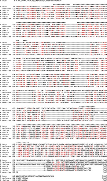
A sequence homology search with the PSI-BLASTP program (Lipman and Pearson, Reference Lipman and Pearson1985) revealed that TbVPS4l was approximately 22–24% identical to sequences from other organisms, including VPS41s of Arabidopsis thaliana (plant; 24% amino acid identity), Mus musculus (metazoa; 22% identity) and Cryptococcus neoformans (fungus; 24% identity) (Fig. 1). The deduced amino acid sequence of TbVPS4l contains the conserved WD40 domain and clathrin heavy chain (CLH) repeat domain found in other VPS41s, which was identified by Pfam search.
Multiple amino acid sequence alignment of TbVPS41 with other VPS41 homologues. Multiple sequence alignment of VSP41 from C. neoformans (fungus, GenBank Accession no. EAL19143), A. thaliana (plant, NP-172297), A. gambiae (insect, XP-313397), M. musculus (AAH23243), H. sapiens (AAB47563) and S. cerevisiae (Z74376) was performed using the GENETYX-MAC computer program. Identical amino acids in at least 4 of the 7 sequences are shown in red. *, WD40 domain; +, CLH domain.

Complementation analysis of TbVPS41 in ΔScVPS41 yeast cells
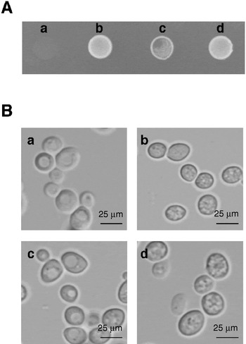
To study the function of TbVPS41, we expressed TbVPS41 in ΔScvps41 mutant yeast cells. RT-PCR analysis confirmed that the expression of TbVPS41 in TbVPS41-transformed ΔScvps41 cells and ScVPS4l expression in ScVPS41-transformed ΔScvps41 cells was equal (data not shown). ΔScvps41 cells did not grow on S-LIM, but the ScVPS41-transformed ΔScvps41 cells had a full recovery of growth on S-LIM. In contrast, the TbVPS41-transformed ΔScvps41 yeast cells showed only a partial recovery of growth on S-LIM, indicating that TbVPS4l partially compensates for VPS41 in the mutant yeast cells (Fig. 2A).
Complementation analysis of ΔScVPS41 yeast cells with TbVPS41. (A) The growth phenotype of △vps41 (a), ScVPS41-transformed △vps41 (b), TbVPS41-transformed △vps41 (c) and wild type (d) yeast on S-LIM. The △vps41 yeast cells could not grow on S-LIM medium, but the ScVPS41-transformed △vps41 yeast cells recovered the ability to grow. The TbVPS41-transformed △vps41 yeast cells partially recovered the ability to grow normally. (B) Differential interference contrast microscopy of wild-type (a), △vps41 (b), ScVPS41-transformed △vps41 (c), and TbVPS41-transformed △vps41 (d) yeast cells.

In addition to an inability to grow on S-LIM, the ΔScvps41 yeast cells had fragmented vacuoles (Zheng et al. Reference Zheng, Wu, Schober, Lewis and Vida1998) (Fig. 2B(b)). Thus, we subsequently analysed whether TbVPS41 can suppress the fragmented vacuole phenotype of ΔScvps41 yeast cells. Differential interference microscopy showed that, despite the partial recovery of growth on S-LIM, there was no suppression of the fragmented vacuoles in the TbVPS41-transformed ΔScvps41 yeast cells and no restoration of a normal large vacuole phenotype (Fig. 2B(d)). However, more than 70% of ScVPS41-transformed ΔScvps41 yeast cells showed a normal large vacuole phenotype (Fig. 2B(c)), which was equal to the level observed in wild type cells (Fig. 2B(a)), suggesting that VPS4l molecules function to mediate growth in low-iron conditions and maintain a normal vacuole phenotype.
TbVPS41 gene knock-down analysis
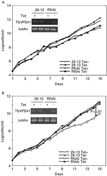
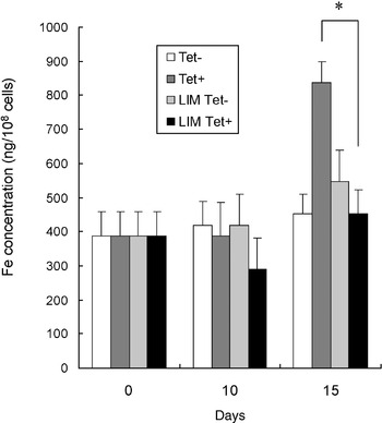
To analyse the physiological function of TbVPS4l, we performed RNAi-mediated gene knock-down analysis of TbVPS41. For these studies, we used T. b. brucei procyclic 29-13 cells, in which RNAi can be induced by the addition of tetracycline. RT-PCR analysis 7 days after the induction of RNAi demonstrated a marked reduction of TbVPS41 transcripts (Fig. 3A, B). The cells grew normally when 0·8 μm of the iron chelator deferoxamine was added to the culture medium (LIM) (data not shown). As expected, there was no significant change in growth when the cells were cultured in normal (iron-rich) medium. The doubling time of the RNAi-induced (TbVPS41 knock-down) cells was 17·1 h, whereas the doubling time of uninduced cells was 16·8 h (Fig. 3A). This suggests the presence of a TbVPS41-independent iron uptake system. On the contrary, in LIM, the doubling time of the RNAi-induced (TbVPS41 knock-down) cells was 20·5 h, whereas the doubling time of uninduced cells was 15·9 h (P<0·01). This indicated that inhibition of TbVPS41 expression caused a significant reduction in the growth rate in LIM. Since trypanosome cells in LIM would mostly depend on intracellular storage of iron molecules, TbVPS41 is considered to possess an intracellular iron-utilization function, including extraction of iron molecules from intracellular storage. To verify this hypothesis, intracellular iron concentrations were subsequently measured. A larger iron concentration was detected in TbVPS41 knock-down cells at 15 days after RNAi induction in normal culture medium, while no significant difference in iron concentration was detected in TbVPS41 knock-down cells in LIM (Fig. 4). These data suggest that when TbVPS41 function is suppressed, extracellular iron uptake is increased to compensate for the intracellular lack of usable iron molecules. Consequently, TbVPS41 knock-down cells could grow normally in normal medium, but could not in LIM.
Growth of control and TbVPS41 knock-down procyclic-form cells. RNAi-mediated gene knock-down was induced with tetracycline. (A) Growth in normal medium. RNAi Tet−, RNAi-uninduced control cells; RNAi Tet+, TbVPS41 knock-down cells; 29-13 Tet−, parental 29-13 control cells; 29-13 Tet+, 29-13 cells with tetracycline. There was no significant difference in the growth of the cell lines. The inset in A shows the RT-PCR analysis of the gene encoding TbVPS41 with tubulin as a control. (B) Growth in LIM. RNAi Tet−, RNAi-uninduced control cells; RNAi Tet+, TbVPS41 knock-down cells; 29-13 Tet−, parental 29-13 control cells; 29-13 Tet+, 29-13 cells with tetracycline. The TbVPS41 knock-down cells grew at a significantly lower rate than the other cells; their doubling time was significantly different than the others (P<0·01). The inset in B shows the RT-PCR analysis of TbVPS41 in LIM with tubulin as a control.

Iron concentration of TbVPS41 knock-down and RNAi-uninduced control cells, in normal medium and LIM. In each analysis, 108 cells were collected at 0 days, 10 days, and 15 days after tetracycline induction. Tet−, control cells in normal medium; Tet+, TbVPS41 knock-down cells in normal medium; Tet− LIM, control cells in LIM; Tet+ LIM, TbVPS41 knock-down cells in LIM. Iron concentration of TbVPS41 knock-down cells at 15 days after tetracycline induction, in normal medium was significantly higher than that in LIM (*: P<0·01).

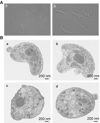
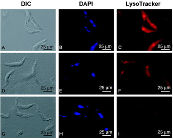
We then analysed whether TbVPS41 knock-down caused intracellular morphological change in trypanosome cells. Differential interference microscopy showed that larger numbers of vesicles were accumulated inside TbVPS41 knock-down cells. Further observation by TEM demonstrated that this was due to a marked increase in the number of small vesicles, which was similar to the fragment vacuole phenotype of ΔScvps41 yeast cells (Fig. 5). Fig. 5B(c, d) shows a representative phenotype of TbVPS41 knock-down cells, which was observed in more than 50% of the transverse sections of TbVPS41 knock-down cells. Since vacuoles and lysosomes are homologous organelles and transferrin accumulates in lysosomes (Grab et al. Reference Grab, Shaw, Wells, Verjee, Russo, Webster, Naessens and Fish1993), lysosome structure was further examined with LysoTracker (Invitrogen), which can specifically accumulate inside lysosomes. Although DAPI-stained nuclear structure was maintained in TbVPS41 knock-down cells, the LysoTracker signal could not be clearly detected (Fig. 6), indicating that TbVPS41 function is essential for maintaining intracellular lysosome structure.
Effect of TbVPS41 knock-down on intracellular morphology. RNAi-mediated gene knock-down was induced with tetracycline. +, With tetracycline; −, no tetracycline. (A) Differential interference contrast microscopy of an RNAi-uninduced control cells (a) and a TbVPS41 knocked-down cells at 15 days after RNAi induction (b). (B) TEM analysis of a parental 29-13 control cell (−) (a), an RNAi-uniduced control cell (−) (b), and TbVPS41 knock-down cells at 15 days after RNAi induction (+) (c, d).

Effect of TbVPS41 knock-down on lysosomal morphology. Parental 29-13 control cells (A, B, C), RNAi-uninduced control cells (D, E, F) and TbVPS41 knocked-down cells at 15 days after RNAi induction (G, H, I), were observed with differential interface contrast (DIC), DAPI-stained (DAPI), and LysoTracker-stained analyses. Clear LysoTracker signal could be detected both in parental 29-13 and RNAi-uninduced control cells, but could not be detected in TbVPS41 knocked-down cells.

DISCUSSION
Several essential enzymes of T. b. brucei have been reported to contain iron, including aconitase, cytochrome oxidase, ribonucleotide reductase, alternative oxidase, and superoxide dismutase (Le Trant et al. Reference Le Trant, Meshnick, Kitchener, Eaton and Cerami1983; Clarkson et al. Reference Clarkson, Bienen, Pollakis and Grady1989; Dormeyer et al. Reference Dormeyer, Schoneck, Dittmar and Krauth-Siegel1997; Saas et al. Reference Saas, Ziegelbauer, von Haeseler, Fast and Boshart2000). A lack of iron inhibits the proliferation of the parasite due to a low rate of DNA synthesis and diminished oxygen consumption. Thus, as in other organisms, iron is an essential molecule for cell growth in T. b. brucei. In addition, these parasites must adapt to different iron conditions when they switch between insect and mammalian hosts.
Procyclic-form cells propagate in iron-rich conditions and indeed require more iron than other forms for optimal growth in culture. Therefore, we studied the procyclic form to explore the unique features of iron utilization. In the course of our extensive survey of genes involved in iron metabolism, we found a potential VPS41-encoding gene. We cloned the full-length gene and investigated its role in iron utilization. To our knowledge, this is the first characterization of a VPS41 gene from a protozoan species.
Knock-down of TbVPS41 caused a delayed growth of T. b. brucei in LIM, while TbVPS41 knock-down had no effect on the growth in iron-rich normal culture medium. Therefore, iron uptake can be performed in the absence of TbVPS41 and normal trypanosomes could continue to grow in LIM because of a TbVPS41-dependent intracellular iron-utilization system that extracts iron molecules from intracellular storage.
In the bloodstream form of trypanosomes, iron-associated transferrin is taken up by receptor-mediated endocytosis (Steverding et al. Reference Steverding, Stierhof, Fuchs, Tauber and Overath1995). From there it is delivered to lysosomes where the iron is dissociated from the transferrin and stored (Grab et al. Reference Grab, Wells, Shaw, Webster and Russo1992). Rates of endocytosis in the bloodstream form are much higher than rates in the procyclic form (Hall et al. Reference Hall, Smith, Langer, Jacobs, Goulding and Field2005). In the procyclic from, although there are some reports of receptor-mediated endocytosis (Liu et al. Reference Liu, Qiao, Du and Lee2000; Garcia-Salcedo et al. Reference Garcia-Salcedo, Perez-Morga, Gijon, Dilbeck, Pays and Nolan2004), the mechanism by which extracellular iron is taken up remains unknown. In the present study, we showed that iron molecules could be taken up by TbVPS41-independent endocytosis and that endocytosis seems to be accelerated by TbVPS41 knock-down in order to compensate for the lack of usable iron molecules. However, the observed data regarding increased iron concentration is an indirect result of endocytosis and an effect of residual TbVPS41 molecules could not be excluded. Thus, the direct analysis of endocytosis with TbVP41 null mutant is necessary for further validation of accelerated endocytosis for compensation of usable iron molecules.
In the course of the search for genes involved in iron metabolism in the T. b. brucei genome database, we found a possible homologue encoding the sequence of CCC1 (t_brucei|chr_3|RPCI93|27F10|27 in the TIGR database), a vacuole membrane transporter gene in yeast cells. Analysis of this gene might provide valuable information regarding the mechanism of iron uptake in procyclic-form cells.
Trypanosome cells in which TbVPS41 was knocked-down showed larger numbers of intracellular vesicles, which is similar to the fragmented vacuoles found in ΔScvps41 yeast cells. Moreover, intracellular lysosome structure was impaired by TbVPS41 knock-down. Thus, while it remains unclear where, if any, iron accumulates in T. b. brucei cells, the lysosome, which is an organelle that is evolutionally homologous to the vacuole, or a lysosome-related organelle might be an iron storage organelle of T. b. brucei.
S. cerevisiae cells in S-LIM appear to be fully dependent on iron molecules stored intracellularly. Since a lack of ScVPS41 function causes a defect in the sorting of vacuole proteins and an inability to grow in yeast cells (Li et al. Reference Li, Chen, McVey Ward and Kaplan2001), the survival of TbVPS41-transformed ΔScvps41 cells clearly demonstrates the evolutionarily conserved functions of VPS41 molecules. However, TbVPS41 could not suppress the fragmented vacuole phenotype of ΔScvps41 cells, indicating that VPS molecules function to utilize iron and maintain intracellular morphology. However, whether those functions are mutually independent or originated from one function of VPS41 molecule is still unclear. Since TbVPS41 could only partially suppress the growth phenotype of ΔScvps41 cells, which might be due to poor homology in the C-terminal region, the fragmented vacuole phenotype might also be partially suppressed. Thus, phenotypes of TbVPS41-transformed and -untransformed ΔScvps41 cells might be indistinguishable. Alternatively, VPS4l proteins might possess two distinct functions possibly derived from their domain structures. Like other homologues of VPS41, TbVPS41 possesses an N-terminal WD40 domain and a C-terminal CLH domain, both of which are thought to be required for protein interaction (Darsow et al. Reference Darsow, Katzmann, Cowles and Emr2001). These two distinct domains may interact with different proteins; consequently, VPS protein may possess different functions. Thus, to further clarify the role of TbVPS41 in iron utilization and the maintenance of intracellular morphology in T. b. brucei, it will be important to perform domain swapping analysis between TbVPS41 and ScVPS41.
This study was supported in part by a Grant-in-Aid for Young Scientists (B), 16790247, from the Ministry of Education, Science, Culture and Sports. This work was also supported in part by the Japan–China Sasagawa Medical Fellowship grant. We thank Dr George Cross for the generous gift of 29-13 strains and Dr Paul T. Englund for the generous gift of PZJM vector.