Introduction
Calcium is an intracellular messenger regulating many cellular functions (Berridge et al., Reference Berridge, Bootman and Lipp1998). Cyclic ADP-ribose (cADPR) and Nicotinic acid adenine dinucleotide phosphate (NAADP+), two metabolites of NAD+, act as second messengers in Ca2+ signaling pathways (Higashida et al., Reference Higashida, Hashii, Yokoyama, Hoshi, Chen, Egorova, Noda and Zhang2001; Rutter, Reference Rutter2003). While cADPR elicits calcium extrusion from endoplasmic reticulum ryanodine-sensitive stores (Guse, Reference Guse1999) homologue to the inositol 1,4,5-trisphosphate (IP3) receptors (Higashida et al., Reference Higashida, Hashii, Yokoyama, Hoshi, Chen, Egorova, Noda and Zhang2001), NAADP+ acts on a receptor whose identity is still unknown (Rutter, Reference Rutter2003).
cADPR is synthesized by a family of highly conserved proteins. These enzymes synthesize cADPR (ADPR-cyclase activity) and/or ADPR (NAD+-glycohydrolase or NADase activity) from β-NAD+ (Schuber & Lund, Reference Schuber and Lund2004). In lower metazoa, for example, in the mollusk Aplysia californica, ADPR-cyclase activity is cytosolic (Glick et al., Reference Glick, Hellmich, Beushausen, Tempst, Bayley and Strumwasser1991). In mammals, enzymes with ADPR-cyclase activity are the ectoenzymes CD38 and CD157 (or BST-1), a membrane-associated (Yamada et al., Reference Yamada, Mizuguchi, Otsuka, Ikeda and Takahashi1997) and membrane-anchored protein (Itoh et al., Reference Itoh, Ishihara and Tomizawa1994), respectively, sharing about 30% sequence identity with the Aplysia cyclase. CD38 is expressed in the nervous system (Yamada et al., Reference Yamada, Mizuguchi, Otsuka, Ikeda and Takahashi1997), in fact, cADPR is abundant in the brain (Walseth et al., Reference Walseth, Aarhus, Zeleznikar and Lee1991; Partida-Sanchez et al., Reference Partida-Sanchez, Cockayne, Monard, Jacobson, Oppenheimer, Garvy, Kusser, Goodrich, Howard and Harmsen2001). Recently, it was shown that a membrane-bound intracellular ADPR-cyclase is expressed in neural cells from Cd38 −/− mice (Ceni et al., Reference Ceni, Muller-Steffner, Lund, Pochon, Schweitzer, De Waard, Schuber, Villaz and Moutin2003, Reference Ceni, Pochon, Villaz, Muller-Steffner, Schuber, Baratier, De Waard, Ronjat and Moutin2006). This enzyme produces intracellular cADPR and ADPR and is involved in brain development and functioning (Ceni et al., Reference Ceni, Pochon, Villaz, Muller-Steffner, Schuber, Baratier, De Waard, Ronjat and Moutin2006), synaptic transmission, and neuronal plasticity (Mothet et al., Reference Mothet, Fossier, Meunier, Stinnakre, Tauc and Baux1998; Reyes-Harde et al., Reference Reyes-Harde, Empson, Potter, Galione and Stanton1999).
Vertebrate retina, with its laminar composition, has been considered a model of central nervous system, from which it derives (1987).
Calcium ions play a pivotal function in phototransduction in the vertebrate retinal rod outer segment (ROS), mediating 3′,5′-cyclic guanosine monophosphate (cGMP) metabolism (Stryer, Reference Stryer1991; Palczewski et al., Reference Palczewski, Kumasaka, Hori, Behnke, Motoshima, Fox, Le Trong, Teller, Okada, Stenkamp, Yamamoto and Miyano2000; Pepe, Reference Pepe2001), and light adaptation in rods and cones (Pepe, Reference Pepe2001). We have shown the existence of a sarco-endoplasmic reticulum Ca2+-ATPase (Panfoli et al., Reference Panfoli, Morelli and Pepe1994; Pepe et al., Reference Pepe, Panfoli, Notari and Morelli2000; Notari et al., Reference Notari, Morelli and Pepe2003) in the ROS, as well as of a Ca2+-sensitive adenylate kinase activity associated to the disks (Notari et al., Reference Notari, Morelli and Pepe2003). Disks would then be organelles able to store and release calcium ions, so that most of the ROS calcium would be sequestered within the disks, consistently with the presence of large-capacity/slow release Ca2+ pool previously documented in rods (Fain & Schröder, Reference Fain and Schröder1985; Panfoli et al., Reference Panfoli, Ravera, Fabiano, Magrassi, Diaspro, Morelli and Pepe2007).
We have described the existence of a cADPR-dependent calcium signaling pathway in bovine ROS, which would possess a cADPR synthetic activity, and cADPR-elicited Ca2+ release influenced by the external calcium concentration (Panfoli et al., Reference Panfoli, Ravera, Fabiano, Magrassi, Diaspro, Morelli and Pepe2007). This synthetic enzyme may be novel because it is membrane bound, endocellular, and it lacks NADase activity (Panfoli et al., Reference Panfoli, Ravera, Fabiano, Magrassi, Diaspro, Morelli and Pepe2007). In this respect, the disk-associated ADPR-cyclase activity of ROS is similar to the intracellular activity described in mouse CD38-deficient brain and synaptosomes (Ceni et al., Reference Ceni, Muller-Steffner, Lund, Pochon, Schweitzer, De Waard, Schuber, Villaz and Moutin2003, Reference Ceni, Pochon, Villaz, Muller-Steffner, Schuber, Baratier, De Waard, Ronjat and Moutin2006), rather than to CD38 or CD157. We had proposed that Ca2+ is released from the disks by cADPR at various rates in response to change in free Ca2+ concentrations caused by light versus dark adaptation.
Here, we report a biochemical characterization of the ROS disk ADPR-cyclase using the substrate nicotinamide guanine dinucleotide (NGD+), which is specific for this enzyme. Overall results suggest that retina, which originates from CNS, shares with brain the property to express an intracellular membrane-bound ADPR-cyclase.
Material and methods
Materials
Ficoll, protease inhibitor cocktail, leupeptin, ampicillin, and NGD+ were purchased from Sigma-Aldrich (St. Louis, MO). Monoclonal antibodies (Abs) used were mouse monoclonal anti-bovine rhodopsin (Rh) (C-terminus, last nine amino acids, catalogue number MAB5356; Chemicon Int., Temecula, CA) and mouse monoclonal Ab against α1 rabbit Na+/K+-ATPase (catalogue number sc-21712). Secondary Abs were anti-mouse conjugated with Cy3 or Cy5 fluorochromes (Molecular Probes, Invitrogen Corporation, Carlsbad, CA) and horseradish peroxidase–conjugated anti-mouse IgG (Amersham Biosciences, Piscataway, NJ). Ultrapure water (Milli-Q; Millipore, Billerica, MA) was used throughout. Safety precautions were taken for chemical hazards in carrying out the experiments described below. All other reagents were of analytical grade.
Preparation of whole-retinal homogenates
All operations were carried out at 4°C. Retinas were extracted in dim red light from freshly enucleated bovine eyes coming from a local abattoir within 1.5 h of animal death. Retinas were significantly deprived of ROS in dim red light according to Schnetkamp and Daemen (Reference Schnetkamp and Daemen1982). Briefly, five retinas were resuspended in 5 ml of mammalian Ringer (extracellular saline) containing (in millimolars) 157 NaCl, 5 KCl, 1 MgCl2, 7 K2HPO4, 8 KH2PO4 (pH 7.3), 5 mM dithiothreitol, and protease inhibitor cocktail (Sigma-Aldrich) in the presence of 35% sucrose (w/v), vortexed for 30 s and centrifuged at 4,000 × g. Operation was repeated three times, and each time retinal pellet was retained and the “ROS-containing” supernatant discarded. Twenty such retinal pellets were homogenized by Potter-Elvehjem system in 20 ml of hypotonic homogenizing buffer containing 10 mM Tris–HCl (pH 8.0), 0.2 mM Phenylmethylsulfonyl fluoride (PMSF), according to Hall and Kuhn (Reference Hall and Kuhn1986). In order to check for the absence of ROS, absorption spectrum from 280 to 700 nm was analyzed and absorbance A280/A500 ratio was significantly higher than 2.5 (21.6), as expected for retinas consistently devoid of ROS.
Preparation of bovine ROS
Purified intact bovine ROS were prepared under dim red light from 20 retinas (from freshly enucleated cattle eyes) at 4°C by sucrose/Ficoll continuous gradient centrifugation (Schnetkamp and Daemen, Reference Schnetkamp and Daemen1982, Panfoli et al., Reference Panfoli, Musante, Bachi, Ravera, Calzia, Cattaneo, Bruschi, Bianchini, Diaspro, Morelli, Pepe, Tacchetti and Candiano2008) in the presence of protease inhibitor cocktail (Sigma-Aldrich) and ampicillin (100 μg/ml).
Osmotically intact disk preparation
Osmotically intact disks were obtained from ROS (Smith & Litman, Reference Smith and Litman1982), as previously reported (Panfoli et al., Reference Panfoli, Musante, Bachi, Ravera, Calzia, Cattaneo, Bruschi, Bianchini, Diaspro, Morelli, Pepe, Tacchetti and Candiano2008). Briefly, ROS were burst for 3 h in distilled water, 70 μg/ml leupeptin and 100 μg/ml ampicillin, then the suspension layered over a solution of 5% (w/v) Ficoll in distilled water with 100 μg/ml ampicillin, and centrifuged for 2 h at 75,000 × g. Disks were collected at the interface between water and Ficoll.
Rhodopsin determination
Rh concentration was determined spectrophotometrically with a dual-beam spectrometer (UNICAM UV2; Analytical S.n.c., Parma, Italy) by measuring the difference in 500 nm absorption, molar extinction coefficient of 41,000 cm−1 M−1 (Wald & Brown, Reference Wald and Brown1954), between spectra recorded before and after exhaustive bleaching (green light, for 5 min) of disk suspensions (0.06 mg/ml, in distilled water). Rhodopsin concentration in the original preparation of disks was 0.8 mM.
Skeletal muscle preparations
Three grams of bovine muscle was homogenized in 10 ml of 0.25 M sucrose, 5 mM HEPES buffer, 1 mM EDTA (pH 7.2), and protease inhibitor cocktail (Sigma-Aldrich). Homogenate was centrifuged for 10 min at 500 × g, the precipitate discarded, and the supernatant centrifuged for 20 min at 20,000 × g (Heraeus centrifuge, Hanau, Germany). A microsomal pellet was collected after the 20,000 × g supernatant was centrifuged for 60 min at 100,000 × g (SW40 Rotor; Beckman, Fullerton, CA).
Transmission electron microscopy of osmotically intact disk preparations
For transmission electron microscopy (TEM) analysis, disks (1.4 mg total protein) were treated as previously described (Stryer, Reference Stryer1991; Panfoli et al., Reference Panfoli, Musante, Bachi, Ravera, Calzia, Cattaneo, Bruschi, Bianchini, Diaspro, Morelli, Pepe, Tacchetti and Candiano2008). Ultrathin sections were analyzed with a Tecnai 12 G2 (FE1, Eindhoven, The Netherlands) electron microscope.
Confocal laser scanning microscopy
Immunohistochemical treatment of disk samples for confocal laser scanning microscopy (CLSM) was conducted in Eppendorf vials (50 μl final volume) on disks (60 μg total protein/sample), as previously described (Ravera et al., Reference Ravera, Calzia, Bianchini, Diaspro and Panfoli2007). Primary Abs were 2 μg/ml of mouse monoclonal Ab against bovine Rh and 2 μg/ml mouse monoclonal Ab against α1 rabbit Na+/K+-ATPase. Secondary anti-mouse Abs were diluted in 10 mM phosphate-buffered saline (PBS). In controls, disks were treated with secondary Abs only, which yielded negligible immunoreactivity, confirming the specificity of disk–surface interaction with the protein target (data not shown). Fluorescence of Cy3 and of Cy5 was excited at 543.5 and 647 nm, respectively. Look-up table liner was used to collect all the acquisitions. The spectral window used for collecting fluorescence was 560–660 nm for Cy3 and conjugates were excited maximally at 650 nm and fluoresced maximally at 660–780 nm for Cy5.
Image acquisition was performed using a confocal microscope (Leica Microsystems S.p.A., Milan, Italy) with an internal spectrophotometer function and equipped with a 20-mW laser diode. The resulting images were acquired and elaborated with the Leica confocal software.
One-dimensional denaturing gel electrophoresis
Denaturing sodium dodecyl sulfate–polyacrylamide gel electrophoresis (SDS-PAGE) was performed according to Laemmli (Reference Laemmli1970) using a Mini Protean IV (Bio-Rad, Hercules, CA) (60 × 80 × 1.5 mm) as described (Graeff et al., Reference Graeff, Walseth, Fryxell, Branton and Lee1994). Separating gel was a gradient from 8 to 14% (w/v) SDS-PAGE. Polypeptides were then transferred electrophoretically onto 0.2 μM nitrocellulose membranes.
Semiquantitative Western blot analysis and densitometric analysis
Nitrocellulose membranes were probed with the same mouse monoclonal Abs against bovine Rh and Na/K-ATPase used for CLSM, diluted in 3% skimmed milk in PBS (1:500 for anti-Rh; 1:1000 for anti-Na+/K+-ATPase). Secondary Abs were diluted (1:5000) in 1% skimmed milk in PBS. Quantitative densitometry was performed using the ImageJ 1.31v software (http://rsb.info.nih.gov/ij/). Each band was converted into a densitometric trace allowing calculations of intensity. Enhanced chemiluminescent band signals were compared with the signal of whole protein pattern. For quantification, densitometric values were compared and results expressed as Relative Optical Density (R.O.D.)
Assay for ADPR-cyclase activity
Putative ADPR-cyclase was assayed by using NGD+, a β-NAD+ analogue, as a substrate, according to Graeff et al. (Reference Graeff, Walseth, Fryxell, Branton and Lee1994) and with the experimental protocol previously reported (Panfoli et al., Reference Panfoli, Ravera, Fabiano, Magrassi, Diaspro, Morelli and Pepe2007). The conversion of NGD+ to cyclic GDP-Ribose (cGDPR) was followed spectrophotometrically at 300 nm. Optical densities were converted to rates of cGDPR production (molar extinction coefficient of 3,758 cm−1 M−1). In fact, the presence of retinal does not allow to conduct a fluorimetric measurement.
Aliquots of bovine-purified disk suspensions (0.10 mg protein) were incubated in a reaction mixture (RM) consisting of 30 mM HEPES (pH 7.2), 120 mM KCl, 10 mM MgCl2 (Panfoli et al., Reference Panfoli, Ravera, Fabiano, Magrassi, Diaspro, Morelli and Pepe2007). For the determination of optimum pH value, aliquots of purified disks were incubated in the presence of 50 μM NGD at 37°C for 30 min in the RM in which a pH gradient (4.5–8.5) was generated with HEPES. The subsequent experiments were performed at the pH optimum for cGDPR production. To study the kinetic properties of ADPR-cyclase, 0.10 mg of disks were incubated for 30 min at 37°C at pH 6.5 in RM with increasing concentrations (5, 10, 20, 30, 50, 75, and 100 μM) of NGD+. K m and V max were calculated from the double-reciprocal plots. To study the effect of Zn2+, Cu2+, and Ca2+, disks (0.20 mg of protein) were incubated 30 min at 37°C with RM, at pH 6.5, with 50 μM NGD+ in the presence of increasing concentrations of ZnSO4 (0, 0.1, 0.5, 1.0, 5.0, and 10.0 μM), CuSO4 (0, 1.0, 2.5, 5.0, 7.5, and 10.0 μM), and CaCl2 (0, 1, 10 and 50 μM) ions.
To verify whether EDTA was able to revert the inhibitory effect of Zn2+ and Cu2+ on disk ADPR-cyclase, samples (0.20 mg of protein) were preincubated at pH 6.5 with RM containing 10 μM Zn2+ or Cu2+ (which elicited maximal inhibition) in the presence of 0.5 mM EDTA for 10 min at room temperature. Then reaction was started adding 50 μM NGD+, and samples were incubated for 30 or more min at 37°C.
Finally to study the effect of light- and dark-adapted conditions on ADPR-cyclase activity, purified intact ROS were utilized (0.2 mg of protein). ROS were preincubated in physiological conditions: 2 mM glucose, 1 mM CaCl2, and 0.025 mg/ml ampicillin in Mammalian Ringer, at room temperature for 10 min. Then, ROS were homogenized and immediately added to the RM and the reaction started by addition of 50 μM NGD+. Samples were incubated for 30 min at 37°C, either in darkness or in saturating room light.
Protein assay
Protein content was determined by bicinchoninic acid protein assay kit from Pierce Biotechnology, Inc. (Rockford, IL), with BSA as the standard.
Results
Characterization of the disk fractions
The disk fractions are composed of swollen vesicles that were defined “osmotically intact” (Smith & Litman, Reference Smith and Litman1982) as disks swell but do not burst by hypotonic shock thank to their peculiar phospholipid composition (Lamba et al., Reference Lamba, Borchman and O’Brien1994), therefore they can be collected at the interface between 5% Ficoll solution and water, after ultracentrifugation. Purity of the disk fractions was assayed by TEM and immunofluorescence CLSM imaging and immunoblot analyses.
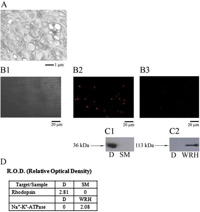
Panel A in Fig. 1 is a TEM image of the disk fraction, representative of three different experiments. Flattened disks and sealed vesicles were observed. No melanosomes or rod inner segment (RIS) organelles, such as mitochondria or endoplasmic reticulum were observed. Vesicles of other nature would not float up to the disk band (Smith & Litman, Reference Smith and Litman1982). By a new technique that we have recently developed (Ravera et al., Reference Ravera, Calzia, Bianchini, Diaspro and Panfoli2007), allowing to image disks with CLSM by indirect immunohistochemistry, experiments were performed to monitor with specific Abs the presence on disks of two proteins: the visual pigment Rh and the sodium/potassium ATPase pump (Na+/K+-ATPase) as an example of a RIS plasma membrane protein. The proteins are stained by indirect immunocytochemistry (in red or green being secondary Abs coupled to Cy5 or Cy3). Panel B1 is a phase-contrast CLSM image of disks. Panel B2 shows that the visual pigment is uniformly distributed on disks. Panel B3 shows the fluorescence of a Cy3-labeled secondary Ab bound to an Ab against α1 subunit of rabbit Na+/K+-ATPase. Spectral analysis of fluorochromes showed that immunofluorescence was specific of Cy3- and Cy5-labeled secondary Abs (data not shown). Semiquantitative Western immunoblot analyses were conducted by means of the same Abs utilized for CLSM. Panel C1 shows that Rh is present in isolated disks fractions (D). Apparent molecular weight (MW) was 36 ± 2 kDa. Bovine skeletal muscle (SM) fractions were used as negative control. Panel C2 shows the Western immunoblot analysis of disks Na+/K+-ATPase. Signal was present in whole-retinal homogenate fractions, used as positive control, and absent in isolated disks fractions. Apparent MW was 113 ± 10 kDa. Results suggest negligible RIS contamination in disks, similarly to what reported in Panfoli et al. (Reference Panfoli, Ravera, Fabiano, Magrassi, Diaspro, Morelli and Pepe2007). Relative quantification, shown in Panel D of chemiluminescent band signals, was calculated as the ratio of densitometric value of the band to that of total protein in each lane of the SDS-PAGE gel.
Morphological analysis of osmotically intact ROS disk membranes. (A) Osmotically intact purified disk suspensions observed by TEM. Mitochondria and other evident RIS subfractions are absent. The length of the membrane profiles ranged about 1 μm. (B1) CLSM phase-contrast image of intact disks. (B2) CLSM fluorescence image of intact disks stained with primary monoclonal Ab against bovine rhodopsin and a Cy3 secondary Ab. (B3) CLSM fluorescence image of intact disks stained with primary monoclonal Ab against Na+/K+-ATPase and a Cy5-bound secondary Ab. (C1) Western blot analysis using the same primary Ab performed on intact disks fractions (D) and bovine SM fractions, used as negative control. (C2) Western blot analysis using the same primary Ab performed on intact disks fractions (D) and whole-retinal homogenate (WRH) used as positive control. (D) Relative quantification of protein band density, expressed as the ratio of relative optical densities (R.O.D.) of chemiluminescent signal in panel (C1) and (C2) to the R.O.D. of total stained protein, subtracted of the background values. Each value in the table shows a densitometric analysis performed on a single blot, representative of three experiments.

Kinetic properties of the putative ADPR-cyclase of bovine ROS disks
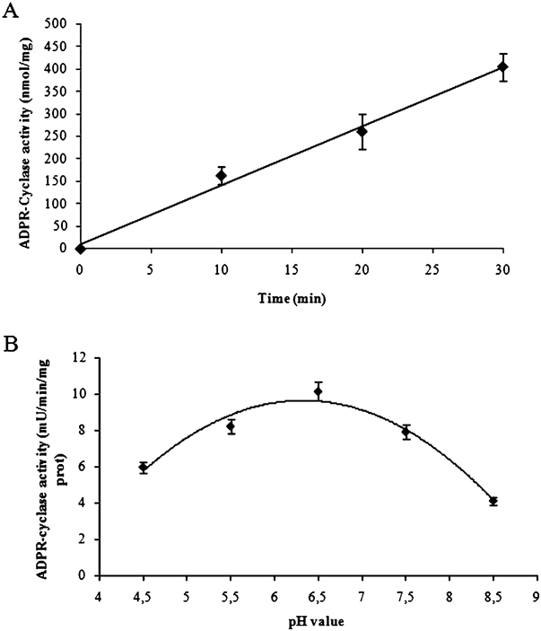
The GDPR-cyclase activity was tested in disk fractions using a spectrophotometric cycling assay, which quantifies the production of cGDPR from NGD+, chosen because this substrate is more specific and has an absorption spectrum different from both NGD+ and GDPR (Graeff et al., Reference Graeff, Walseth, Fryxell, Branton and Lee1994). This method is based on NGD+ conversion by most known ADPR-cyclases into a cyclic derivative, cGDPR, is resistant to hydrolysis (Graeff et al., Reference Graeff, Walseth, Fryxell, Branton and Lee1994). Disks were assayed at 37°C, at pH 6.5. Fig. 2A shows that disks contain readily detected ADPR-cyclase activity, with an average initial rate of 15 ± 2 nmoles cGDPR/min/mg protein. Fig. 2B shows the dependence of disk ADPR-cyclase activity on pH. A pH gradient (4.5–8.5) was built with HEPES. Optimum pH value was around 6.5, while the best pH range was among 6.0 and 7.0. Activity was strongly inhibited at pH 8.5 and 4.5. Such result is very similar to that obtained by Ceni et al. (Reference Ceni, Muller-Steffner, Lund, Pochon, Schweitzer, De Waard, Schuber, Villaz and Moutin2003, Reference Ceni, Pochon, Villaz, Muller-Steffner, Schuber, Baratier, De Waard, Ronjat and Moutin2006) for the brain endocellular ADPR-cyclase. The fluorimetric measurements of cGDPR production were prevented, due to the fluorescence of retinal at 300 nm, as previously discussed (Panfoli et al., Reference Panfoli, Ravera, Fabiano, Magrassi, Diaspro, Morelli and Pepe2007).
Assay of ADPR-cyclase activity in osmotically intact bovine ROS disks. (A) Time course of cGDPR generation by intact ROS disks with 50 μM NGD+. cGDPR production was assayed spectrophotometrically at 300 nm wavelength (filled circles). Each circle represents cGDPR production (mean ± s.d., n = 4), after 10, 20, and 30 min from addition of NGD+, respectively. (B) Effect of pH on ADPR-cyclase activity of ROS disks. Samples were incubated at various pH, as described in material and methods section. Each circle represents cGDPR production (mean ± s.d., n = 3). Optical densities were converted to rates of cGDPR production (ε = 3758/M/cm).

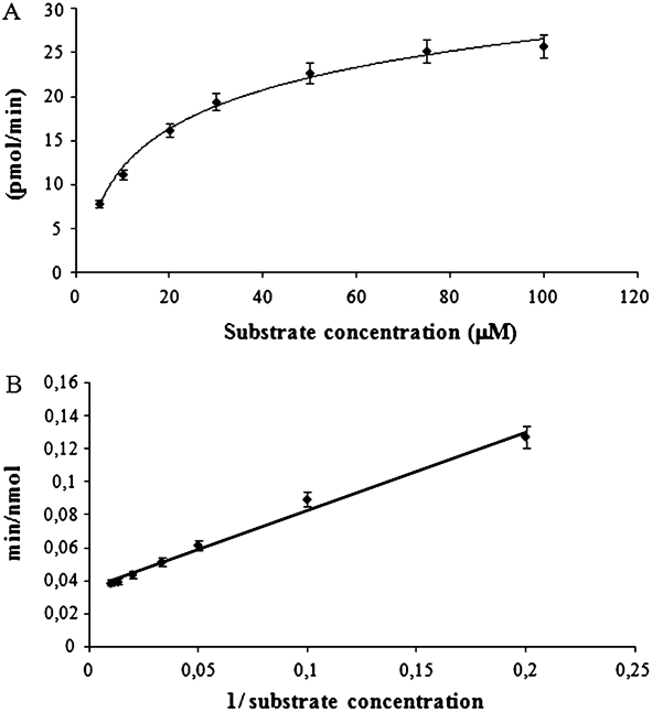
The kinetic properties of the disk cyclase were studied and the apparent K m and V max for the transformation of NGD+ in cGDPR. Purified disks were incubated at pH 6.5 and at 37°C in the presence of increasing concentrations of NGD+, in order to determine the initial rates of cGDPR synthesis. Fig. 3A shows the experimental data fitted assuming a hyperbolic dependence from NGD+ concentration. From the double-reciprocal plot shown in Fig. 3B, a K m of 12.5 ± 0.3 μM (mean ± s.d., n = 5) was estimated for NGD+. This value is within the range of the Michaelis constants determined for the other known mammalian enzymes of the cyclase/NADase family (Price & Pekala, Reference Price, Pekala, Dolphin, Poulson and Avramovic1987; Hirata et al., Reference Hirata, Kimura, Sato, Ohsugi, Takasawa, Okamoto, Ishikawa, Kaisho, Ishihara and Hirano1994; Kukimoto et al., Reference Kukimoto, Hoshino, Kontani, Inageda, Nishina, Takahashi and Katada1996; Cakir-Kiefer et al., Reference Cakir-Kiefer, Muller-Steffner, Oppenheimer and Schuber2001; Ceni et al., Reference Ceni, Muller-Steffner, Lund, Pochon, Schweitzer, De Waard, Schuber, Villaz and Moutin2003). The V max of the putative disk ADPR-cylase, estimated from the plot in Fig. 3B, was 26.50 ± 0.70 (mean ± s.d., n = 5) pmol cGDPR formed/min/mg.
Determination of K m and V max of ADPR-cyclase activity. (A) cGDPR production rate (picomoles/minute) in the presence of increasing concentrations (expressed in micromolars) of the substrate (NGD+). The values of each condition are representative of five experiments (mean ± s.d., n = 5). (B) Double-reciprocal plot of Panel A.

Effect of micromolar concentrations of Zn2+, Cu2+, and Ca2+ ions on the disk ADPR-cyclase
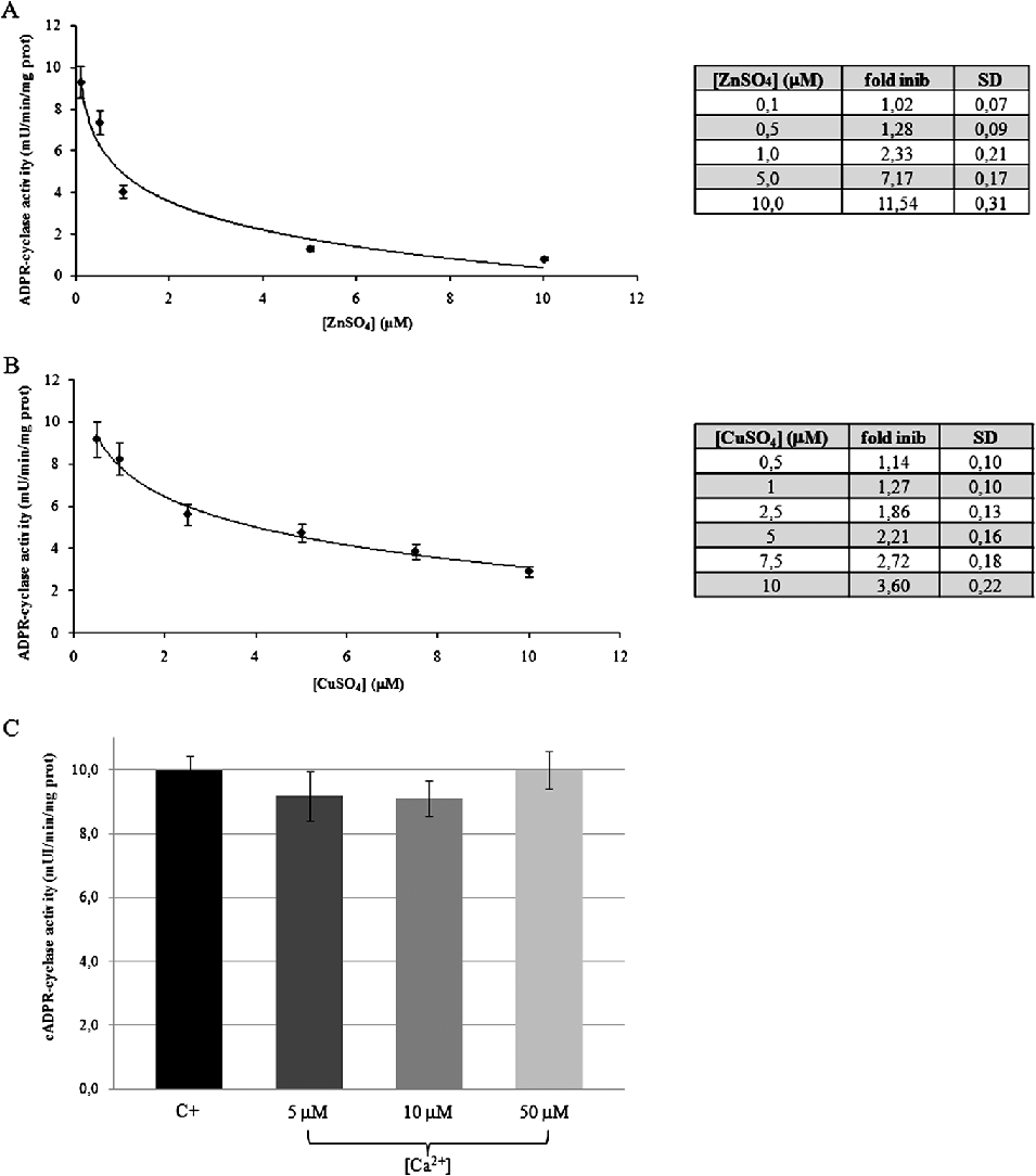
We examined the effect of increasing concentrations of zinc sulfate (ZnSO4), copper sulfate (CuSO4), and calcium chloride (CaCl2), in the micromolar range, on the cGDPR production by the disk cyclase. The cGDPR production by disks decreased with increasing both Zn2+ (Fig. 4A) and Cu2+ (Fig. 4B) concentration, with an half maximal Inhibitory Concentration (IC50) of 1.1 μM for Zn2+ and 3.6 μM for Cu2+, respectively (very similar to that of the synaptosomal ADPR-cyclase activity obtained by Ceni et al., Reference Ceni, Pochon, Villaz, Muller-Steffner, Schuber, Baratier, De Waard, Ronjat and Moutin2006). Ten micromolar Zn2+ and Cu2+ caused 91% and 72% inhibition of the cGDPR synthesis, respectively (see Fig. 4, insets). By contrast, the effect of Ca2+ ions on cGDPR production, tested at 1, 10, and 50 μM Ca2+ was negligible (Fig. 4C).
Zn2+, Cu2+, and Ca2+ effect on ADPR-cyclase activity of intact ROS disks. (A) Effect of increasing concentrations of zinc ions on ADPR-cyclase activity. Each fitted circle represents the ADPR-cyclase activity (mean ± s.d., n = 4) at the various Zn2+ concentrations. Table in the inset shows the fold inhibition (mean ± s.d., n = 4) of the cyclase activity. (B) Effect of increasing concentration of copper ions on ADPR-cyclase activity. Each fitted circle represents the activity (mean ± s.d., n = 4) at the various Cu2+ concentrations. Table in the inset shows the fold inhibition (mean ± s.d., n = 4) of the correspondent ADPR-cyclase activity. (C) ADPR-cyclase showing the effect of increasing concentration of calcium ions on ADPR-cyclase activity. Each bar represents the activity (mean ± s.d., n = 4) at the various Ca2+ concentrations.

EDTA reverts inhibitory action of Zn2+ and Cu2+ ions on the disk ADPR-cyclase
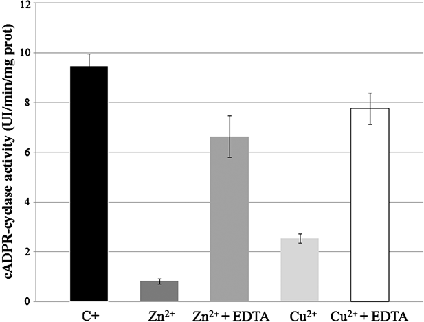
The reverting of inhibition of disk GDPR-cyclase by 10 μM Zn2+ and Cu2+ was tested incubating disks with 0.5 mM EDTA and either of the two ions for 10 min at room temperature, before addition of NGD+. Then, the reaction was started and, after 30 min of incubation with substrate, data showed a good, albeit not total (Fig. 5), reversion of the inhibitory effect of Zn2+ and Cu2+ ions, with respect to the controls.
Reversion of Zn2+ and Cu2+ ions action on the disk ADPR-cyclase by EDTA. Histogram shows the ability of EDTA to revert Zn2+ and Cu2+ action on the disk ADPR-cyclase. The first bar represents activity (mean ± s.d., n = 4) of ADPR-cyclase without inhibitors; the second and the fourth bar represent, respectively, the activity (mean ± s.d., n = 4) of ADPR-cyclase treated with 10 μM ZnSO4 or CuSO4; the third and fifth bar represent the activity (mean ± s.d., n = 4) ADPR-cyclase treated at 10 μM ZnSO4 or CuSO4, respectively, in the presence of EDTA (0.5 mM).

Effect of light conditions on GDPR-cyclase activity
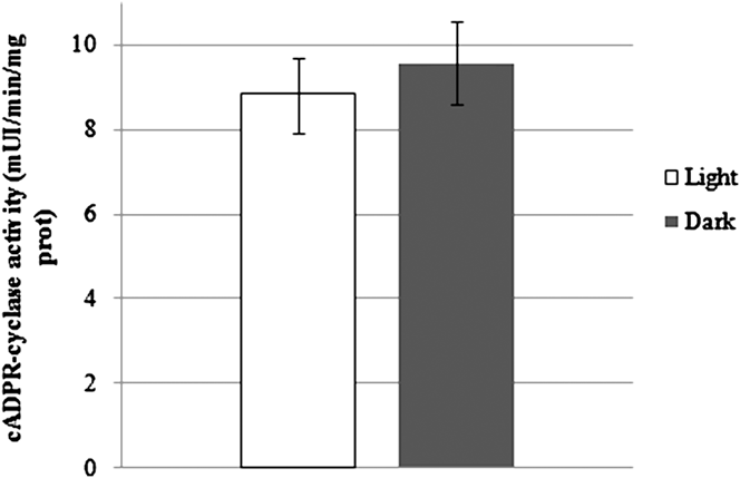
The effect of light- or dark-adapted conditions on GDPR-cyclase activity was tested on purified intact ROS, in physiological ionic conditions, as described in the material and methods section. ROS were first incubated for 10 min either in the dark or in saturating light conditions in Mammalian Ringer containing 2 mM glucose and 1 mM CaCl2. Then, ROS were homogenated and additioned to RM and NGD+. Fig. 6 shows that there was no significant difference between samples assayed in light- or dark-adapted conditions.
Effect of light and dark action on the disk ADPR-cyclase. Histogram representative of light and dark action on the disk ADPR-cyclase. The first bar represents the activity value (mean ± s.d., n = 6) of ADPR-cyclase at the light; the second bar represents the activity value (mean ± s.d., n = 6) of ADPR-cyclase at the dark.

Discussion
In the present paper, we characterized in the purified ROS disks the kinetic properties of a putative ADPR-cyclase activity that we had previously localized, intracellularly, in the bovine retinal ROS (Panfoli et al., Reference Panfoli, Ravera, Fabiano, Magrassi, Diaspro, Morelli and Pepe2007), using its specific substrate, NGD+ (a NAD+ analogue). In fact, disk ADPR-cyclase is able to transform NGD+ into cGDPR.
The catalytic activity of the disk GDPR-cyclase functions best between pH 6.0 and 7.0, with a pH optimum at 6.5. Affinity for NGD+ was in the micromolar range (K m = 12.5 ± 0.3 μM) and V max was 26.50 ± 0.70 pmol cGDPR formed/min/mg. In many respects, the disk cyclase is similar to the novel cyclase described in the CD38-deficient mouse brain membranes and synaptosomes (Ceni et al., Reference Ceni, Muller-Steffner, Lund, Pochon, Schweitzer, De Waard, Schuber, Villaz and Moutin2003, Reference Ceni, Pochon, Villaz, Muller-Steffner, Schuber, Baratier, De Waard, Ronjat and Moutin2006) rather than of the other known enzymes of the family (the mammalian CD38 and CD157, and the Aplysia cyclase) (Glick et al., Reference Glick, Hellmich, Beushausen, Tempst, Bayley and Strumwasser1991; Yamada et al., Reference Yamada, Mizuguchi, Otsuka, Ikeda and Takahashi1997). However, the synaptosomal cyclase was unable to transform NGD+ into the cyclic compound, which acted as an inhibitor (Ceni et al., Reference Ceni, Pochon, Villaz, Muller-Steffner, Schuber, Baratier, De Waard, Ronjat and Moutin2006), a major difference among the bovine disk and the mouse brain cyclase.
The disk ADPR-cyclase was sensitive to Zn2+ and Cu2+, being inhibited by micromolar concentrations of these ions. Also, micromolar Cu2+ concentrations were inhibitory, an effect observed at higher concentrations on CD157 activity (Hirata et al., Reference Hirata, Kimura, Sato, Ohsugi, Takasawa, Okamoto, Ishikawa, Kaisho, Ishihara and Hirano1994; Kukimoto et al., Reference Kukimoto, Hoshino, Kontani, Inageda, Nishina, Takahashi and Katada1996). The effect of these divalent ions was almost totally reverted by incubating 0.5 mM EDTA (see Fig. 5). By contrast, the ADPR-cyclase activities of CD38 were activated by Zn2+ and Cu2+, whereas Zn2+ but not Cu2+ activated CD157 (Hirata et al., Reference Hirata, Kimura, Sato, Ohsugi, Takasawa, Okamoto, Ishikawa, Kaisho, Ishihara and Hirano1994; Kukimoto et al., Reference Kukimoto, Hoshino, Kontani, Inageda, Nishina, Takahashi and Katada1996). This opposing effect of zinc and copper ions on the disk cyclase, with respect to CD38 and CD157, is common to the novel intracellular membrane-bound synaptosomal enzyme (Ceni et al., Reference Ceni, Pochon, Villaz, Muller-Steffner, Schuber, Baratier, De Waard, Ronjat and Moutin2006).
Zinc, a trace element, has been recognized as an essential heavy metal for plants and animals (Prasad, Reference Prasad1995). In fact, it functions in many physiological pathways and has critical roles in the central nervous system, both on the synaptic activity and during brain development and aging (Hyun et al., Reference Hyun, Sohn, Ahn, Shin, Koh and Yoon2000; Grahn et al., Reference Grahn, Paterson, Gottschall-Pass and Zhang2001). Zinc is present in high concentrations in the nervous system (about 150 μM) (Prasad, Reference Prasad1995) and in the ocular tissue, particularly in retina (Hyun et al., Reference Hyun, Sohn, Ahn, Shin, Koh and Yoon2000), which is more sensitive to the lack of dietary zinc than the liver. The physiological functions of Zn2+ have been studied predominantly in retina and retinal pigment epithelium where it is believed to interact with taurine and vitamin A, modify photoreceptor plasma membranes, regulate the light–rhodopsin reaction, modulate synaptic transmission, and serve as an antioxidant (Miceli et al., Reference Miceli, Tate, Alcock and Newsome1999). It was reported that in the presence of Zn2+ the thermal stability of overall secondary structure content of rhodopsin is increased (Balem et al., Reference Balem, Yanamala and Klein-Seetharaman2009). This was also noted with Cu2+ but not with other metal ions (Balem et al., Reference Balem, Yanamala and Klein-Seetharaman2009). Zn2+ deficiency causes retinal dysfunctions, such as night blindness and neurodegeneration, probably affecting the retinol–retinal conversion. In a murine model, Zn, Cu, Mn, and vitamins A and E deficiencies caused photoreceptor cells to degenerate and disappear (Amemiya, Reference Amemiya2000). Zinc deficiency has also been associated to the development and progression of several chronic eye diseases, among which age-related macular degeneration (Krishnadev et al., Reference Krishnadev, Meleth and Chew2010), through an as yet uncertain pathogenic mechanism that may include increase in oxidative stress (Miceli et al., Reference Miceli, Tate, Alcock and Newsome1999).
Copper is an important nutrient for normal brain and retinal homeostasis serving as a cofactor of key metabolic enzymes involved in mitochondrial function, neurotransmitter biosynthesis, and oxidative stress defense (Krishnadev et al., Reference Krishnadev, Meleth and Chew2010). Copper deficiency has been associated to the pathogenesis of retinopathy of prematurity, a major cause of blindness in very low-birth weight preterm infants (Caddell, Reference Caddell1995). Ceruloplasmin, a retinal ferroxidase, is upregulated at the messenger RNA and protein levels upon light damage (Chen et al., Reference Chen, Dentchev, Wong, Hahn, Wen, Bennett and Dunaief2003). Ceruloplasmin is considerably more abundant in retina than in brain. Patients with aceruloplasminemia display retinal degeneration (Chen et al., Reference Chen, Dentchev, Wong, Hahn, Wen, Bennett and Dunaief2003). Abnormal copper homeostasis is the primary cause of Menkes disease and Wilson disease, severe metabolic disorders that are associated with retinal degeneration (Barnes et al., Reference Barnes, Tsivkovskii, Tsivkovskaia and Lutsenko2005; Krajacic et al., Reference Krajacic, Qian, Hahn, Dentchev, Lukinova and Dunaief2006).
Therefore, the retinal rod disk ADPR-cyclase may be a new intracellular target of Zn2+ and Cu2+ depletion or toxic action. By acting on this enzyme, zinc and copper ions may modify ROS calcium homeostasis, affecting the crucial role of the disk ADPR-cyclase in Ca2+-dependent processes pivotal for phototransduction.
The presence of an ADPR-cyclase activity in ROS carries great potential significance in revealing additional complexity to internal Ca2+ dynamics in ROS. It may provide some explanation for the transient light-induced increase in intracellular Ca2+ observed under some conditions and for the failure of rods to attain the theoretical Ca2+ minimum when all cGMP channels are closed (Fain & Schröder, Reference Fain and Schröder1985). In fact, disk ADPR-cyclase was not affected by change in light conditions during an incubation of intact ROS in physiological conditions (Fig. 6) nor by change of calcium concentration (Fig. 4C), suggesting that the enzyme has a basal activity, which is indirectly regulated by cytosolic calcium ion levels, in turn eliciting and calcium release from disks ryanodine receptors (Panfoli et al., Reference Panfoli, Ravera, Fabiano, Magrassi, Diaspro, Morelli and Pepe2007).
Acknowledgments
We thank I. Mario Pepe (University of Genoa, Italy) for his invaluable contribution. The work was supported by grants from Italian Ministry for Research and Technology (MIUR).