Introduction
Bipolar disorder (BD) is a severe psychiatric disease characterized by recurrent periods of mania and depression (BD I) or hypomania and depression (BD II). It is a leading cause of disability in young people as they can lead to cognitive and functional impairment and increased mortality, particularly from suicide and cardiovascular disease (Vieta et al., Reference Vieta, Berk, Schulze, Carvalho, Suppes, Calabrese and Grande2018). Specifically, anxiety and depression together with alexithymia may be considered as risk factors for negative outcomes such as suicidal behavior, even simply enhancing vulnerability to depressive symptoms or per se (De Berardis et al., Reference De Berardis, Fornaro, Orsolini, Valchera, Carano, Vellante and Di Giannantonio2017). Additionally, most of the depressed patients demonstrate psychomotor retardation including slowed speech, decreased movement, and impaired cognitive function (Buyukdura, McClintock, & Croarkin, Reference Buyukdura, McClintock and Croarkin2011). Although abnormalities in specific brain regions and connections in BD have been reported by growing structural and functional neuroimaging studies, the neuropathology of BD is still not fully understood.
Resting-state functional magnetic resonance imaging (rs-fMRI) is a non-invasive neuroimaging technique that has been used to investigate the abnormal regional function and functional connectivity (FC) in BD (Vargas, López-Jaramillo, & Vieta, Reference Vargas, López-Jaramillo and Vieta2013). Two methods widely used in FC studies include seed-based correlation approach (Anticevic et al., Reference Anticevic, Brumbaugh, Winkler, Lombardo, Barrett, Corlett and Glahn2013; Chai et al., Reference Chai, Whitfield-Gabrieli, Shinn, Gabrieli, Castanon, McCarthy and Oenguer2011) and independent component analysis (ICA) (Khadka et al., Reference Khadka, Meda, Stevens, Glahn, Calhoun, Sweeney and Pearlson2013; Oenguer et al., Reference Oenguer, Lundy, Greenhouse, Shinn, Menon, Cohen and Renshaw2010). The ICA is a data-driven approach that does not require prior knowledge of predefined brain regions or voxels when estimating functional networks (Du, Fu, & Calhoun, Reference Du, Fu and Calhoun2018). Several studies reported altered FC in the default mode network (DMN) (Wang et al., Reference Wang, Gao, Tang, Lu, Zhang, Bu and Huang2020b; Zhong et al., Reference Zhong, Wang, Gao, Xiao, Lu, Jiao and Lu2019), limbic network (Chen et al., Reference Chen, Wang, Niu, Zhong, Hu, Chen and Huang2018; Wang et al., Reference Wang, Zhong, Jia, Sun, Wang, Liu and Huang2016b; Wessa, Kanske, & Linke, Reference Wessa, Kanske and Linke2014), somatomotor network/sensorimotor network (SMN) (Ishida et al., Reference Ishida, Donishi, Iwatani, Yamada, Takahashi, Ukai and Kaneoke2017; Khadka et al., Reference Khadka, Meda, Stevens, Glahn, Calhoun, Sweeney and Pearlson2013), and dorsal and ventral attention network (Son, Han, Kim, Min, & Renshaw, Reference Son, Han, Kim, Min and Renshaw2017) in BD. Reasons for the inconsistent findings are as following: heterogeneity of the patients included, different states and subtypes of BD, illness duration, drug administration, multiple comparison correction strategies, and metrics of fMRI applied.
Studies investigating systemic inflammation and immune dysregulation have provided important insights into the pathophysiologic processes underlying BD (Felger, Reference Felger2018; Frey et al., Reference Frey, Andreazza, Houenou, Jamain, Goldstein, Frye and Young2013; Goldsmith, Rapaport, & Miller, Reference Goldsmith, Rapaport and Miller2016; Goldstein, Kemp, Soczynska, & McIntyre, Reference Goldstein, Kemp, Soczynska and McIntyre2009). Pro-inflammatory cytokines have been shown to access the brain (Miller, Maletic, & Raison, Reference Miller, Maletic and Raison2009) and interact with neurocircuits to influence the risk for depression (Miller & Raison, Reference Miller and Raison2016) and are implicated in many neurobiological processes including neuromodulator effects, neurotransmitter-like effects, regulation of neurogenesis that is potentially relevant to psychiatric disorders such as BD (Stuart & Baune, Reference Stuart and Baune2014). Previous studies examined the relationship between inflammation and BD focusing on abnormal peripheral pro-inflammatory cytokines levels, and found significantly elevated levels of interleukin 6 (IL-6) (Brietzke et al., Reference Brietzke, Stertz, Fernandes, Kauer-Sant'anna, Mascarenhas, Escosteguy Vargas and Kapczinski2009; Modabbernia, Taslimi, Brietzke, & Ashrafi, Reference Modabbernia, Taslimi, Brietzke and Ashrafi2013; Remlinger-Molenda, Wojciak, Michalak, & Rybakowski, Reference Remlinger-Molenda, Wojciak, Michalak and Rybakowski2012; Wu et al., Reference Wu, Zheng, Tian, Lai, Hu, Zhang and Hu2017) and interleukin 8 (IL-8) (Lu et al., Reference Lu, Rao, Mou, Chen, Lou, Zhang and Fang2019; Strawbridge et al., Reference Strawbridge, Hodsoll, Powell, Hotopf, Hatch, Breen and Cleare2019; Wang et al., Reference Wang, Lee, Chen, Chung, Li, Chang and Lu2016a) in BD. Higher baseline IL-6 and IL-8 levels have been found to be related to poorer antidepressant responses in BD (Benedetti et al., Reference Benedetti, Poletti, Hoogenboezem, Locatelli, de Wit, Wijkhuijs and Drexhage2017). Moreover, inflammation was associated with cognitive dysfunction such as poor performance in the memory, speed domains (information processing speed and psychomotor speed) and motor function in the older population (Baune et al., Reference Baune, Ponath, Golledge, Varga, Arolt, Rothermundt and Berger2008; Chi et al., Reference Chi, Fitzpatrick, Sharma, Jenny, Lopez and DeKosky2017; Heringa et al., Reference Heringa, van den Berg, Reijmer, Nijpels, Stehouwer, Schalkwijk and Biessels2014). Another study indicated that inflammation (modulated by IL-6) causes mood deterioration through alterations in emotional processing related neural activity (Harrison et al., Reference Harrison, Brydon, Walker, Gray, Steptoe and Critchley2009).
Some previous findings have demonstrated associations between systemic inflammation and FC disruption within the DMN and the dorsal attention network (Magalhães et al., Reference Magalhães, Weiler, Teixeira, Hayata, Moraes, Boldrini and Balthazar2018; Marsland et al., Reference Marsland, Kuan, Sheu, Krajina, Kraynak, Manuck and Gianaros2017; Walker et al., Reference Walker, Gross, Moghekar, Soldan, Pettigrew, Hou and Walston2020). However, the correlation between systemic inflammation and FC in the limbic network and SMN remains largely unknown. The limbic network, which includes the hippocampus, para-hippocampal gyrus, amygdala, orbitofrontal cortex (OFC), cingulate gyrus (Catani, Dell'acqua, & Thiebaut de Schotten, Reference Catani, Dell'acqua and Thiebaut de Schotten2013), involves in various functions such as emotional processing and regulation (Delvecchio et al., Reference Delvecchio, Fossati, Boyer, Brambilla, Falkai, Gruber and Frangou2012; Hariri, Bookheimer, & Mazziotta, Reference Hariri, Bookheimer and Mazziotta2000) and reward processing (Dutra, Man, Kober, Cunningham, & Gruber, Reference Dutra, Man, Kober, Cunningham and Gruber2017; Phillips & Swartz, Reference Phillips and Swartz2014). Previous studies have suggested altered FC of limbic network may contribute to emotional dysregulation in BD (Houenou, Perlini, & Brambilla, Reference Houenou, Perlini and Brambilla2015; Wessa et al., Reference Wessa, Kanske and Linke2014). In addition, the involvement of sensory perception in emotional processes and clinical outcomes of individuals with psychiatric disorders such as BD has been clearly reported (Engel-Yeger et al., Reference Engel-Yeger, Muzio, Rinosi, Solano, Geoffroy, Pompili and Serafini2016). More importantly, psychomotor retardation and reduced daily activities are core features of the depressive syndrome including BD (Bracht et al., Reference Bracht, Steinau, Federspiel, Schneider, Wiest and Walther2018). Besides, recent research suggested abnormal FC of SMN may result in psychomotor alterations in BD (Martino et al., Reference Martino, Magioncalda, Conio, Capobianco, Russo, Adavastro and Northoff2020). Given these considerations, we tried to focus on the FC patterns of limbic network and SMN and their correlations with systemic inflammation.
In this study, we tried to detect brain FC alterations in limbic network and SMN based on ICA approach. Because medication use and different subtypes of patients could confound the findings, all the BD II patients recruited in the present study were unmedicated. We hypothesized that patients with BD II would show disrupted FC patterns in regions of the limbic network or SMN compared to healthy controls (HCs), and elevations of pro-inflammatory markers IL-6 and IL-8 would exist in BD groups. Additionally, we sought to determine whether inflammation (as identified by changes in pro-inflammatory markers) is correlated to FC disruption in limbic network and SMN.
Materials and methods
Participants
A total of 42 right-handed, currently depressed individuals (7 outpatients, 35 inpatients) diagnosed with BD II were recruited from the psychiatry department, First Affiliated Hospital of Jinan University, Guangzhou, China. The patients were aged from 18 to 55 years. All patients met Diagnostic and Statistical Manual of Mental Disorders, Fifth Edition (known as DSM-V) criteria for BD II according to the diagnostic assessment by the Structured Clinical Interview for DSM-V Patient Edition (SCID-P) by two experienced psychiatrists (Y.J. and S.Z., with 21 and 6 years of experienced clinical psychiatry, respectively). The clinical state was assessed by using the 24-item Hamilton Depression Rating Scale (HDRS) and the Young Mania Rating Scale (YMRS) during the 3-day period prior to the imaging session. The inclusion criterion for the depressed patients with BD II was a total HDRS-24 score > 21 and YMRS score <7. The exclusion criteria were patients with other Axis-I psychiatric disorders, a history of electroconvulsive therapy, neurological disorders, any history of organic brain disorder, mental retardation, pregnancy, alcohol/substance abuse, cardiovascular diseases or any presence of a concurrent and major physical illness. At the time of testing, all patients were either medication-naïve, or were not medicated for at least 6 months. Among 42 BD II depression patients, 25 patients were at suicide risks (13 subjects with a positive history of suicide attempts, 12 patients with active suicidal ideation). In addition, 69 right-handed HC were recruited via local advertisements. They were carefully screened through a diagnostic interview, the SCID Nonpatient Edition, to rule out the presence of the current or past history of any psychiatric illness. Further exclusion criteria for HCs were any history of psychiatric illness in first-degree relatives, current or past significant medical or neurological illness.
MR Imaging Data Acquisitions and Preprocessing
All MRI data were obtained on a GE Discovery MR750 3.0 T System with an eight-channel phased-array head coil. The participants were scanned in a supine, head-first position with symmetrically placed cushions on both sides of the head to decrease motion. During the scanning, the participants were instructed to relax with their eyes closed without falling asleep. After the experiment, each participant confirmed not having fallen asleep.
The rs-fMRI data were acquired using a gradient-echo echo-planar imaging sequence with the following parameters: time repetition (TR)/time echo (TE) = 2000/25 ms; flip angle = 90°; voxel size = 3.75 × 3.75 × 3 mm3; field of view (FOV) = 240 × 240 mm2; matrix = 64 × 64; slice thickness/gap = 3.0/1.0 mm; 35 axial slices covering the whole brain; and 210 volumes acquired in 7 min. In addition, a three-dimensional brain volume imaging (3D-BRAVO) sequence covering the whole brain was used for structural data acquisition with the following parameters: TR/TE = 8.2/3.2 ms; flip angle = 12°; bandwidth = 31.25 Hz; slice thickness/gap = 1.0/0 mm; matrix = 256 × 256; FOV = 240 × 240 mm2; NEX = 1; and acquisition time = 3 min 45 s. Routine MRI examination images were also collected for excluding any anatomic abnormality. All participants were found by two experienced neuroradiologists (YM and YS, with 8 and 3 years of experience in neuroimaging, respectively) to confirm the absence of any brain structural abnormalities.
Functional image preprocessing
The preprocessing was carried out using Data Processing Assistant for Resting-State fMRI (DPABI_V3.0, http://restfmri.net/forum/DPABI) (Yan, Wang, Zuo, & Zang, Reference Yan, Wang, Zuo and Zang2016) which is based on Statistical Parametric Mapping (SPM12, http://www.fil.ion.ucl.ac.uk/spm/). For each subject, the first 10 images of the rs-fMRI dataset were discarded to ensure steady-state longitudinal magnetization. The remaining 200 images were first slice-time corrected and then were realigned to the first image for correcting for inter-TR head motion. This realignment correction provided a record of the head motion within the rs-fMRI scan. All subjects should have no more than 2 mm maximum displacement in any plane, 2° of angular motion as well as 0.2 mm in mean frame-wise displacement (FD) (Jenkinson, Bannister, Brady, & Smith, Reference Jenkinson, Bannister, Brady and Smith2002). The individual T1 structural images were segmented (white matter, gray matter, and cerebrospinal fluid) using a segmentation toolbox. Then, the DARTEL toolbox was used to create a study-specific template for the accurate normalization. Then, resting-state functional images were coregistered to the structural images and transformed into standard Montreal Neurological Institute space, resliced to a voxel size of 3 × 3 × 3 mm3 resolution and smoothed using a 6 mm full width at half maximum Gaussian kernel. The data were removed linear trend and passed through band-pass filter of 0.01–0.1 Hz.
Component selection
Referring to previous work (Liemburg et al., Reference Liemburg, Vercammen, Ter Horst, Curcic-Blake, Knegtering and Aleman2012; Tang et al., Reference Tang, Liao, Song, Gao, Zhou, Tan and Chen2013; Wang et al., Reference Wang, Zhang, Lin, Zhou, Du and Dong2018), the present study also chose the ICA for further analysis. Following preprocessing, images were processed in the Group ICA FMRI Toolbox (GIFT) (http://icab.sourceforge.net). Data were first prewhitened and dimensions reduced via a 2-step principal component analysis (Calhoun et al., Reference Calhoun, Adali, McGinty, Pekar, Watson and Pearlson2001). Estimation showed an estimate of 82 components. Then, images of all subjects were decomposed into a set of 82 spatially independent components by the Infomax algorithm. Each independent component depicted a distinct network of brain regions that have the same pattern of homodynamic change over time, and robustness of the component was achieved by running ICASSO (Himberg, Hyvarinen, & Esposito, Reference Himberg, Hyvarinen and Esposito2004) (http://www.cis.hut.fi/projects/ica/icasso) for 100 iterations. We then chose the components using component labeler in GIFT. The component labeler option is provided to label components given the templates of interest. Each component is correlated with the given templates and the best template is selected based on the maximum correlation value. The 7-network templates used for sorting were created by yeo (https://surfer.nmr.mgh.harvard.edu/fswiki/CorticalParcellation_Yeo2011), and the networks including visual network, SMN, dorsal attention network, ventral attention network, limbic network, frontoparietal network, and default network. After this procedure, nine components and eight components of 82 components were respectively identified as components of the SMN and limbic network. Moreover, all components that excluded from the templates of the two networks of interest were removed (Lois et al., Reference Lois, Gerchen, Kirsch, Kanske, Schönfelder and Wessa2017; Tang et al., Reference Tang, Liao, Song, Gao, Zhou, Tan and Chen2013). The components of SMN and limbic network were then converted to z values for further analysis.
Individual maps of each group were subjected to a random effect one sample t test in SPM12. Deemed significant at uncorrected p < 0.01 with a minimum extent threshold of 10 contiguous voxels and created a group-specific component map. These two maps of BD patients and HCs were combined as a mask for group analyses within the SMN components or limbic network components. Thus, results are not biased by SMN and limbic network maps defined from HCs or BD patients only.
Pro-inflammatory cytokines measures
Blood samples from BD patients and HCs were obtained in the morning under fasting condition, abstained from alcoholic beverages for at least 1 day prior to testing and processed (then frozen) by technicians. Fasting serum samples were collected in serum tubes, clotted for 30 min, and stored at −80deg; until use. Levels of pro-inflammatory cytokines, including IL-6 and IL-8 levels, were determined from serum by the Bio-Plex Pro Human Cytokine Assay kit (Bio-Rad). According to manufacturer's directions using a Bio-Plex 200 array reader (Bio-Rad). Bio-Plex Manager Software, version 6.1, was used for data acquisition (Bio-Rad).
Group comparison and correlation analyses
Independent-sample t test (normal variable) and Mann–Whitney U test (skewed variables) were used to compare demographic data (except gender) and levels of serum pro-inflammatory cytokines between the two groups with SPSS 19.0 software (SPSS, Chicago, IL, USA). A chi-squared test was performed to compare gender distribution. All tests were two-tailed and p < 0.05 was considered statistically significant. Individual SMN component or limbic network component GIFT maps were entered into SPM12 for group analyses. The z values in these individual maps represent the fit of a specific voxel blood-oxygenation-level-dependent time course to the group averaged component's time course. Thus, group analyses test the connectivity strength (i.e. signal synchronization) of each voxel to the whole spatial component. Random effects two-sample t test was performed to assess the significant differences of each interest component (including nine SMN components and eight limbic network components) between BD II patients and HCs within the union mask of one-sample t test results of both groups. The union mask is the study-specific general map of the relevant component to explore results within this network only. Age, gender, years of education and the mean FD were included as nuisance covariates in the comparisons. Statistical maps were thresholded using permutation tests (PTs) as implemented in Permutation Analysis of Linear Models (PALM) and integrated into Data Processing & Analysis of Brain Imaging (DPABI). The threshold-free cluster enhancement (TFCE) and voxel-wise correction (VOX) with PT were tested at two-tailed p < 0.05 for multiple comparisons. The number of permutations was set at 1000. The previous study observed that permutation test with TFCE, a strict multiple comparison adjustment strategies, reached the best balance between family-wise error rate (under 5%) and test-retest reliability and replicability (Gong et al., Reference Gong, Chen, Jia, Zhong, Zhao, Luo and Wang2019).
When statistically significant group differences were observed in brain regions and pro-inflammatory cytokines levels, partial correlation analysis was used to compute the correlation between FC values and inflammatory cytokines levels in BD II depression whilst controlling for the effect of gender, age and years of education. Also, the partial correlation coefficients were calculated between the clinical variables and abnormal FC values, abnormal inflammatory cytokines levels in BD II group. These clinical variables included onset age of illness, number of episodes, duration of illness, 24-item HDRS scores, and YMRS scores.
Results
Demographic and clinical characteristics
Table 1 presents the demographic and clinical data of all participants. No significant differences were found between the BD II group and the HCs group in sex, age, or FD parameters (p > 0.05). The HCs group received a significantly higher level of education than the BD II group (p < 0.001).
Demographic and clinical data for patients with Bipolar II disorder and healthy controls

HDRS, Hamilton Depression Rating Scale; YMRS, Young Mania Rating Scale; FD, framewise displacement for in-scanner head motion.
*The p values were obtained by Mann–Whitney U tests.
†The p value for sex distribution was obtained by χ2 test.
Independent components and group differences
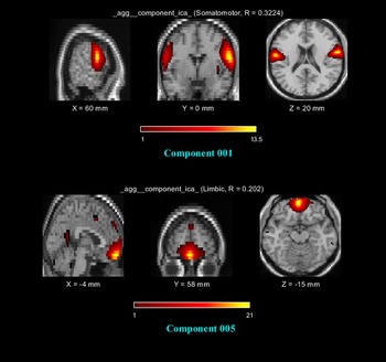
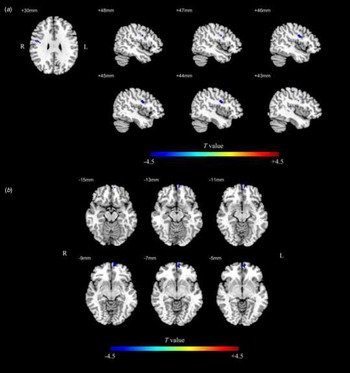
Of the interest components, two independent components (corresponding to the SMN and limbic network respectively) were interpreted as meaningful neural network components (Fig. 1). The significant SMN component consisted of the bilateral precentral and postcentral gyrus, and the significant limbic network component consisted of the bilateral medial frontal gyrus and the bilateral posterior cingulate. The significant SMN component showed decreased FC in the right precentral gyrus, and the significant limbic network component showed decreased FC in the left OFC in BD II group compared to the HCs (p < 0.05, TFCE corrected) (Table 2 and Fig. 2).
Of the interest components, two independent components (corresponding to the SMN and limbic network, respectively) were interpreted as meaningful neural network components between BD II and HCs. BD, bipolar disorder; HCs, healthy controls.

The FC patterns of the left orbitofrontal cortex (OFC) and the right precentral gyrus in BD patients and HCs (p < 0.05, TFCE corrected). The color bar indicates the t values. FC, functional connectivity; BD, bipolar disorder; HCs, healthy controls.

The areas of significantly different FC between the BD II patients and the HCs (p < 0.05, TFCE corrected)

FC, functional connectivity; BD, bipolar disorder; HCs, healthy controls; TFCE, threshold-free cluster enhancement; L (R), left (right) hemisphere.
Group differences between peripheral cytokine levels
The levels of IL-6 and IL-8 of the two groups are shown in Table 1. Compared with the HCs group, the BD II group showed increased IL-6 (p = 0.039) and IL-8 (p = 0.002) levels.
Correlation analyses
The IL-8 levels were significantly inversely correlated with FC in the right precentral gyrus in the patients with BD II (r = −0.458, p = 0.004) (Fig. 3). No significant correlation result was found between FC in the left OFC and pro-inflammatory cytokines. In addition, there was no significant correlation between cytokines levels and clinical variables, or between FC and clinical variables (24-item HDRS score, YMRS score, number of episodes, onset age of illness, duration of illness) in the patients with BD II (p > 0.05).
The correlation between abnormal FC values in the right precentral gyrus and IL-8 (p < 0.05) levels. FC, functional connectivity; L (R), left (right) hemisphere.

Discussion
The main findings are the following: (i). decreased FC in the left OFC implicating the limbic network, and in the right precentral gyrus implicating the SMN in unmedicated BD II depression. (ii). Increased IL-6 and IL-8 levels in BD II depression. (iii). Negative correlation between the right precentral gyrus FC and IL-8 levels in BD II depression. This study is among the first to investigate the FC in unmedicated BD patients using ICA methods combined with inflammation markers, which may improve our understanding of the neuropathological mechanisms underlying BD from the perspective of neuro-inflammation.
Decreased FC in the left OFC
In this study, the BD II patients showed hypoconnectivity in the left OFC compared with the HCs, suggesting disrupted FC in the limbic network in BD. The OFC is a part of the limbic network which is known to be involved in emotion processing (Stalnaker, Cooch, & Schoenbaum, Reference Stalnaker, Cooch and Schoenbaum2015) and regulation (Phillips, Ladouceur, & Drevets, Reference Phillips, Ladouceur and Drevets2008), reward value encoding (Grabenhorst & Rolls, Reference Grabenhorst and Rolls2011), and decision making including the vulnerability to suicidal behavior (Jollant et al., Reference Jollant, Lawrence, Olie, O'Daly, Malafosse, Courtet and Phillips2010). A previous rs-fMRI study found decreased FC between the left OFC and left anterior cingulate cortex in BD using seed-based correlation approach (Chen et al., Reference Chen, Wang, Niu, Zhong, Hu, Chen and Huang2018), which may be associated with dysfunction in emotion processing and decision making in BD. In addition, several studies found reduced regional homogeneity (ReHo) (Qiu et al., Reference Qiu, Chen, Chen, Jia, Gong, Luo and Wang2019; Xiao et al., Reference Xiao, Zhong, Lu, Gao, Jiao, Lu and Su2013), amplitude of low-frequency fluctuations (Lui et al., Reference Lui, Yao, Xiao, Keedy, Reilly, Keefe and Sweeney2015), and regional blood flow (Blumberg et al., Reference Blumberg, Stern, Ricketts, Martinez, de Asis, White and Silbersweig1999) in the OFC in BD. Task-based fMRI research also demonstrated hypoactivation in the OFC during emotion processing and reward-related tasks in BD (Sharma et al., Reference Sharma, Satterthwaite, Vandekar, Katchmar, Daldal, Ruparel and Wolf2016; Vizueta et al., Reference Vizueta, Rudie, Townsend, Torrisi, Moody, Bookheimer and Altshuler2012). Structural MRI studies also showed reduced gray matter volume (GMV) (Eker et al., Reference Eker, Simsek, Yilmazer, Kitis, Cinar, Eker and Gonul2014; Neves et al., Reference Neves, Albuquerque, Malloy-Diniz, Nicolato, Neves, de Souza-Duran and Correa2015) and gray matter density (James et al., Reference James, Hough, James, Burge, Winmill, Nijhawan and Zarei2011) in the left OFC in BD. Moreover, BD patients with a prior suicide attempt had lower fractional anisotropy (FA) within the left OFC white matter (Frazier et al., Reference Frazier, Breeze, Papadimitriou, Kennedy, Hodge, Moore and Makris2007; Mahon, Burdick, Wu, Ardekani, & Szeszko, Reference Mahon, Burdick, Wu, Ardekani and Szeszko2012). These findings may reflect a potential structural basis for functional abnormalities in the left OFC in BD. Several studies reported that the OFC was associated with affective liability (Perez et al., Reference Perez, Vago, Pan, Root, Tuescher, Fuchs and Stern2016), emotional dysregulation (Petrovic et al., Reference Petrovic, Ekman, Klahr, Tigerström, Rydén, Johansson and Landén2016), and reward processing (Nusslock et al., Reference Nusslock, Almeida, Forbes, Versace, Frank, Labarbara and Phillips2012). Taken together, our findings suggested that abnormal FC in the left OFC could contribute to the neuropathology of BD and result in characteristic behavioral abnormalities associated with BD such as emotional lability, emotional dysregulation and heightened reward sensitivity.
Decreased FC in the right precentral gyrus
We observed significantly decreased FC in the right precentral gyrus in BD II patients, suggesting disrupted FC in SMN in BD. The precentral gyrus, consists of the primary motor cortex, is a sub-region of SMN implicated in volitional control of motor output (Ebbesen & Brecht, Reference Ebbesen and Brecht2017). The growing number of studies have shown the precentral gyrus playing an important role in emotional-affective processing (Leite, Carvalho, Battistella, Caumo, & Fregni, Reference Leite, Carvalho, Battistella, Caumo and Fregni2017; Moseley et al., Reference Moseley, Shtyrov, Mohr, Lombardo, Baron-Cohen and Pulvermüller2015) and motor performance/activity (Cantisani et al., Reference Cantisani, Stegmayer, Bracht, Federspiel, Wiest, Horn and Walther2016; Schröder et al., Reference Schröder, Essig, Baudendistel, Jahn, Gerdsen, Stockert and Knopp1999) in mood disorders. Two rs-fMRI studies have found decreased FC in the precentral gyrus in BD (Singh, Kelley, Chang, & Gotlib, Reference Singh, Kelley, Chang and Gotlib2015; Zhao et al., Reference Zhao, Wang, Jia, Zhong, Sun, Qi and Huang2017), which supported our findings. Previous neuroimaging studies found decreased neural activities (Jiang et al., Reference Jiang, Fu, Yin, Kang, Wang, Zhou and Tang2020; Qiu et al., Reference Qiu, Chen, Chen, Jia, Gong, Luo and Wang2019; Russo et al., Reference Russo, Martino, Magioncalda, Inglese, Amore and Northoff2020), structural abnormalities in the precentral gyrus in BD (Eker et al., Reference Eker, Simsek, Yilmazer, Kitis, Cinar, Eker and Gonul2014; Fung et al., Reference Fung, Deng, Zhao, Li, Qu, Li and Chan2015; Jørgensen et al., Reference Jørgensen, Nerland, Norbom, Doan, Nesvåg, Mørch-Johnsen and Agartz2016), and the genetic risk to develop BD was associated with reduced gray matter volumes (Nery, Monkul, & Lafer, Reference Nery, Monkul and Lafer2013) and decreased grey matter density (Lyoo et al., Reference Lyoo, Kim, Stoll, Demopulos, Parow, Dager and Renshaw2004; van der Schot et al., Reference van der Schot, Vonk, Brouwer, van Baal, Brans, van Haren and Kahn2010) in the right precentral gyrus, further suggesting the involvement of the precentral gyrus in the pathogenesis of BD. A task-based fMRI study examined the emotionality of autobiographical memory in BD found decreased activation in the precentral areas compared with the control group (Oertel-Knoechel et al., Reference Oertel-Knoechel, Reinke, Hornung, Knoechel, Matura, Knopf and Linden2012). In another task-based fMRI study, decreased activation in the precentral gyrus was found in BD during self-paced finger tapping, which may result in impairments in preparation, control, and execution of movement in BD (Liberg et al., Reference Liberg, Adler, Jonsson, Landén, Rahm, Wahlund and Wahlund2013). Additionally, motor retardation is a characteristic feature of BD (Bowden, Reference Bowden2005). Therefore, in combination with our findings, disrupted FC in SMN may contribute to the typical symptoms of emotion dysregulation and motor retardation in BD II patients.
Correlation between FC in the right precentral gyrus and IL-8 levels
In this study, we found increased pro-inflammatory cytokine levels of IL-6 and IL-8 in BD group when compared to HC, which was consistent with previous studies (Benedetti et al., Reference Benedetti, Poletti, Hoogenboezem, Locatelli, de Wit, Wijkhuijs and Drexhage2017; Lu et al., Reference Lu, Rao, Mou, Chen, Lou, Zhang and Fang2019; Modabbernia et al., Reference Modabbernia, Taslimi, Brietzke and Ashrafi2013; Wang et al., Reference Wang, Zhong, Jia, Sun, Wang, Liu and Huang2016b; Wieck et al., Reference Wieck, Grassi-Oliveira, do Prado, Viola, Petersen, Porto and Bauer2016). Furthermore, we found elevated levels of IL-8 correlated with the reduction of FC in the right precentral gyrus within SMN in BD. IL-8 is a pro-inflammatory cytokine that facilitates blood-brain barrier migration of leukocytes and may sustain brain neuroinflammation resulting in neurotoxic effects (Willette et al., Reference Willette, Coe, Birdsill, Bendlin, Colman, Alexander and Johnson2013). Additionally, IL-8 is associated with blood-brain barrier dysfunction and nerve growth factor production (Kossmann et al., Reference Kossmann, Stahel, Lenzlinger, Redl, Dubs, Trentz and Morganti-Kossmann1997). Recent structural MRI studies showed that IL-8 was associated with reduced GMV in the left OFC in schizophrenia (Wu et al., Reference Wu, Lv, Li, Zhang, Fu, Dai and Lui2019) and bilateral hippocampus in old rhesus macaques (Willette et al., Reference Willette, Coe, Birdsill, Bendlin, Colman, Alexander and Johnson2013), which implicated inflammation might contribute to structural abnormalities in the brain. Another structural MRI study showed that GMV in the precentral gyrus negatively correlated with pro-inflammatory cytokine levels in BD patients (Bai et al., Reference Bai, Chen, Hsu, Huang, Tu, Chang and Tsai2020), further supporting the idea that inflammation might contribute to structural abnormalities in the right precentral gyrus. A diffusion tensor imaging study found that higher levels of IL-8 were associated with lower FA values across the brain including the corticospinal tract (Rodrigue et al., Reference Rodrigue, Knowles, Mollon, Mathias, Koenis, Peralta and Glahn2019), which suggested inflammation might contribute to white matter abnormalities and possibly impair the neural function of the precentral gyrus, for the reason that outputs of voluntary movements are controlled by the precentral gyrus through the corticospinal tract (Carper, Solders, Treiber, Fishman, & Müller, Reference Carper, Solders, Treiber, Fishman and Müller2015). However, the relationship between IL-8 and neural function is little known. In the present study, increased IL-8 levels were associated with reduced FC in the right precentral gyrus in unmedicated BD II patients, which suggested that inflammation may be related to disrupted neural function in BD. Moreover, it was noted that IL-8 concentrations were associated with Beck Depression Inventory score (Suarez, Krishnan, & Lewis, Reference Suarez, Krishnan and Lewis2003) as well as specific neuropsychological domains of cognitive functioning (Baune et al., Reference Baune, Ponath, Golledge, Varga, Arolt, Rothermundt and Berger2008) in healthy individuals. And the association of IL-8 with psychiatric disorders such as depression and BD has been clearly reported (Stuart & Baune, Reference Stuart and Baune2014). Taken together, increased levels of IL-8 possibly impair the FC in the precentral gyrus, thus may contribute to severer depressive symptoms and cognitive dysfunction such as poorer performance in the memory, reduced information processing speed and psychomotor speed in BD.
Limitations
The findings of this study should be interpreted in light of several limitations. First, the sample size was relatively small. Also, patients at suicide risks had not been excluded in this study. Recent studies reported BD may show distinct functional connectivity patterns among patients with and without suicide risks (Cheng et al., Reference Cheng, Chen, Zhang, Zhang, Wu, Ma and Cao2020; Wang et al., Reference Wang, Zhu, Dai, Tian, Shao, Wang and Lu2020a). Therefore, future study should take this in considerations and improve the homogeneity of BD patients. Second, owing to the cross-sectional study, we could not further study the causality between aberrant FC and inflammation. Hence, longitudinal research is warranted to confirm the role cytokines play in brain function. Third, for ethical reasons, we did not measure pro-inflammatory cytokines level in cerebrospinal fluid. However, peripheral cytokines level remains the main stay of assessment because of the ethical considerations involved in obtaining the cerebrospinal fluid or brain tissue. Peripheral cytokine levels may not exactly reflect the brain cytokine levels but can affect the production of cytokines in the brain. In addition, peripheral inflammatory cytokines may also be affected by other factors, such as stress and physical activities, although we have informed the patients to minimize their physical activity and stress a week before scanning. Therefore, further methods may be still needed to measure and control these factors of individuals. Besides, additional studies are needed to explore the role of the pathological ‘state’ (i.e. current mental disorders) and its influence on the brain immune relationships in conditions of social stress. Lastly, we did not assess the cognitive function of BD patients in this study. So, more research is needed to confirm the role of FC abnormalities play in cognitive dysfunction in BD.
Conclusion
In conclusion, our study provided preliminary evidence of the association between brain FC and pro-inflammatory cytokines in unmedicated BD II depression. The current study demonstrated disrupted FC of the left OFC within the limbic network and the right precentral gyrus within the SMN in BD. Moreover, our results suggest disrupted FC of the precentral gyrus may be associated with pro-inflammatory cytokines in BD, specifically the IL-8 levels. Given that pro-inflammatory cytokines may sustain neuroinflammation resulting in neurotoxic effects, the results provided preliminary evidence that higher levels of serum cytokines may be associated with severer FC abnormalities implicated in BD. Further studies with a larger sample or longitudinal design may confirm the role of pro-inflammatory cytokines in aberrant FC in BD[Q4].
Authors contribution
Ying Wang designed the study; Guanmao Chen, Ying Wang contributed to data sources and study selection; Guanmao Chen, Guixian Tang, Pan Chen, Jiaying Gong, Yanbin Jia, Shuming Zhong, Feng Chen, Jurong Wang, Zhenye Luo, Zhangzhang Qi contributed to data acquisition; Guanmao Chen, Pan Chen contributed to data analysis; Guixian Tang, Pan Chen, wrote the manuscript; Guanmao Chen, Guixian Tang, Pan Chen, Jiaying Gong, Li Huang, Ying Wang revised the manuscript. All authors contributed to and have approved the final manuscript.
Acknowledgements
The study was supported by grants from the National Natural Science Foundation of China (81671670, 81501456, and 81971597). The funding organizations played no further role in study design, data collection, analysis and interpretation, paper writing, and decision to publish.
Conflict of interest
The authors have declared that no competing interest exists.
Ethical standards
This study was approved by the Ethics Committee of First Affiliated Hospital of Jinan University, Guangzhou, China. All subjects were right-handed and signed a written informed consent form after a full written and verbal explanation of the study. Two senior clinical psychiatrists confirmed that all subjects had the ability to consent to participate in the examination.
Availability of data and material
The data supporting our findings will not be shared.