Introduction
The carangid fishes, commonly known as “jack”, comprise 32 genera and 146 species (Froese and Pauly, Reference Froese and Pauly2017), occurring worldwide in tropical and temperate waters, where they form large schools (Mohan et al., Reference Mohan2017). They represent one of the most numerous groups of fishes commonly caught by small-scale fishers, and are the most economically important pelagic fishes in the world (Madrid-Vera et al., Reference Madrid-Vera, Amezcua and Morales-Bojórquez2007; Mukherjee et al., Reference Mukherjee2017).
During a survey of three species of carangid fishes off Rio de Janeiro and Santa Catarina states, species of Mazocraeidea Bychowsky, 1957 were found on the gills. The fish Selene vomer (Linnaeus, 1758), distributed in the western Atlantic from Canada to Uruguay, was found parasitized by a new species of Pseudomazocraes and Pseudomazocraes selene Hargis, 1957; Caranx latus Agassiz, 1831, which occurs in the western and eastern Atlantic, also harboured both species of Pseudomazocraes and Cemocotyle carangis (MacCallum, 1919). Previous studies of P. selene and C. carangis off the Brazilian coast were based on morphological and ecological approaches (Kohn et al., Reference Kohn, Santos and Baptista-Farias1992; Luque et al., Reference Luque, Alves and Silveira São Sabas2000; Luque and Alves, Reference Luque and Alves2001; Luque and Cezar, Reference Luque and Cezar2004). Seriola lalandi Valenciennes, 1833, which has a circumglobal distribution in subtropical waters, was found parasitized by Zeuxapta seriolae, a species that has been recorded from the same fish host in Chile, Australia, Japan, New Zealand, Mexico and California (Bravo-Hollis, Reference Bravo-Hollis1978; Ogawa and Yokoyama, Reference Ogawa and Yokohama1998; Sharp et al., Reference Sharp2003; Hutson et al., Reference Hutson2007; Stuart and Drawbridge, Reference Stuart and Drawbridge2013; Sepúlveda and Gonzáles, Reference Sepúlveda and González2015; Mendoza-Garfias et al., Reference Mendoza-Garfias, García-Prieto and Pérez-Ponce de León2017). Other host records include Seriola dumerili (Risso, 1810) in the Mediterranean (Grau et al., Reference Grau2003; Montero et al., Reference Montero2004; Lia et al., Reference Lia2007) and Caranx hippos (Linnaeus, 1766) in Mexico (Lamothe-Argumento, Reference Lamothe-Argumedo1970).
The species P. selene, C. carangis and Z. seriolae have controversial taxonomic status, being placed by different authors in different families and subfamilies. The aim of the present study was to describe a new species of Pseudomazocraes and clarify the taxonomical status of these species, using integrative taxonomy with morphological and molecular analyses. Additionally, we discuss their phylogenetic position.
Materials and methods
Sample collection and morphological analysis
Collections in this study were authorized by the Brazilian Institute of Environment and Renewable Natural Resources (IBAMA, license no. 15898-1). The carangid fishes were collected in two localities, along Praia da Enseada (Enseada Beach), Santa Catarina (26°12′50.3″S, 48°30′46.0″W) during 2015 and in a local market in Rio de Janeiro during 2017. The fishes were measured and weighed, and their gills removed and examined in a saline medium under a stereomicroscope. The monogeneans were fixed in 70% or 100% ethanol. Some specimens were mounted in Berlese medium to study the sclerotized structures. Alternatively, other specimens were stained in Mayer's paracarmine or Gomori's trichrome and mounted in Canada balsam. Measurements are presented in micrometers, with the mean followed by the range in parentheses, unless otherwise stated. Drawings were made with the aid of a drawing tube. Prevalence and intensity data follow Bush et al. (Reference Bush1997). Representative specimens and hologenophores (Pleijel et al., Reference Pleijel2008) were deposited in the Helminthological Collection of Instituto Oswaldo Cruz (CHIOC), Rio de Janeiro, Brazil. For comparative purposes micrographs of specimens deposited at Colección Nacional de Helmintos (CNHE), Instituto de Biología, Universidad Nacional Autónoma de México (UNAM), Mexico were studied: holotype and paratypes of Pseudomazocraes monsivaisae Caballero & Bravo-Hollis, 1955 (CNHE 258, 260, 333), paratype of P. selene (CNHE 247, 249), holotype of Pseudomazocraes riojai (Caballero & Bravo-Hollis, 1963) (CNHE 283) and holotype of Oaxacotyle oaxacensis (Caballero & Bravo-Hollis, 1963) (= Pseudomazocraes oaxacensis) (CNHE 289).
Genetic analysis
DNA extraction was performed using the phenol–chloroform method, as described by Billings et al. (Reference Billings1998), and a set of primers were used to amplify different regions of the DNA. The rDNA region 28S was amplified by polymerase chain reaction (PCR) using the primers C1 (5′-ACCCGCTGAATTTAAGCAT-3′) and D2 (5′-TGGTC CGTGT TTCAA GAC-3′) (according to Chisholm et al., Reference Chisholm2001). The partial 18S, ITS1 and 5.8S were amplified using the primers S1 (5′-TTCCG ATAAC GAACG AGACT-3′) and H7 (5′-GCTGC GTTC TTCAT CGATA CTCG-3′) (Sinnappah et al., Reference Sinnappah2001). The primers BD1 and BD2 (Chen et al., Reference Chen2010) were used to amplify the ITS1, 5.8S and ITS2 regions of the rDNA. The primers JB3 (TTTTTTGGGCATCCTGAGGTTTAT)/JB4 (TAAAGAAAGAACATAATGAAAATG) (Bowles et al., Reference Bowles1993) were used to amplify the mtDNA cox1 region. Polymerase chain reactions were carried out using cycling parameters as previously described by these authors. The PCR products were analysed by electrophoresis in 1.5% agarose in Tris-borate EDTA gels, stained with SyberGreen (Invitrogen, Eugene, Oregon, USA) and photographed under UV transillumination. Amplified PCR products were purified with ExoSap-IT (USB® Products Affymetrix Inc., Cleveland, Ohio, USA). DNA cycle sequencing reactions were performed using BigDye Terminator v.3.1 (Applied Biosystems, Foster City, CA, USA) and automated sequencing was done using the Sequencing Platform at Fundacão Oswaldo Cruz (PDTIS/Fiocruz) in Brazil. Sequences of both strands were generated, edited and aligned using the MEGA version 7.0 software (Kumar et al., Reference Kumar, Stecher and Tamura2016). Sequences were compared to others available in the GenBank database using the BLASTN program from the National Center for Biotechnology Information (NCBI) server (http://www.ncbi.nlm.nih.gov/BLAST) (Altschul et al., Reference Altschul1990).
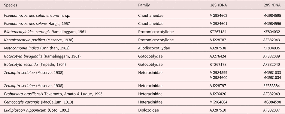
The datasets of different taxa were analysed separately using the MEGA 7.0 package (Thompson et al., Reference Thompson, Higgins and Gibson1994). Additionally, a concatenated analysis of the 18S and 28S genes resulted in a maximum likelihood (ML) phylogenetic tree (Felsenstein, Reference Felsenstein1981) utilizing the Kimura 2 parameters and gamma distribution with invariant sites (G), selected in MEGA 7.0; Eudiplozoon nipponicum (Goto, 1891) was used as outgroup for rooting the tree. In addition, uncorrected p-distance was also inferred to establish the genetic divergence among species. The tree was resampled by 5000 bootstrap replicates to evaluate the reliability of the groups. Taxa for which sequences from GenBank were downloaded and used for the phylogenetic analysis are listed in table 1.
List of the species of Monogenea and outgroups used in the phylogenetic analyses of Pseudomazocraes sulamericana n. sp., Pseudomazocraes selene, Cemocotyle carangis and Zeuxapta seriolae using sequences of the partial 18S and partial 28S rDNA genes.

Results
A total of 22 fishes from two localities were examined. The 18 fish from Rio de Janeiro (RJ) included 14 Caranx latus, measuring 23.8 (21–33.5) cm long, three Selene vomer, measuring 22.5 (22–24) cm long, and one Seriola lalandi 60 cm long. From Praia da Enseada in Santa Catarina, two C. latus, 20.5 and 23 cm long, and two S. vomer, 19.1 and 19.7 cm long, were studied. The monogeneans recovered are presented below.
Chauhaneidae Euzet & Trilles, 1960
Pseudomazocraes sulamericana n. sp.
Taxonomic summary
Type host. Caranx latus (Agassiz, 1831).
Other host. Selene vomer (Linnaeus, 1758).
Type locality. Praia da Enseada (SC), Brazil.
Site. Gills.
Prevalence and intensity. Two Caranx latus with 4 and 8 parasites. From two Selene vomer examined only one was infected, with 3 specimens.
Specimens deposited. CHIOC numbers 39042 (a–holotype, b–paratype), 39081a–c (paratypes) and 39039 (hologenophore) from Caranx latus. Numbers 39082 and 39083 (vouchers) from Selene vomer.
Representative DNA sequences. Sequences deposited in GenBank for partial 18S rDNA included accession numbers MG984602 from C. latus (451 bp); for partial 28S rDNA, MG 984594 (908 bp) from S. vomer and MG984595 (839 bp) from C. latus.
Etymology. The new species is named based on the geographical area.
Description
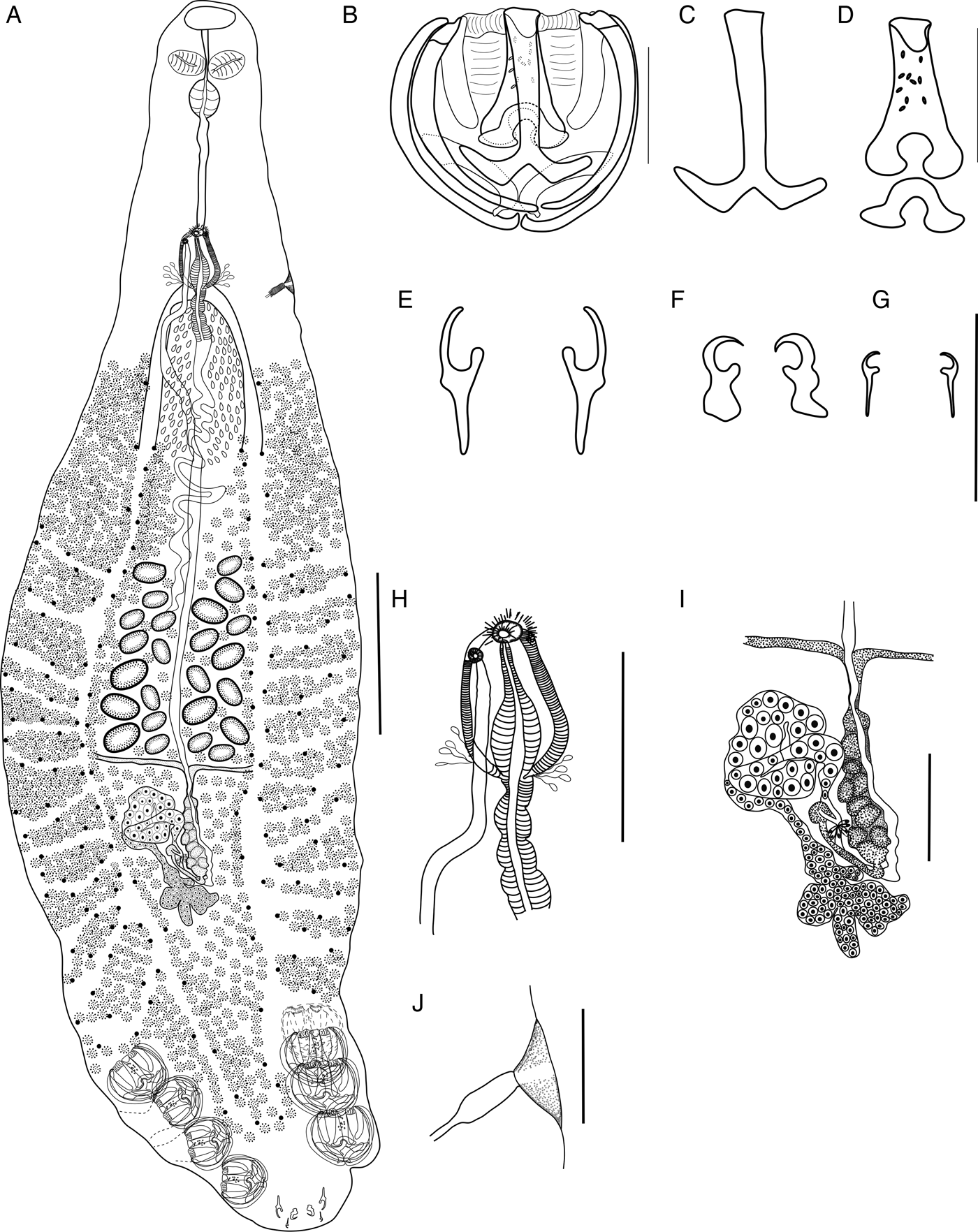
Measurements based on six adult specimens. Body lanceolate, with haptor poorly differentiated from the body. Total length 1.70 (1.30–2.90) mm × 0.45 (0.29–0.54) mm in width at level of ovary (fig. 1A). Prohaptor a pair of oval oral suckers, 45 (45–55) × 55 (50–60). Haptor 382 (340–410) × 408 (320–480), armed with four pairs of clamps, three pairs of larval hooks and gland vesicles. Clamps unequal in size with 8–10 lateral ribs; clamps on the right side larger, 97 (93–103) long × 103 (95–110) (fig. 1B); smaller clamps slightly pedunculated, 86 (80–93) × 77 (70–80). Clamp sclerites: antero and posterolateral thin; basal accessory wall sclerite curved; mid-sclerite with ventral piece bifurcated at end, with large and right-angled edges of almost the same length (fig. 1C) articulated with dorsal piece perforated, which gradually widens and bifurcates forming rounded tips connected with mid-accessory piece (fig. 1D). Lappet short and round. Larger larval hooks 35 (30–43) long with thin handle, thumb-shaped gard and elongate blade (fig. 1E); median larval hooks 21 (18–25) long with handle deformed, small thumb-shaped gard and blade strongly curved with sharp tips (fig. 1F); smallest larval hooks 12 (10–13) long (fig. 1G). Mouth subterminal, ventral, inside of buccal funnel. Pharynx ovoid 55 × 51 (45–55); oesophagus long, bifurcates into two ramified ceca at level of genital atrium extending into haptor up to middle level of third clamps. Testes preovarian, 31 (26–32) in number; vas deferens running sinuously in the midline of the body, prostatic glands well developed, cirrus muscular 42 (33–45) (fig. 1H). Genital pore ventral opens at genital atrium, which has papillae at base. Ovary postequatorial, irregular in shape; anterior portion lobed and posterior tubular, looping towards midline. Vitellaria follicular, extending from mid-level of prostatic glands to mid-level of haptor. Vitelloducts Y-shaped forming reservoir; Mehlis gland present around ootype (fig. 1I). Genito-intestinal canal not observed. Uterus coursing anteriorly in midline up to genital atrium. Vagina muscular; vaginal pore lateral, sinistral at level of intestinal bifurcation (fig. 1J). Eggs were not observed.
Pseudomazocraes sulamericana n. sp. (A) Holotype, ventral view; (B) clamp; (C) details of mid-ventral sclerite; (D) detail of mid-ventral dorsal sclerite and its accessory piece; (E) larger larval hook; (F) median hook; (G) small hook; (H) genital atrium with male copulatory organ and genital pores; (I) female reproductive system; (J) vagina. Scale bars: A = 200 μm; B–G, I–J = 50 μm; H = 100 μm.

Measurements based on two specimens from Selene vomer. Body elongate, with haptor differentiated from body. Total length 2.78 and 3.35 mm × 0.52 and 0.72 mm in width at level of ovary. Oral suckers, 35–45 × 50–55. Haptor 540 and 490 × 490 and 400; larger clamps 90–110 × 90–90, smaller clamps pedunculated 78–85 × 78–85; largest larval hooks 33–38; median hooks 20–25 long; smallest hooks 15–17 long. Pharynx 55 and 60 × 50 and 55. Testes 25 and 27 in number.
Remarks
Pseudomazocraes sulamericana n. sp. is the fourth species described in the genus. The main diagnostic characters of the new species include the shape of mid-sclerite of the clamps, with ventral piece bifurcated at end, with large and right-angled edges of almost the same length, size and shape of larval hooks and rounded terminal lappet slightly striated and poorly differentiated from the body. The closest species is Pseudomazocraes monsivaisae Caballero & Bravo-Hollis, 1955, from which it differs by the size and shape of mid-dorsal and ventral sclerites, shape of larval hooks and terminal lappet apart from the geographical distribution (Pacific Ocean). Pseudomazocraes selene can be differentiated by the shape of body, absence of papillated genital atrium, shape of larval hooks, median sclerites and accessory median piece of clamps. Pseudomazocraes riojai (Caballero & Bravo-Hollis, 1963) is readily differentiated by the shape of clamps, with longer basal accessory piece and mid-ventral sclerite, larger and robust larval hooks and very long terminal lappet.
Pseudomazocraes selene Hargis, 1957
Taxonomic summary
Hosts. Caranx latus (Agassiz, 1831) and Selene vomer (Linnaeus, 1758).
Locality. Local market in Rio de Janeiro, Brazil.
Site. Gills.
Prevalence and intensity. Caranx latus – 71.4% (10 fishes infected/14 examined), 1–37 parasites per fish; Selene vomer – all three fish were parasitized, with one specimen in each fish examined.
Specimens deposited. CHIOC numbers 39041 and 39040 (hologenophore) from C. latus, and number 39083 from S. vomer.
Representative DNA sequences. Sequences deposited in GenBank for partial 18S rDNA included accession numbers MG984601 (490 bp) from S. vomer; for partial 28S rDNA, MG984596 (839 bp) and MG984597 (879 bp) from C. latus.
Redescription
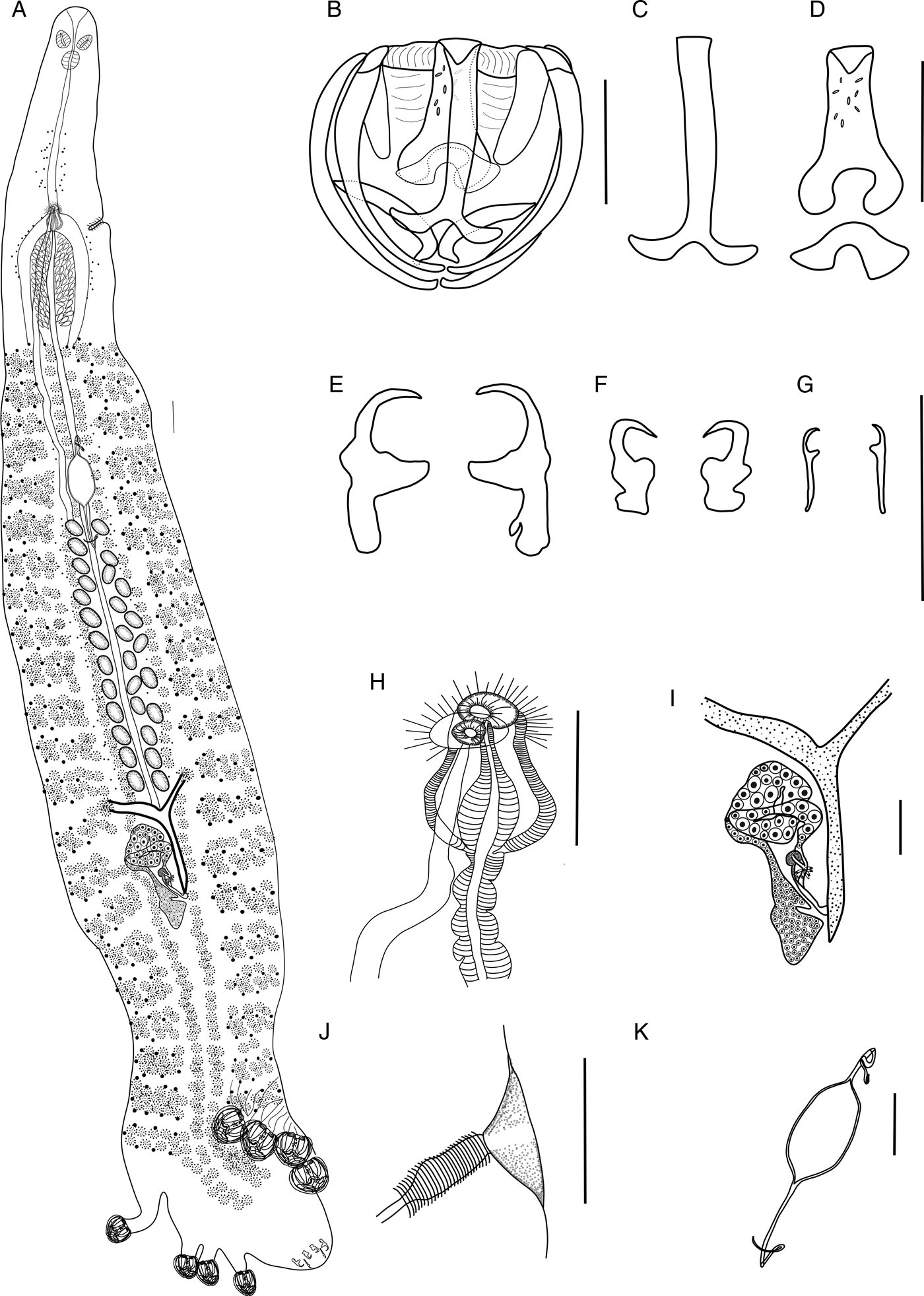
Measurements based on 10 adult specimens from C. latus. Body elongate, with haptor differentiated from the body. Total length 3.40 (2.90–4.32) mm × 0.57 (0.39–0.80) mm in width at level of ovary (fig. 2A). Prohaptor a pair of oval oral suckers, 46 (35–55) × 54 (45–60). Haptor 585 (510–720) × 515 (420–680), armed with four pairs of clamps and three pairs of larval hooks. Clamps unequal in size; sinistral clamps larger, 100 (93–105) × 100 (90–105); smaller clamps on the right side pedunculated, 75 (70–77) × 74 (70–78) (fig. 2B). Antero and posterolateral sclerites of clamps stout; basal accessory wall sclerite curved; mid-ventral sclerite bifurcated, with double pointed tips different in size and shape (fig. 2C), articulated with mid-dorsal perforated piece, which widens and bifurcates to end as pointed tips directed inwards; mid-accessory piece thin with irregular tips (fig. 2D). Larger larval hooks 41 (33–45) long with elongate and strong roots, gard thick, expanded and tips pointed and curved; median hooks 24 (20–25) long with irregular roots, gard short, round and tips strongly curved; smallest hooks 19 (18–20) long (fig. 2E–G). Mouth subterminal, ventral, inside of buccal funnel. Pharynx 54 (45–60) × 54 (45–60); oesophagus long, bifurcates into two ramified ceca extending into haptor up to middle level of clamps. Testes preovarian, 27 (23–30) in number; vas deferens thick, running sinuously in midline towards the genital atrium, forming at the end a pear-shaped male copulatory organ 73 (58–93) long. Genital pore ventral, opens at genital atrium (fig. 2H). Ovary irregular in shape; anterior portion lobed and posteriormost portion tubular, looping towards midline. Vitellaria follicular, extending from posterior level of prostatic glands to mid-level of haptor. Vitelloducts Y-shaped, forming reservoir; Mehlis gland small around ootype (fig. 2I). Uterus coursing anteriorly in midline genital atrium. Vagina muscular, extracecal; vaginal pore lateral, sinistral at level of intestinal bifurcation (fig. 2J). Eggs fusiform, 190 (175–200) × 84 (75–95), with long filaments at both ends, being anterior 168 (115–213) and posterior 209 (180–235) (fig. 1K).
Pseudomazocraes selene. (A) Body, ventral view; (B) clamp; (C) details of medial ventral piece; (D) details of medial dorsal piece and accessory; (E) larger anchor; (F) median anchor; (G) smallest larval hooks; (H) genital atrium with male copulatory organ and genital pores; (I) female reproductive system; (J) vagina; (K) egg. Scale bars: A = 100 μm; B–J = 50 μm; K = 100 μm.

Measurements based on one specimen of Selene vomer. Total length 4.35 × 0.47 mm in width at level of ovary; oral suckers, 35 × 45; haptor 640 × 490; larger clamps 100 × 100; smaller clamps pedunculated, 73 × 75; largest larval hooks, 43 long; median ones 25 long; smallest larval hooks 15 long. Pharynx 45 × 35; testes not observed.
Molecular analysis of P. sulamericana n. sp. and P. selene
The BLAST result for the partial 18S rDNA sequences of P. sulamericana n. sp. indicated 98% identity with Neomicrocotyle pacifica (AJ228787) and 97% with Bilaterocotyle carangis (KT267184). For P. selene the identity with N. pacifica (AJ228787) and B. carangis (KT267184) was 97%. The p-distance between the 18S rDNA sequences of P. sulamericana n. sp. from C. latus off SC and P. selene from S. vomer off RJ was 1.3%. We were not successful in obtaining sequences of mtDNA cox1 for comparison between these two species. For partial 28S rDNA of both species there was 96% identity with Bilaterocotyle madrasensis (KF804029) and 95% with Neomicrocotyle pacifica (AF382043). There was no divergence among the 28S rDNA sequences from different hosts and areas.
Heteraxinidae Unnithan, 1957
Cemocotyle carangis (MacCallum, 1913)
Taxonomic summary
Host. Caranx latus (Agassiz, 1831).
Site. Gills.
L ocality. Fish market in Rio de Janeiro, Brazil.
Specimens deposited. CHIOC numbers 39037 a–c and 39036 (hologenophore).
Prevalence and intensity. 21.4% (3 fishes infected/14 examined), 1–11 parasites per fish.
Representative DNA sequences. Sequences were deposited in GenBank under the accession numbers MH043592 for the partial 18S, ITS1 and 5.8 rDNA with 1207 bp; MG984604 for partial 18S rDNA with 498 bp; MH037238 for the ITS1, 5.8S and ITS2 rDNA with 1247 bp; MG984598 for the partial 28S rDNA with 932 bp, and MH090371 for the mtDNA cox1 with 464 bp.
Redescription
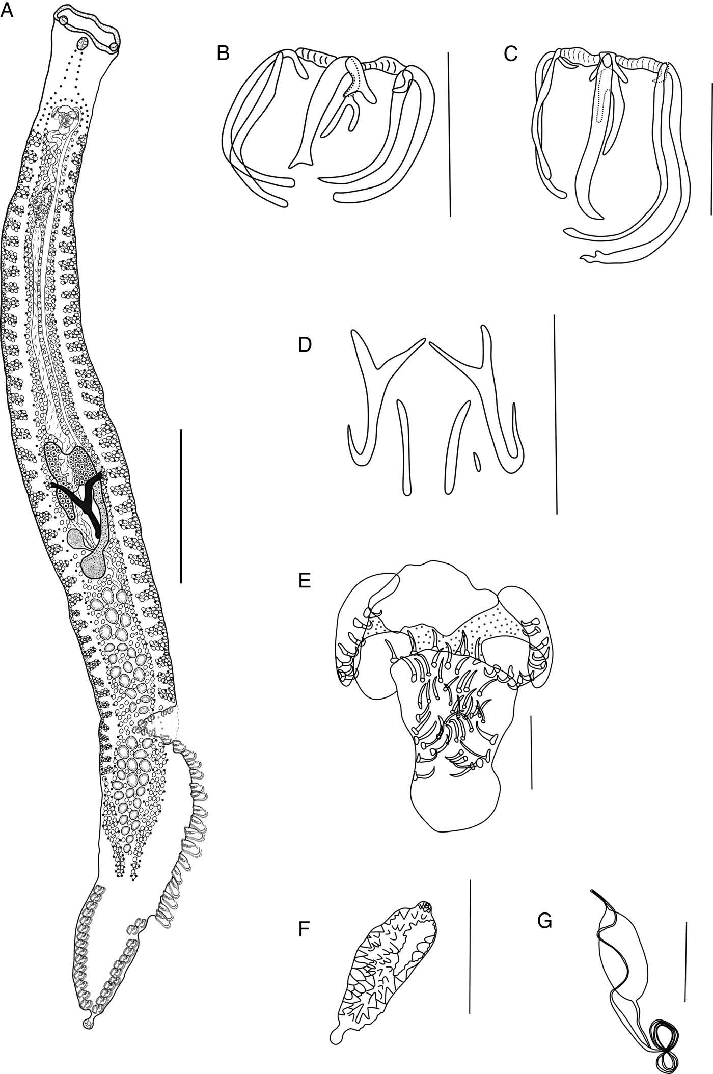
Measurements based on 10 adult specimens. Body elongated 3.97 (2.95–4.76) mm × 0.39 (0.26–0.48) mm (fig. 3A). Prohaptor a pair of widely separated buccal suckers 36 (30–40) × 38.5 (30–45) inside of a muscular enlarged area. Haptor asymmetric, armed with 52 unequal clamps; left side greater than the right side; clamps 1–9 measure 30 (28–33) × 32 (25–38) (fig. 3B); clamps 10–25 with lateral sclerites different in size and shape: 74 (63–85) × 49 (40–53) (fig. 3C) and clamps 26–52 similar in shape with the first ones, 44 (40–50) × 50 (38–55). Terminal lappet armed with three pairs of larval hooks; the outer pair 37 (27–43) long, inner pair 29 (25–33) long, and intermediate pair 8 (8–9) long (fig. 3D). Pharynx oval 40 (33–50) × 35 (30–40); oesophagus short. Intestinal ceca bifurcating at level of genital atrium, ramified, extending into haptor. Testes postovarian, 35 to 47 in number; seminal receptacle oval; vas deferens runs upwards to join the muscular male copulatory organ, armed with curved spines 12 (10–13) long (fig. 3E). Genital atrium triangular 403 (300–480) long, provided with “cuplike pocket” on each side, armed with spines of 10 (9–13) long, at level of intestinal bifurcation. Ovary long, curved, pretesticular. Vitellaria follicular, extending from level of genital atrium to midhaptor. Vitelline reservoir Y-shaped. Uterus extending anteriorly to genital atrium. Vagina 140 (93–175) long with triangular spines (fig. 3F). Eggs 119 (113–125) long and 48 (45–55) wide, with filaments at both ends (fig. 3G).
Cemocotyle carangis. (A) Body, ventral view; (B) small clamp; (C) large clamp; (D) larval hooks; (E) genital atrium with male copulatory organ; (F) vagina; (G) egg. Scale bars: A = 100 μm; B–F, G = 50 μm.

Molecular analysis
The BLAST search revealed for the partial 18S, ITS1 and 5.8S rDNA that C. carangis exhibits close similarity (94%) with Microcotyle erythrinii (AJ287999). However, when only the first 498 bp corresponded to the partial 18S rDNA, the BLAST resulted in 96% identity with Probursata brasiliensis (AJ276426). The analysis using ITS1, 5.8S and ITS2 rDNA sequences indicated 90% identity with Diplostamenides sciaenae (EF452639). For the partial 28S rDNA region there was 92% identity with Probursata brasiliensis (AF382049) and for the mtDNA cox1 sequences, there was similarity (81%) with Pseudochauhanea macrorchis (JN592039). The genetic divergence between C. carangis and Cemocotyle trachuri (AF131726) was 9.3%, with 29 nucleotide difference.
Zeuxapta seriolae (Meserve, 1938)
Taxonomic summary
Host. Seriola lalandi Valenciennes, 1833.
Site. Gills.
Locality. Fish market in Rio de Janeiro, Brazil. New locality.
Specimen deposited. CHIOC number 39043.
Prevalence and intensity. One fish with three monogenean specimens.
Representative DNA sequences. Sequences were deposited in GenBank under the accession numbers MG984599 and MG984600 with 1066 bp for the partial 18S, ITS1 and 5.8 rDNA; MG981033 (448 bp) and MG981034 (704 bp) for the partial 28S rDNA.
Redescription
Based on one adult specimen. Body elongated 15 × 0.87 mm at level of ovary. Prohaptor a pair of buccal suckers 95 × 130. Haptor asymmetric, with 68 clamps (37 and 31). Anterior clamps medium sized 75 × 110 progressively increase in size, reaching 125 × 195, and then as they approach the posterior part progressively decrease, the last ones measuring 35 × 50. Pharynx oval; oesophagus short. Intestinal ceca ramified extending into haptor. Testes postovarian, numerous; vas deferens winding. Genital atrium broad, muscular, 185 × 175. Ovary long and curved. Vitelline reservoir Y-shaped; uterus extending anteriorly to genital atrium. The vaginal transverse lateral tubes join the median, dorsal genital pore. Vitellaria follicular extending from near the level of genital atrium into the haptor. Eggs not observed.
Molecular analysis
The closest sequence for partial 18S, ITS1 and 5.8 rDNA according to BLAST was Z. seriolae (AJ228797), with a p-distance of 0.4%. For the partial 28S rDNA, the p-distance with two Z. seriolae sequences (EF653384 and AF026103) ranged from 1.9–2.3%.
Phylogenetic analyses of the Mazocraeidea
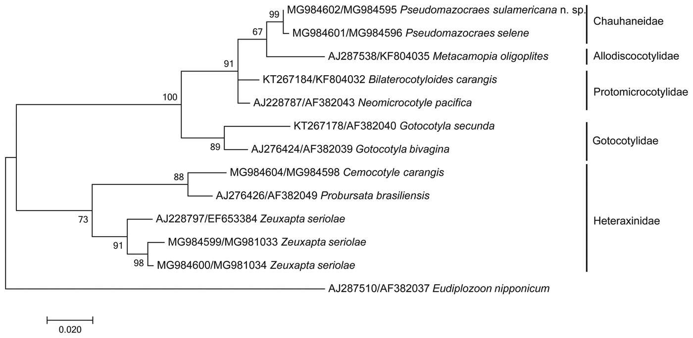
The concatenated analysis of our partial sequences of the 18S and 28S rDNA genes of Pseudomazocraes sulamericana n. sp., P. selene, C. carangis and Z. seriolae showed two main clades within the order Mazocraeidea. The first clade, represented by species of Heteraxinidae with 89% bootstrap support, included C. carangis close to P. brasiliensis (69%) and species of Z. seriolae from Brazil and Australia (99%). The second clade, with 100% bootstrap support, comprised species of Chauhaneidae, Protomicrocotylidae, Allodyscocotylidae and Gotocotylidae. The Chauhaneidae, represented by Pseudomazocraes sulamericana n. sp. and P. selene showed a bootstrap of 99% and appeared as the sister clade of Allodiscocotylidae with 67% and Protomicrocotylidae with 91% bootstrap (fig. 4).
Phylogenetic tree obtained from ML of the concatenated data (partial 18S and 28S rDNA gene) with representative species of order Mazocraeidea, families Heteraxinidae, Chauhaneidae, Protomicrocotylidae, Allodiscocotylidae and Gotocotylidae. Numbers (%) on the branches indicate 5000 bootstrap replicates. GenBank accession numbers are shown, and the scale bar indicates the nucleotide mutations per site.

Discussion
Historically, representatives of the genus Pseudomazocraes Caballero & Bravo-Hollis, 1955 were included in the family Gastrocotylidae Price, 1943, subfamily Gastrocotylinae Sproston, 1946. Hargis (Reference Hargis1957) confirmed the family but changed this genus to the subfamily Vallisiinae Price, 1943. Tripathi (Reference Tripathi1959) reassigned it in the Allodiscocotylidae Tripathi, 1959, Allodiscocotylinae Tripathi, 1959. In the same year, Price (Reference Price1959) proposed a reclassification of the gastrocotylids with Pseudomazocraes and Vallisiinae into the Protomicrocotylidae Poche, 1925. Unnithan (Reference Unnithan1962) erected Opisthogynidae, Opisthogyninae to accommodate Pseudomazocraes, whereas Yamaguti (Reference Yamaguti1963) considered the genus as Allodiscocotylinae but rearranged it into the Discocotylidae Price, 1936. Lebedev (Reference Lebedev1972) proposed Chauhaneidae, Pseudomazocraeinae, but based on the priority rule, he later recognized Chauhaneidae Euzet & Trilles, 1960 (syn. Opisthogynidae), and erected Pseudomazocraeidae Lebedev, 1972 with a unique genus (Lebedev, Reference Lebedev1986, Reference Lebedev1988). At present, Pseudomazocraes is considered a member of Chauhaneidae, as also stated in the WoRMS database (WoRMS Editorial Board, 2018).
The genus Pseudomazocraes now includes four species that parasitize marine fishes: Pseudomazocraes monsivaisae, the type species described from three different hosts, Balistides polylepsis Steindachner, 1876, Citula dorsalis Gill, 1863, and Argyreiosus brevoorti Gill, 1863, accepted as Selene brevoorti (Gill, 1863) off the Pacific coast of Mexico. Hargis (Reference Hargis1957) reported that Caballero and Bravo-Hollis (Reference Caballero and Bravo-Hollis1955) probably included more than one species in their description of P. monsivaisae and proposed P. selene from the Atlantic Ocean. Pseudomazocraes selene has been reported parasitizing fish from Mexico, USA and Brazil (Hargis, Reference Hargis1957; Caballero and Bravo-Hollis, Reference Caballero and Bravo-Hollis1965; Lamothe-Argumedo, Reference Lamothe-Argumedo1970, Kohn et al., Reference Kohn, Santos and Baptista-Farias1992; Cezar et al., Reference Cezar, Luque and Chaves2000; Luque and Alves, Reference Luque and Alves2001; Mendoza-Garfias et al., Reference Mendoza-Garfias, García-Prieto and Pérez-Ponce de León2017). The third species of the genus is P. riojai, described by Caballero and Bravo-Hollis (Reference Caballero and Bravo-Hollis1963) originally as Vallisia riojai from Caranx hippos of Mexico; Lebedev (Reference Lebedev1970) accommodated it in Pseudomazocraes. Pseudomazocraes longicauda Mamaev, 1984 is no longer a valid member of the Chauhaneidae because the clamps and male copulatory organ with spines are typical of the Mazocraeidae. Pseudomazocraes sulamericana n. sp. with distinct shape of mid-sclerite, larval hooks and terminal lappet is the second species to be described from the Atlantic Ocean. Therefore, P. selene, the only known species occurring in the Atlantic Ocean, also reported in this study, can be readily differentiated from P. sulamericana n. sp. not only by morphological characters but also by the new rDNA sequences, which gives a more comprehensive profile of the species for future comparative studies in different geographical areas.
Cemocotyle carangis (MacCallum, 1913) was described within the Microcotylidae Taschenberg, 1879. Price in 1962 proposed Cemocotylinae within the Heteraxinidae Unnithan, 1957 to include the genus Cemocotyle. Yamaguti (Reference Yamaguti1963) erected Cemocotylinae to family level. However, Mamaev (Reference Mamaev1990) suggested that Cemocotyle should return to the subfamily Cemocotylinae in the Heteraxinidae as proposed by Price (Reference Price1962), and as is currently accepted. Cemocotyle carangis was previously reported in different species of carangids from USA, Mexico and Brazil (MacCallum, 1913; Price, Reference Price1962; Bravo-Hollis and Salgado Maldonado, Reference Bravo-Hollis and Salgado-Maldonado1982; Kohn et al., Reference Kohn, Santos and Baptista-Farias1992; Cezar et al., Reference Cezar, Luque and Chaves2000; Luque and Alves, Reference Luque and Alves2001; Montoya-Mendoza et al., Reference Montoya-Mendoza, Salgado-Maldonado and Mendoza Palmero2008; Mendoza-Garfias et al., Reference Mendoza-Garfias, García-Prieto and Pérez-Ponce de León2017).
Zeuxapta seriolae, considered a cosmopolitan parasite highly pathogenic to farmed Seriola spp. (Montero et al., Reference Montero2004; Hutson et al., Reference Hutson2007; Sepúlveda and Gonzalez, Reference Sepúlveda and González2015), is now reported for the first time in Brazil. The taxonomical position of the species changed from Axinidae, Heteraxininae Unnithan, 1957 when Price (Reference Price1962) erected the subfamily to Heteraxinidae with Z. seriolae (syn. Axine seriolae, Microcotyle seriolae and Z. zyxivaginata). In GenBank, Z. seriolae is reported either as a member of Axinidae or Heteraxinidae.
The concatenated phylogenetic reconstruction of the 18S + 28S rDNA genes exhibited consistent data with representatives of the families Heteraxinidae, Chauhaneidae, Protomicrocotylidae, Allodiscocotylidae and Gotocotylidae. Boeger and Kritsky (Reference Boeger and Kritsky1993), in their study on the phylogenetic relationship of monogeneans based on morphology, showed the Chauhaneidae as a sister family of ‘Pseudomazocraeidae’, followed by Protomicrocotylidae and Allodiscocotylidae. The present sequences of P. sulamericana n. sp. and P. selene represent the first genetic data for the genus considered herein as Chauhaneidae, which in the ML reconstruction is closer to Allodiscocotylidae, although with low nodal support. Protomicrocotylidae appeared as a sister clade with high statistical support, corroborating the molecular results of Olson and Littlewood (Reference Olson and Littlewood2002). These authors did not use representatives of the Chauhaneidae due to the absence of data in GenBank at that time. The phylogenetic position of C. carangis based on 18S and 28S rDNA genes was confirmed within the Heteraxinidae, grouping with P. brasiliensis. The sequences of the mitochondrial gene cox1 and of ITS1-5.8-ITS2 rDNA of C. carangis did not yield a well-supported phylogenetic analysis due to the reduced number of sequences deposited in GenBank.
The species Z. seriolae nested within the Heteraxinidae, a relationship supported with high bootstrap value. Sepúlveda and González (Reference Sepúlveda and González2015) evaluated the pattern of genetic variation of Z. seriolae through cox1 DNA sequences by comparing one population parasitizing S. lalandi that approaches the coast in the summer, and the other from the oceanic area in the south-eastern Pacific Ocean; they found a genetic divergence varying from 0.4–0.9%. The comparison of our sequences with the sequences from Australia showed a genetic distance of 0.4% for 18S rDNA and 1.9–2.3% for 28S rDNA. According to Sepúlveda and González (Reference Sepúlveda and González2015), the parasite population's genetic structure could be influenced by oceanographic barriers to parasite dispersion and/or by host population segregation. As there is little information about intraspecific distances to compare using 18S and 28S rDNA, we encourage efforts to better support a different DNA barcoding protocol for monogeneans using ribosomal markers, as suggested by Vanhove et al. (Reference Vanhove2013).
The morphology described in this study presented similarity as previously described by other authors for all the species, including samples from Brazil. Perkins et al. (Reference Perkins2009) emphasized the necessity to combine morphological and molecular approaches in parasite studies, as the taxonomy should provide tools to better understand the diversity of organisms and the historical processes that generate biological diversity. Our study, based on morphological and molecular data, provided an integrative taxonomic overview of four Mazocraeid species, thus confirming their taxonomic position. The new sequences were important to show that C. carangis and Z. seriolae belong to Heteraxinidae. However, molecular data for species of Chauhaneidae are still scarce in GenBank, with only mitochondrial DNA sequences available for comparison, thus requiring further studies using different molecular markers.
Author ORCIDs
C.P. Santos 0000-0001-5165-0853.
Acknowledgements
The authors are grateful to Dr Gerardo Pérez Ponce de Leon and Dr Luis Garcia from UNAM, México for providing micrographs of CNHE type material. This study is part of the PhD thesis of the first author as part of the Programa de Pós-Graduação em Ciências Veterinárias, Universidade Federal Rural do Rio de Janeiro.
Financial support
The study was financially supported by Conselho Nacional de Desenvolvimento Científico e Tecnológico (CNPq- Universal no. 449658/2014-7), Coordenação de Aperfeiçoamento de Pessoal de Ensino Superior ─ Brasil (CAPES-Parasitologia 247/2012, Finance code 001) and Fundação Oswaldo Cruz (PAEF no. IOC-008-FIO-04). C.P. Santos was sponsored by a CNPq fellowship.
Conflict of interest
None.