Implant-associated infections
Implant-associated infections are a significant healthcare problem and a major cause of post-surgical morbidity and mortality in patients. Over half of the two million cases of nosocomial infections that occur in the United States annually are associated with indwelling devices.Reference Darouiche1, Reference Costerton, Stewart and Greenberg2 Catheters alone account for hundreds of thousands of infections each year resulting in a significant cost and burden to the healthcare system.Reference Dunne3 Although less common than catheter-based bloodstream infection, infections established on surgical implants and devices are considerably harder to treat because they require longer antibiotic regiments and repeated surgical procedures.Reference Whitehouse, Friedman, Kirkland, Richardson and Sexton4 In the case of orthopedic trauma surgeries, up to 10% of fracture fixators and 85% of external fixators typically become infected and have to be managed with several weeks of systemic antibiotic therapy.Reference Davis5–Reference Malik, Harwood, Diggle and Khan7 Surgical treatments usually involve the removal of the infected nail or pin followed by insertion of a replacement. Infection of implanted devices with particularly virulent multiresistant strains, such as methicillin-resistant Staphylococcus aureus (MRSA), may necessitate multiple, and often traumatic, surgical procedures that can be psychologically devastating in older patients, with additional risk and cost. Furthermore, infections are likely to play a larger role than is currently appreciated because low-level infections are difficult to detect radiographically and because biofilm bacteria resist culture, hindering traditional detection methods.Reference Hendriks, van Hornm, van der Meim and Busscherm8, Reference Flemming, Wingender, Griebe, Mayer and Evans146
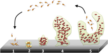
The treatment of device-associated infections is far more complex than the simple administration of antibiotics. When an implant is inserted into the host tissue, small biomolecules, including extracellular matrix (ECM) proteins (e.g., fibronectin, fibrinogen, and collagen), adsorb onto the material surface to form a conditioned protein layer conducive to the adherence of free-floating planktonic bacteria. The adhered bacteria then rapidly proliferate, recruit other cells, and produce sticky secretions to form dense three-dimensional communities of attached (sessile) cells called biofilmsReference Darouiche1, Reference Vinh and Embil9 (Figure 1). Antibiotics and other antimicrobial compounds show greatly reduced antimicrobial activity against organisms living within biofilm communities. Consequently, bacterial biofilms are notoriously difficult to eradicate.
Schematic representation of biofilm development and progression. This process occurs in five stages: (1) Individual free-floating (planktonic) cells populate the surface. (2) Extracellular polymeric substance is produced, and attachment becomes irreversible. (3 and 4) Biofilm architecture develops and matures. (5) Detachment and dispersion of individual cells from the biofilm occur to repeat the process. Adapted from Peg Dirckx and David Davies, 2003. Center for Biofilm Engineering, Montana State University.

Despite decades of prophylactic antibiotic use, high infection rates continue, particularly with the emergence of lethal multi-drug-resistant strains. This is due, in part, to the remarkable ability of bacteria to mutate and acquire resistance genes from other organisms, thereby developing resistance to an antimicrobial drug (because selective pressure favors organisms that are resistant to the drug’s action). In fact, antimicrobial resistance has become a rapidly growing problem of national importance. Almost all strains of Staphylococcus aureus (S. aureus) in the United States are now resistant to penicillin, with many also resistant to newer methicillin-related drugs.Reference Panlilio, Culver, Gaynes, Banerjee, Henderson, Tolson and Martone10 Strains of S. aureus with decreased susceptibility to vancomycin, for many years the only uniformly effective treatment, have also been reported.Reference Hiramatsu, Hanaki, Ino, Yabuta, Oguri and Tenover11, Reference Smith, Pearson, Wilcox, Cruz, Lancaster, Robinson-Dunn, Tenover, Zervos, Band, White and Jarvis12 If this problem is left unchecked, effective and dependable treatment of infections may no longer be possible. Clearly, there is a pressing clinical need for novel approaches to prevent and treat antibiotic-resistant infections.
Therapeutic strategies
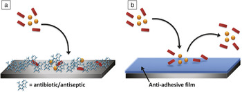
A large variety of molecules and macromolecules, both organic and inorganic, have been investigated as antimicrobial coatings that are capable of limiting or preventing bacterial growth and colonization on a surface. These coatings can be broadly classified as either taking an active or passive route toward preventing infection (Figure 2). Bactericidal and bacteriostatic agents have been extensively explored as active components of infection-resistant coatings. Bactericidals (e.g., many antibiotics, antiseptics, disinfectants) directly kill pathogens, whereas bacteriostatic agents inhibit bacteria growth and reproduction to allow the immune system to clear them from the body. An alternative, and more passive, approach is to block adhesion itself to the implant by altering its surface properties. The principal function of these adhesion-resistant coatings is to keep bacteria numbers sufficiently low to impede the development of a biofilm so that the host defensive mechanism can be effective.
Prevention of microbial colonization of implant can be achieved through the use of (a) “active” bactericidal and bacteriostatic coatings that actively impair pathogens (spheres and rods) and (b) “passive” bacteriophobic coatings that block microbial adhesion onto surfaces.

Traditional approaches
Prevention and treatment of post-surgical infections following implantation typically involve systemic antibiotic prophylaxis. Antibiotics are a particular class of antimicrobials that demonstrate potent antagonistic activity against selective microorganisms. However, systemic administration of antibiotics has the disadvantages of relative low drug concentration at the target site as well as potential toxicity upon tissue accumulation. Thus, starting from the introduction of antibiotic-loaded bone cement in the 1970s, there has been growing interest in localized antibiotic prophylaxis through the use of antibiotic-releasing surface coatings.Reference Buchholz and Engelbrecht13 In the past decade alone, antibiotic-laden silica sol-gels, porous hydroxyapatite (HA), and biodegradable polylactide coatings have been investigated as means to deliver broad spectrum antibiotics in a controlled fashion from titanium (Ti) implants.Reference Alt, Bitschnau, Osterling, Sewing, Meyer, Kraus, Meissner, Wenisch, Domann and Schnettler14–Reference Adams, Antoci, Harrison, Patal, Freeman, Shapiro, Parvizi, Hickok, Radin and Ducheyne16 Although these coatings exhibit some infection prophylaxis, they release their antibiotic payloads in a matter of hours to days, far too quickly for long-term clinical efficacy. Efforts have been made, therefore, to covalently immobilize antibiotics directly on implant surfaces. Vancomycin-grafted Ti has been shown to be stable in aqueous solutions and retains antibacterial activity against S. aureus and Staphylococcus epidermidis (S. epidermidis) for at least 11 months, even upon multiple challenges.Reference Antoci, Adams, Hickok and Shapiro17–Reference Antoci, Adams, Parvizi, Davidson, Composto, Freeman, Wickstrom, Ducheyne, Jungkind, Shapiro and Hickok19 It is important to note that the covalently bound vancomycin is not released from the surface upon exposure to bacteria, which may contribute to long-term bactericidal activity and possibly circumvent side effects caused by drug leaching into body fluids and tissues. However, the in vivo efficacy of covalently bonded antibiotics on implants has yet to be conclusively determined.
These strategies that involve the administration of antibiotics directly to the implant site may be simple and straightforward, but there are still many outstanding issues that limit their widespread adoption. It has been challenging to optimize a delivery system capable of sustained antibiotic release over therapeutically relevant periods (i.e., weeks to months), all the while maintaining antibiotic levels above the minimum inhibitory concentration (MIC). Long-term exposure to sub-lethal doses of antibiotics has been linked to the development of drug-resistance in bacteria.Reference van de Belt, Neut, Schenk, van Horn, van der Mei and Busscher20, Reference Zhao, Chu, Zhang and Wu21 Due, in part, to the rapidly escalating threat of antimicrobial resistance, non-antibiotic approaches have received much attention in recent years. The remainder of this article reviews the current status of non-antibiotic surface treatments against implant-associated infections, with an emphasis on treatment methods that are highly versatile and easily administered onto implant material surfaces.
Exploring alternative antimicrobials
With the inherent difficulties of maintaining prolonged antimicrobial activity and the diminishing effectiveness of many systemic antibiotics, various other classes of antimicrobials, including silver ions,Reference Berger, Spadaro, Chapin and Becher22 nitric oxide,Reference Nablo, Chen and Schoenfisch23 bioactive antibodies,Reference Rojas, Slunt and Grainger24 and naturally occurring biocidal compounds,Reference Nuryastuti, van der Mei, Busscher, Iravati, Aman and Krom25 are now being considered as promising alternatives. These non-antibiotic compounds, which include antiseptics and disinfectants, exhibit bactericidal or bacteriostatic activity against bacteria, and, in some cases, work in synergy with other existing antimicrobials to enhance efficacy.
Inorganic dopants are attractive alternatives due to their broad bactericidal activity, excellent biocompatibility, adequate stability, and reduced likelihood of antimicrobial resistance.Reference Zhao, Chu, Zhang and Wu21, Reference Percival, Bowler and Russell26 Among the various dopants, silver (Ag) has been the most widely investigated for use as infection-resistant implant coatings for a number of reasons. Silver shows long-lasting efficacy against Gram-positive, Gram-negative, and some drug resistant organisms, even at low ppb concentrations.Reference Berger, Spadaro, Chapin and Becher22, Reference Melaiye and Youngs27 The relative stability of Ag also allows it to be introduced by many established techniques such as physical vapor deposition (PVD) and magnetron sputtering. Coatings have been shown to effectively inhibit in vitro bacterial adhesion and growth with minimal toxicity.Reference Hardes, Ahrens, Gebert, Streitbuerger, Buerger, Erren, Gunsel, Wedemeyer, Saxler, Winkelmann and Gosheger28–Reference Gosheger, Hardes, Ahrens, Streitburger, Buerger, Erren, Gunsel, Kemper, Winkelmann and von Eiff30 Titanium/Ag hard coatings introduced via PVD showed significant antimicrobial potency against S. epidermidis and Klebsiella pneumoniae (K. pneumoniae) strains. At the same time, no cytotoxic effects of the coatings on osteoblast (bone-forming cell) and epithelial (cells that line tissue surface) cells were observed.Reference Ewald, Gluckermann, Thull and Gbureck31 Polymeric urinary catheters impregnated with Ag showed substantial reduction of S. epidermidis adhesion and proliferation on the surface of the catheter. Biofilm production was also substantially reduced by the elution of free Ag+ ions from the catheter matrix.Reference Bechert, Boswald, Lugauer, Regenfus, Greil and Guggenbichler32 Despite encouraging results in vitro, Ag coatings have shown poor antibacterial activity in vivo Reference Raad, Darouiche, Hachem, Mansouri and Bodey33 and have been found to be considerably less effective than antibiotic coatings at reducing microbial colonization in an analysis of clinical data.Reference Gilbert and Harden34 The increased use of Ag itself has also raised some concerns about the development of silver-resistant bacterial strains.
Many natural and naturally derived substances also possess some inherent antimicrobial activity and may play a part in host defense against parasites or infection. A comprehensive study by Inoyue et al. on the antibacterial activity of 14 plant-derived essential oils in the gaseous state against Haemophilus influenzae, Streptococcus pneumoniae, Streptococcus pyogenes, and S. aureus suggests that a maximal vapor level of 0.1–0.9 mg/L in air could suppress the growth of the bacterial pathogens of respiratory infection.Reference Inouye, Takizawa and Yamaguchi35 As judged from minimum inhibitory concentration values, essential oils containing aldehyde or phenol as a major constituent (e.g., cinnamon bark, lemongrass, and thyme oils) were the most active, followed by those containing alcohol components. Farnesol, a natural terpene alcohol, has been shown to inhibit biofilm formation and to compromise bacterial membrane integrity in a number of streptococcal and staphylococcal strains.Reference Inoue, Shiraishi, Hada, Hirose, Hamashima and Shimada36 In fact, farnesol concentrations above 200 μM showed the same or higher bactericidal effect as vancomycin at peak serum conditions in both planktonic and biofilm S. epidermidis cells.Reference Gomes, Teixeira, Azeredo and Oliveira37 Such molecules have also been adapted for use as antibacterial surface treatments. Polyterpenol thin films were prepared from terpinen-4-ol, a monoterpene alcohol constituent of tea tree oil with known antibacterial properties, using radio frequency plasma-enhanced chemical vapor deposition (RF-PECVD).Reference Bazaka, Jacob, Truong, Wang, Pushpamali, Wang, Ellis, Berndt, Crawford and Ivanova38 Polyterpenol films on glass substrates demonstrated an inhibitory effect on the growth of P. aeruginosa, which was attributed to the presence of intact terpinen-4-ol on the surface of the thin films. Coatings of berberine alkaloids, cationic antimicrobials produced by a variety of medicinal plants, have showed an inhibitory effect on S. epidermidis adhesion and biofilm formation on the surface of titanium alloy disks.Reference Wang, Qiu, Yao, Tang, Dai and Zhu39 More significantly, berberine has been found to have an additive effect with ampicillin and a synergistic effect with oxacillin against various tested strains of MRSA and could potentially restore the effectiveness of β-lactam antibiotics against MRSA.Reference Yu, Kim, Cha, Kim, Lee, Choi and You40
Antimicrobial peptides (AMPs), or host defense peptides, are another group of naturally occurring molecules produced as part of the innate immune response.Reference Hancock and Chapple41, Reference Boman42 The best peptides show activity against a broad spectrum of Gram-negative and Gram-positive bacteria, including some of the most difficult to treat, antibiotic-resistant pathogens.Reference Komatsuzawa, Ouhara, Yamada, Fujiwara, Sayama, Hashimoto and Sugai43, Reference Zasloff44 Magainin 2 greatly reduced the viable cell counts of several tested organisms (S. aureus, E. coli, P. aeruginosa, K. pneumoniae, Bacillus subtilis, Candida albicans, and Aspergillus niger) when immobilized on polymer surfaces.Reference Haynie, Crum and Doele45 Besides their excellent antimicrobial activity, the ability of AMPs to rapidly kill bacteria and the relative difficulty with which bacteria develop resistance are two reasons why these peptides and their mimics are attractive alternatives to antibiotics. However, these natural “peptide antibiotics” are expensive to prepare and difficult to produce on a large scale, hampering their widespread therapeutic acceptance.Reference Hancock and Chapple41 In response to this, a new class of antimicrobial polymers that mimics the physical and biological properties of AMPs, while showing ease of preparation and improved stabilities, has been developed.Reference Liu, Choi, Chen, Doerksen, Clements, Winkler, Klein and DeGrado46, Reference Mowery, Lee, Kissounko, Epand, Epand, Weisblum, Stahl and Gellman47 DeGrado et al. first reported the de novo design of antibacterial β-peptides, short polymers of β-amino acids. This class of formed secondary structures is analogous to those found in proteins composed of α-amino acids but with increased resistance to enzymatic degradation. Tew et al. later reported selective antimicrobial activity and low toxicity with a series of polymer-based oligomers. These classes of facially amphiphilic arylamide and phenylene ethynylene oligomers showed potent antimicrobial activity against a large panel of pathogenic bacteria with low MIC values.Reference Tew, Liu, Chen, Doerksen, Kaplan, Carroll, Klein and DeGrado48, Reference Tew, Scott, Klein and DeGrado49, Reference Rotem and Mor147
Other non-antibiotic-based approaches include the impregnation of chitosan nanoparticles into materials such as bone cementReference Shi, Neoh, Kang and Wang50 and the development of coatings that release nitrous oxide (NO).Reference Hetrick and Schoenfisch51, Reference Nablo, Rothrock and Schoenfisch52 Chitosan, a biopolymer derived from shells of crustaceans, demonstrates low toxicity toward mammalian cells as well as moderate antimicrobial properties, while NO is involved in chemical reactions that produce highly reactive species such as peroxynitrite (-OONO) that have been implicated in bacterial membrane damage. Membrane-destabilizing, branched polycations chemically grafted to silica nanoparticles and silicon rubber surfaces have also been shown to kill up to 99% of bacteria on contact.Reference Lin, Qiu, Lewis and Klibanov53, Reference Gottenbos, van der Mei, Klatter, Nieuwenhuis and Busscher54 Tiller et al. reported that covalent attachment of N-alkylated poly(4-vinylpyridine) groups (N-alkyl PVP) to glass surfaces made those surfaces lethal on contact to both Gram-positive and Gram-negative microorganisms. This work has since been extended to a number of other commonly employed medical polymers, including polyethylene, polypropylene, nylon, and poly(ethylene terephthalate).Reference Tiller, Liao, Lewis and Klibanov55, Reference Tiller, Lee, Lewis and Klibanov56
These alternative antimicrobial agents offer exciting possibilities in the face of the declining efficacy of conventional antibiotics owing to the rise of antibiotic-resistant organisms. A surface treatment utilizing a combination of antibiotics and other antibacterial agents could potentially offer a means to enhance overall therapeutic efficacy while minimizing the side effects of each agent. Nonetheless, better immobilization strategies and suitable coating materials for sustained delivery of these non-antibiotic agents are still urgently needed. More comprehensive studies to elucidate tissue toxicity and long-term biocompatibility within the body will also be essential for their eventual clinical acceptance.
Adhesion-resistant coatings: A passive approach
Anti-adhesive coatings provide a relatively simple and potentially cost-effective means to reduce bacteria colonization of a surface without the need for antibiotics or other antibacterial agents. Implant-based infections develop primarily through the attachment of bacteria, which may already be present in the patient’s skin or body, or introduced to the implant site through the procedure itself. An adhesion-resistant coating, by repelling bacteria from the surface, can prevent the development of an established biofilm that may otherwise challenge the host defense mechanisms.Reference Campoccia, Montanaro and Arciola57
The surface characteristics of implants such as surface morphology, roughness, wettability, and chemistry mediate the formation and properties of a protein adlayer. This resultant conditioning film, in turn, influences the ensuing bacteria adhesion and biofilm formation.Reference Zhao, Chu, Zhang and Wu21 An adhesion-resistant surface can be obtained by altering these surface characteristics (see the Renner and Weibel article). For example, surface topography was shown to influence the extent of staphylococcal adhesion on polyurethane central venous catheters (CVCs). CVCs with rougher surfaces (as determined from profile peak values using atomic force microscopy and scanning electron microscopy) exhibited significantly higher bacteria colonization than their smooth counterparts.Reference Tebbs, Sawyer and Elliott58 This relationship holds true for a number of other substrates, including polymethyl methacrylate,Reference Taylor, Verran, Lees and Ward59 titanium,Reference Wu, Zitelli, TenHuisen, Yu and Libera60 acrylic resin, and silicone, although only when the root-mean-square roughness was higher than 200 nm.Reference Verran and Maryan61, Reference Tang, Cao, Liang, Wang, Salley, McAllister and Ng148 Other physical properties, such as substrate resistivityReference Koerner, Butterworth, Mayer, Dasbach and Busscher62 and crystallinity,Reference Del Curto, Brunella, Giordano, Pedeferri, Valtulina, Visai and Cigada63 have also been related to antibacterial activity. Liu et al. reported another novel approach to inhibit bacterial adhesion, whereby a 10 μA electric current was applied to polymeric catheters pre-doped with electroconductive carbon particles. A zone of inhibition was observed around the cathode for both S. epidermidis and S. aureus, which was attributed to the establishment of electrostatic repulsions between the negatively charged bacterial cell wall and the polymer.Reference Liu, Tebbs, Byrne and Elliott64
The influence of specific surface functional groups on bacterial adhesion has also been extensively studied. It has been widely accepted that electrostatic interactions play an important role in the adhesion process, with surface charge and hydrophilicity strongly influencing the extent of microbial adhesion (see the Renner and Weibel article). Harkes and co-workers examined the adhesion of three E. coli strains on six poly(methacrylates) with varying hydrophobicity and surface charge.Reference Harkes, Feijen and Dankert65 Significantly more bacteria adhered to positively charged surfaces than to uncharged hydrophilic and negatively charged substrates. In another study by Yang et al., the grafting ratio of acrylic acid onto polypropylene fabric was found to influence bacterial adhesion.Reference Yang, Lin, Wu and Chen66 Cell numbers decreased with increasing acrylic acid grafting content, indicating that increased hydrophilicity and surface charge enhanced antibacterial activity. Similarly, Baumgartner and co-workers found that the introduction of hydrophilic phosphonate groups in polyurethanes, by the use of glycerophosphorylcholine as a polymer chain extender, resulted in substantially reduced S. aureus attachment on surfaces.Reference Baumgartner, Yang and Cooper67 These experimental findings, plus others on the hydrophobic nature of bacterial surfaces, suggest that hydrophilic polymer coatings could present a promising strategy for reducing microbial adhesion on surfaces.Reference Van Der Mei, Léonard, Weerkamp, Rouxhet and Busscher68 In the past two decades, various coating molecules and techniques have been developed and explored to generate hydrophilic bacteria-resistant implant surfaces.
Hydrophilic polymer coatings
Natural or naturally derived polymers with inherent hydrophilicity are obvious candidates for use as anti-adhesive surface coatings. Hyaluronic acid (HA), a highly hydrated natural polysaccharide with excellent lubricity and biocompatibility, has been shown to be inhibitory to bacterial growth.Reference Pitt, Morris, Mason, Hall, Luo and Prestwich69 Hyaluronan coatings were determined to greatly reduce S. epidermidis adhesion on polyurethane catheters.Reference Morra and Cassineli70 Heparin has also been extensively investigated as an anti-adhesive vascular catheter coating both in vitro and in vivo, with mixed results. Heparin imparts an overall negative charge to catheter surfaces, while at the same time reducing fibronectin deposition and has had some success at reducing thrombosis and microbial colonization.Reference Ruggieri, Hanno and Levin71–Reference Appelgren, Ransjo, Bindslev, Espersen and Larm73 However, a recent study highlighted the inability of heparin coatings to prevent bacteria adherence on ureteral stents, which suggests that the anti-adhesiveness of heparin may be limited to certain microorganisms and substrates.Reference Lange, Elwood, Choi, Hendlin, Monga and Chew74
Synthetic hydrophilic polymers have been found to be excellent alternatives to natural macromolecules because of the ability to easily control and fine-tune their physicochemical properties. A polyurethane catheter utilizing a hydrophilic poly-N-vinylpyrrolidone (PVP) coating (Hydrocath) has been commercially available since the early 1990s; the reduction in bacterial attachment to the hydrated surface of this polymer has been well-documented.Reference Bridgett, Davies, Denyer and Eldridge75–Reference Tebbs and Elliott77 Polyacrylamide and phospholipid-based coatings also have received much attention because they are hydrophilic polymers or have a hydrophilic region that yields a hydrated interface.Reference Glasmästar, Larsson, Höök and Kasemo78, Reference Kingshott and Griesser79 Of the various types of hydrophilic polymer coatings that have been studied, poly(ethylene glycol) (PEG) and its derivatives have received the most attention.Reference Ratner80–Reference Shen, Pan, Wagner, Hauch, Castner, Ratner and Horbett82 This particular polymer has been shown to be highly effective at limiting protein fouling and cell adhesion onto coated surfaces. Although the exact mechanism is unclear, it has been suggested that at sufficiently high densities, PEG coatings form hydrated “brushes” that prevent proteins from penetrating the substrate surface while shielding secondary adsorption onto the outer surface.Reference Blättler, Pasche, Textor and Griesser83 Heuberger et al. examined this subject in greater detail.Reference Heuberger, Drobek and Spencer84
Oligo- and poly(ethylene glycol)-based coatings
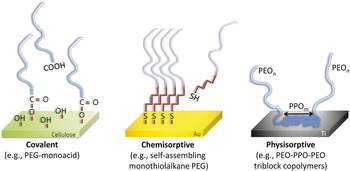
Mori et al. first investigated PEG as an anti-thrombogenic surface coating.Reference Mori, Nagaoka, Takiuchi, Kikuchi, Noguchi, Tanzawa and Noishiki85 Poly(ethylene oxide) (PEO) chains of various chain lengths were photograft co-polymerized onto poly(vinyl chloride) (PVC) surfaces and shown to reduce the adsorption and denaturation of blood components. In the three decades since, marked progress has been made in the development of PEG-based coating techniques for anti-microbial applications. These can be broadly divided into two distinct approaches: covalent attachment or passive adsorption. Covalent strategies rely on the formation of permanent chemical bonds to immobilize a reactive PEG functionality on the surface. Adsorptive strategies, on the other hand, involve chemisorptive or non-covalent (e.g., electrostatic and hydrophobic interactions) means to preferentially anchor PEG chains onto a surface. Figure 3 highlights representative examples of each modification approach.
Representative examples of the three dominant strategies for the immobilization of poly(ethylene glycol) (PEG) onto biomaterial surfaces. Covalent strategies rely on the formation of permanent chemical bonds, whereas chemisorptive and physisorptive strategies involve adsorptive mechanisms to preferentially anchor PEG chains onto a surface. PEO, poly(ethylene oxide); PPO, poly(propylene oxide).

Covalent grafting of PEG onto a surface has been well-studied since the early 1980s and continues to be extensively used for PEGylation of surfaces. This approach calls for specific chemical reactions to occur between terminal functional groups (e.g., epoxide, amine, anhydride, or hydroxide) on PEG and the surface groups present on the substrate, resulting in the formation of strong covalent bonds. Roosjen and co-workers applied PEO chains onto glass slides by reacting surface silanol groups with vinyl-terminated polymers in a polymer melt to yield covalent silyl-ether bonds.Reference Roosjen, de Vries, van der Mei, Willem and Busscher86, Reference Roosjen, van der Mei, Busscher and Norde87 This method has been shown to yield high surface grafting densities, where the PEO chains extend out into aqueous medium, adopting a brush-like conformation. PEO-modified surfaces exposed to various bacteria strains over a 48-hour period yielded more than a 98% reduction in bacterial adhesion for most bacteria strains tested, with a lesser effect on the more hydrophobic P. aeruginosa strain.
Oftentimes, the substrate to be coated lacks the appropriate surface reactive groups for PEG grafting. As a result, chemical procedures and surface pre-treatments are commonly employed to introduce the necessary functionalities—a process that could require multiple steps. Park et al. utilized one such approach to covalently graft PEG chains (molecular weight, Mw, 1000 and 3400) onto polyurethane films.Reference Park, Kim, Han, Kim, Lee, Suh and Choi88 Polyurethane was first treated with hexamethylene diisocyanate (HDI) in toluene with stannous octoate at 40°C for one hour to introduce the appropriate reactive groups for further functionalization with PEG. Surfaces modified with the longer PEG minimized bacteria adhesion regardless of culture medium (tryptic soy broth, brain heart infusion, and human plasma). In another multi-step approach, PEG-aldehyde was grafted onto poly(ethylene terephthalate) (PET) substrates using an intermediate branched poly(ethylenimine) (PEI) linker layer containing free reactive amino groups for subsequent PEG immobilization. PEI was covalently immobilized onto PET after the generation of reactive hydroxyl and carboxyl groups through a wet-chemical method.Reference Kingshott, Wei, Bagge-Ravn, Gadegaard and Gram89 The PET−PEG surface displayed a fourfold decrease in bacterial adhesion after five hours.
Glow discharge (plasma) deposition provides another direct method to lay down a PEG-like coating onto a surface. Highly reactive plasma is bonded directly to the surface to yield a thin film approximately 10–30 nm thick. Lopez et al. first reported the use of a tetra(ethylene glycol) dimethyl ether (TEGDME), also known as tetraglyme, vapor to generate a fouling-resistant ethylene glycol-rich surface coating on glass.Reference Lopez, Ratner, Tidwell, Haycox, Rapoza and Horbett90 Others have expanded this technique to other substrates and have also demonstrated the ability of oligoglyme coatings to effectively inhibit bacteria adhesion.Reference Hendricks, Kwok, Shen, Horbett, Ratner and Bryers81, Reference Denes, Somers, Wong and Denes91 In one study, coated surfaces saw a reduction in P. aeruginosa colonization by 99% relative to uncoated controls. Radio frequency plasma deposition can be particularly useful and attractive in the preparation of large area films or the modification of substrates with more complex three-dimensional structures.
Unlike covalent grafting, adsorptive strategies such as chemisorption and physisorption rely on surface modification through adsorption of the coating molecule from an aqueous solution. Since adsorptive processes can occur under comparatively mild conditions, this approach may be better suited for rapid and robust PEGylation of biomaterial surfaces. One of the most widely used chemisorptive approaches relies on PEG-terminated self-assembled monolayers (SAMs). Alkanethiols consisting of an alkyl tail and a S-H head group have been functionalized with PEG and used to create an anti-adhesive SAM coating on gold (Au) and silver (Ag) through the formation of strong S–Au or S–Ag bonds.Reference Chapman, Ostuni, Liang, Meluleni, Kim, Yan, Pier, Warren and Whitesides92, Reference Ostuni, Chapman, Liang, Meluleni, Pier, Ingber and Whitesides93 TEG-terminated SAMs on Au reduced E. coli biofilm formation by 99.5% for one day compared to the biofilm formation on a bare gold surface.Reference Hou, Burton, Simon, Blodgett, Luk and Ren94 When surfaces were patterned with regions consisting of methyl-terminated SAMs surrounded by TEG-terminated SAMs, biofilms were observed only on methyl-terminated patterns. However, the thiol-metal bond is specific to the noble metals, thus limiting the substrate usage of alkanethiolate SAMs. PEG-silane SAMs have proven to be a good alternative for modification of non-metallic oxides and potentially greater use for medical devices. Yang et al. modified glass surfaces through silylation of surface silanol groups using PEG-modified trimethoxysilanes.Reference Yang, Galloway and Yu95 Cox et al. functionalized silicon dioxide (SiO2) wafers with N-(triethoxysilylpropyl)-O-polyethylene oxide urethane (TESP) SAMs.Reference Cox, Curry, Skirboll, Gourley and Sasaki96 A better understanding of these specific chemistries is imperative to the development of new modification strategies and more robust surface coatings.
MessersmithReference Dalsin, Hu, Lee and Messersmith97 and othersReference Waite and Tanzer98, Reference Yu, Hwang and Deming99 have reported another unique chemisorptive approach based on proteins produced by marine mussels that allow strong adhesion to virtually any inorganic or organic surface. Lysine and catechol 3,4-dihydroxyphenylalanine (DOPA) residues were identified in high amounts at the adhesion interface and found to be responsible for the adhesive and cross-linking characteristics of these proteins. Linear monomethoxy-terminated PEG conjugated to 1–3 DOPA residues (mPEG-DOPA) was found to readily adsorb onto Ti surfaces through the formation of a coordinated DOPA-TiO2 bond, rendering modified surfaces resistant to proteins, cells, and bacteria.Reference Dalsin, Hu, Lee and Messersmith97, Reference Dalsin, Lin, Tosatti, Voros, Textor and Messersmith100 Model surfaces coated with mPEG-DOPA strongly resisted both urinary film formation and bacterial attachment in vitro.Reference Ko, Cadieux, Dalsin, Lee, Elwood and Razvi101 Subsequently, Fan et al. reported the use of a DOPA mimetic initiator as an anchor for surface initiated atom transfer radical polymerization of mPEG onto Ti and stainless steel substrates. This approach resulted in a thicker and denser polymer brush layer with improved antifouling properties.Reference Fan, Lin and Messersmith102 More recently, Lee et al. demonstrated the use of multiple catechols tethered on the backbone of PEG (versus end-functionalization) to obtain a significant enhancement of PEGylation capabilities on various types of material surfaces, including metals, oxides, and polymers such as polytetrafluoroethylene.Reference Lee, Lee, Pyo, Park and Lee103
Physisorption, in contrast to chemisorption, is driven by non-covalent electrostatic and hydrophobic interactions between the exposed surface and the adsorbed species. The ease of application and relatively short coating times make this particular approach highly attractive for use in the clinic. Triblock copolymers of poly(ethylene oxide)-poly(propylene oxide)-poly(ethylene oxide) (PEO-PPO-PEO) are commonly used to passivate surfaces against non-specific adsorption and, in some cases, to tether biological recognition ligands for added surface bioactivity.Reference Neff, Caldwell and Tresco104, Reference Neff, Tresco and Caldwell105 The central PPO block adsorbs strongly via hydrophobic interactions to form a polymer brush layer on hydrophobic base substrates. These coatings effectively resist initial bacterial adhesion and are relatively stable even under fluid shear in physiological environments.Reference Nejadnik, van der Mei, Busscher and Norde106, Reference Liu, Jastromb and Bhatia107 Marchant and co-workers developed surfactant polymers consisting of a poly(vinyl amine) backbone with hydrophilic PEO and hydrophobic hexanal (Hex) side chains (PVAm/PEO:Hex).Reference Vacheethasanee and Marchant108 These polymers undergo spontaneous surface-induced assembly to form a hydrophilic PEO coating on hydrophobic polyethylene and highly oriented pyrolytic graphite substrates. Suppression of S. epidermidis adhesion was achieved for all modified surfaces over the shear range of 0–15 dyn/cm2.
Poly-L-lysine (PLL) has been extensively characterized for use as a physisorptive anchor to graft PEG onto hydrophilic substrates. PLL-grafted-PEG (PLL-g-PEG) rapidly adsorbs through electrostatic attraction onto negatively charged surfaces, such as metal oxides. These molecules orient themselves with the cationic lysine backbone resting on the anionic surface and multiple grafted PEG chains extending into the aqueous environment to create a fully hydrated PEG brush layer. Textor and co-workers investigated the ability of PLL-g-PEG to reduce adhesion of implant-associated pathogens on Ti. An 89–93% reduction in S. aureus, depending on the duration of incubation and length of the PEG chain employed, was reported on coated surfaces.Reference Harris, Tosatti, Wieland, Textor and Richards109, Reference Maddikeri, Tosatti, Schuler, Chessari, Textor, Richards and Harris110 More recently, bi-functional PLL coatings containing both an anti-adhesive PEG moiety and a cell-adhesive RGD (Arginine-Glycine-Aspartic acid) tripeptide were developed. Bacterial adhesion remained minimal even when mammalian cell-binding was recapitulated on these coated Ti surfaces.Reference Subbiahdoss, Pidhatika, Coullerez, Charnley, Kuijer, van der Mei, Textor and Busscher111
The adsorptive approaches just detailed present a simple and rapid means to modify biomaterial surfaces without the need for specific chemical reactions requisite for covalent modification. For instance, stainless steel stents modified via dip-coating in an aqueous PLL-g-PEG solution for a mere 30 s still demonstrated reasonable in vivo efficacy against restenosis.Reference Billinger, Buddeberg, Hubbell, Elbert, Schaffner, Mettler, Windecker, Meier and Hess112 Additionally, the relatively mild reaction conditions for the formation of surface bonds are compatible with sensitive functional groups and biological macromolecules such as polypeptides. However, the stability of bonds between the adsorbed species and the surface can be compromised over time by competing adsorptive and degradative processes, particularly in biological fluids, resulting in eventual desorption and loss of the surface coating.
In recent years, several research groups have turned their interest to the identification of polypeptide sequences that bind strongly to a given material. These peptides, by acting as a physisorptive anchor, offer a novel way to immobilize biomolecules and other functional moieties on material surfaces. Using combinatorial phage and cell surface display techniques, high affinity polypeptides binders have already been identified for several classes of materials, including metals and metal oxides (e.g., Au, Ti, Pt, SiO2 ZnO2),Reference Sarikaya, Tamerler, Jen, Schulten and Baneyx113–Reference Sanghvi, Miller, Belcher and Schmidt116 natural and synthetic polymers,Reference Huang, Zauscher, Klitzman, Truskey, Reichert, Kenan and Grinstaff117, Reference Kenan, Walsh, Meyers, O’Toole, Carruthers, Lee, Zauscher, Prata and Grinstaff118 and even specific biological cellsReference Koivunen, Arap, Rajotte, Lahdenranta and Pasqualini119 and tissues.Reference Nowakowski, Dooner, Valinski, Mihaliak, Quesenberry and Becker120, Reference Rothenfluh, Bermudez, O’Neil and Hubbell121 These high throughput screening methods are extensively used in the investigation of protein-protein, protein-peptide, and protein-DNA interactions, and have been instrumental to the process of drug discovery.Reference Kay, Winter and McCafferty122–Reference Smith125
The development of a library of material-binding polypeptides would be extremely useful, since it would offer researchers a means to easily modify virtually any material of interest, inorganic or otherwise. The compositions of the selected polypeptides could also provide valuable insight into the residues and motifs that contribute greatly to binding affinity and specificity on a given material. These data, combined with additional molecular modeling and bioinformatics screens, could yield a wealth of information to allow for rational design of better and improved peptide anchors for surface modification.
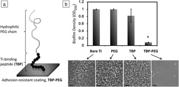
Kenan and co-workers performed a series of phage display screens to identify polypeptides that bind strongly to polystyrene (PS) and Ti, two commonly used implant materials.Reference Meyers, Hamilton, Walsh, Kenan and Grinstaff115, Reference Walsh, Middleton, Davis, Kenan and Grinstaff126 Polypeptides were subsequently assayed for binding affinity to their targets and for cross-binding to other materials. Peptides with the highest binding affinity and lowest cross-reactivity were then coupled to linear PEG (Mw 3400) and used to coat PS and Ti substrates (Figure 4a). Peptides spontaneously adsorbed from a dilute, aqueous solution onto substrates with the attached PEG chains extending into solution to form a non-fouling brush layer. Coated surfaces saw a significant reduction in S. aureus attachment and biofilm formation in the first 16 hours of exposure as compared to uncoated surfaces and surfaces coated with non-PEGylated peptidesReference Kenan, Walsh, Meyers, O’Toole, Carruthers, Lee, Zauscher, Prata and Grinstaff118, Reference Khoo, Hamilton, O’Toole, Snyder, Kenan and Grinstaff127 (Figure 4b). In a subsequent study, PEGylated-peptide anchors containing two and four repeats of the Ti-binding peptides were synthesized and evaluated for bacteria resistance.Reference Khoo, O’Toole, Nair, Snyder, Kenan and Grinstaff128 Not surprisingly, tetravalent analogues showed the greatest S. aureus inhibition as well as significantly improved serum stability. These results suggest that a cooperative multivalent attachment strategy could be used to obtain more robust and long lasting coatings with affinities closer to those of ideal covalent bonds. The modularity of this approach, whereby two or more separate domains are assembled together to create a multifunctional coating, makes it an extremely versatile platform for surface modification. In support of this multifunctional coating idea, other functionalities besides PEG have been conjugated to these peptide anchors, including the cell-adhesive ligand RGD,Reference Meyers, Hamilton, Walsh, Kenan and Grinstaff115 fluorophores,Reference Meyers, Khoo, Huang, Walsh, Grinstaff and Kenan129 and enzymes.Reference Kacar, Zin, So, Wilson, Ma, Gul-Karaguler, Jen, Sarikaya and Tamerler130 These types of peptide coatings can be readily applied to Ti surfaces under mild conditions and demonstrated functionality, thereby lending this platform to the idea of a generalized one-step approach to device modification.
(a) Schematic drawing of a PEGylated-peptide anchor, TBP-PEG, for the facile modification of surfaces; PEG, poly(ethylene glycol); TBP, titanium binding peptide. (b) Evaluation of S. aureus biofilm formation in response to various surface coatings using (top) a crystal-violet colorimetric assay and (bottom) direct phase contrast imaging. Scale bar = 35 μm. (n = sample size = 3, *p < 0.01, p is the p-value). OD595, optical density measured at 595 nm. Adapted from Reference 127.

Cross-linked PEG coatings
In contrast to previously introduced PEG grafting strategies, which typically rely on end point attachment of polymer chains to surfaces, new methods have been developed to obtain thicker and denser PEG coatings for improved coating stability and durability. Elbert and co-workers used larger polymer aggregates (microgels) to achieve thin PEG hydrogel coatings on surfaces. A single incubation of the surface with a microgel solution reduced non-specific cell adhesion to an extent that was much greater than 20 layers of PEG applied via a covalent layer-by-layer method.Reference Scott, Nichols, Cordova, George, Jun and Elbert131, Reference Kim, Wacker and Elbert132 Another approach involves the grafting of interpenetrating networks of a PEG to incorporate larger amounts of polymer onto the material surfaces.Reference Bearinger, Castner, Golledge, Rezania, Hubchak and Healy133, Reference Desai, Hossainy and Hubbell134 A thin cross-linked PEG-based hydrogel coating (OptiChem) has since been introduced commercially and has shown promising in vitro and in vivo efficacy. OptiChem-coated silicone discs remained effective against bacterial adhesion upon in vivo re-implantation, and no bacteria were harvested from coated discs, demonstrating the efficacy of the coating to resist biofilm formation.Reference Harbers, Emoto, Greef, Metzger, Woodward, Mascali, Grainger and Lochhead135, Reference Saldarriaga Fernández, Mei, Metzger, Grainger, Engelsman, Nejadnik and Busscher136
Polyelectrolyte multilayer films
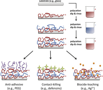
The physisorptive approach has been further extended to the design of multilayer films formed by the layer-by-layer (LbL) deposition of polyelectrolyte films of alternating charge (Figure 5). Boulmedais et al. incorporated PEG into thin polyelectrolyte films on silica surfaces through the sequential electrostatic deposition of PLL and poly(L-glutamic acid)-grafted PEG (PGA-g-PEG).Reference Boulmedais, Frisch, Etienne, Lavalle, Picart, Ogier, Voegel, Schaaf and Egles137 Multilayers topped with three PLL/PGA-g-PEG bilayers saw a 92% reduction in E. coli adhesion, even in the presence of nutrient-rich media. This strategy also allows for the combination of multiple antimicrobial strategies to combat bacteria more effectively. Anti-adhesive, antibiotic, and antiseptic compounds can be simultaneously incorporated into polyelectrolyte multilayer (PEM) coatings to develop a new class of multifunctional infection-resistant coatings. Hyaluronan/chitosan (HA/CH) polysaccharide PEMs with up to 10 bilayers offered an 80% reduction in the number of adherent E. coli on glass surfaces.Reference Richert, Lavalle, Payan, Shu, Prestwich, Stoltz, Schaaf, Voegel and Picart138 PEM coatings containing the insertion of defensin, a potent antimicrobial peptide, were also shown to inhibit the growth of infectious pathogens.Reference Etienne, Picart, Taddei, Haikel, Dimarcq, Schaaf, Voegel, Ogier and Egles139 The addition of leachable bactericides to these contact-killing PEMs can further enhance antibacterial efficacy, as demonstrated by Li et al.Reference Li, Lee, Sheng, Cohen and Rubner140 Silver nanoparticle-PEM composite films showed very high initial bacteria-killing efficiency from the release of Ag+ ions, and retained significant antibacterial activity even after the depletion of embedded Ag due to surface-immobilized quaternary ammonium salts. These dual-acting coatings present a viable strategy to impart long-term infection resistance to modified surfaces.
(top) Polyelectrolyte multilayers (PEMs) formed by the layer-by-layer deposition of polyelectrolyte films of alternating charge onto a substrate. (bottom) Three major classes of antibacterial coatings based on the PEM platform. Combinations of these approaches have been investigated and shown to combat bacteria more effectively. PEG, poly(ethylene glycol).

Stimuli-responsive PEM films have also been extensively researched, with several pH-tunable systems reported in the literature.Reference Pavlukhina, Lu, Patimetha, Libera and Sukhishvili141 Hammond and co-workers developed weak hydrogen-bonded PEMs that disassemble at physiological pHs to release polymeric micelles containing the antimicrobial triclosan.Reference Kim, Park and Hammond142 Release of bactericidal nanosilver from chitosan/heparin PEM films containing photocatalytic titania has also been triggered using UV irradiation.Reference Yuan, Ji, Fu and Shen143 Films were shown to sustain the long-term antibacterial activity in low intensity UV light. This is a rapidly growing field that could benefit applications necessitating temporal and spatial control of anti-microbial activity, for example, devices that penetrate the skin surface by allowing for repeated on-demand “sterilization.”
Summary and outlook
In the past two decades, research and development efforts for infection-resistant coatings have seen considerable progress in the form of numerous publications and a handful of FDA approved devices. Antibiotic therapy continues to be at the forefront of the battle against device infection, although significant efforts have been made to immobilize or confine the antibiotic to a localized region in order to combat systemic toxicity. Likewise, non-antibiotic-based agents have emerged as an attractive class of antimicrobials. Advances in chemical synthesis techniques in recent years have yielded a series of novel polymers and macromolecules, which can be finely tuned to modulate their interaction with bacteria. Clearly, the need for better infection-resistant coatings has resulted in many interesting treatment strategies to replace or supplement the traditional antibiotic regimen.
Although a significant reduction (>99%) in bacteria adhesion has been achieved with existing antibacterial coating strategies, this success has mostly been limited to short-term in vitro environments and less so in the harsher in vivo milieu, where coatings are continuously challenged by conditioning proteins and degradative processes. The antibacterial efficacy of coatings has also been found to vary greatly with bacterial species. One promising strategy that has only very recently emerged utilizes bioactive substances (e.g., quorum-sensing peptides, enzymes, viruses) to either disrupt the integrity or to inhibit the formation of a protective biofilm matrix,Reference Olsen, Pedersen, Laursen, Kiil and Dam-Johansen144, Reference Francolini and Donelli145 thus leaving sessile bacteria susceptible to antibiotics and to host immune defenses.
With the limited in vivo experimental data currently available, it is not yet clear if any one coating strategy will emerge as a superior candidate for the inhibition of implant-associated infection. Careful consideration of the desired drug elution and activity profile is necessary to determine the type of coating material and processing technique most suited for a specific device. A comprehensive understanding of the target pathogen and extensive research into potential synergistic combinations of antibiotic, antiseptic, and anti-adhesive agents will also be crucial to the development of better and improved devices.
However, as with many aspects of device development, there is no “one size fits all” approach for the selection of an infection-resistant implant coating. It is likely that a hybrid coating combining both active and passive strategies will be required in order to obtain a substantial reduction in clinical infection rates. Future advances will likely focus on more sophisticated “smart” surface coatings capable of triggerable and targeted antibacterial activity.
Acknowledgments
We thank the National Institutes of Health (Grant No. AR054872-01) for support. MWG has a conflict of interest, as he is co-founder of Affinergy Inc.—a start-up company that has an interest in device coatings.