Introduction
Cochlear implant (CI) is the most successful state-of-the-art device for the restoration of hearing, especially for those with severe to profound hearing loss.Reference Meng, Li, Zhang, Li and Qin1, Reference Chen, Chen, Tan, Jiang, Wu and Zhang2 In the past, CIs were only considered for adults with bilateral profound hearing loss. With technological advancements, the indications and the minimum age at implantation have expanded liberally. Even cochlear malformations are being considered for implantation with specialised and tailored devices.Reference Shah, Walters, Langlie, Davies, Finberg and Tuset3 Post-operative audiological outcomes vary among recipients of CIs due to surgical technique, inner ear damage, electrode array insertion depth and electrode array stiffness.Reference Schurzig, Timm, Batsoulis, Salcher, Sieber and Jolly4 Customised electrode implantation is one of the novel concepts.Reference Oh, Cheon, Park, Choi, Cho and Lee5 This is because over-insertion is linked to inner ear damage, and under-insertion is linked to poor audiological outcomes. However, completely atraumatic electrode insertion is not guaranteed in every case due to the variability of cochlear size, shape and anatomy and the electrode designs of various brands and insertion methods. This creates a need for an accurate measurement of the cochlear dimensions (CDs) for optimal post-operative results.Reference Chen, Chen, Tan, Jiang, Wu and Zhang2
Cochlear anatomy and orientation have been studied in the past by various methods: direct method, using histologic sections of the cochlea, indirect method, graphically representing the cochlea using plastic casts, 3D reconstructions and modelling the cochlea as a mathematical spiral function. The major disadvantage of the earliest methods was the necessity of a dissected temporal bone for study.Reference Koch, Ladak, Elfarnawany and Agrawal6 High-resolution computerised tomography (HRCT) was a milestone in this regard as it allowed scientists to view the cochlea as it is, in each individual. Further developments in the field led to the development of curved multi-planar reconstruction (cMPR), which visualises the HRCT scans in three perpendicular planes and measures the dimensions of non-linear structures. Otoplan is a multifunctional patient-specific otological planning software that primarily aims to help surgeons during otological surgery.Reference Bhavana, Timmaraju, Kumar, Kumar, Bharti and Prakash7 It can reconstruct HRCT and magnetic resonance Imaging (MRI) images for in-depth pre-operative analysis of CDs as well as other areas of interest such as the internal auditory canal (IAC), facial nerve, vestibular structures and Chorda tympani.Reference Bhavana, Timmaraju, Kumar, Kumar, Bharti and Prakash7, Reference Paouris, Kunzo and Goljerová8 Although the images are subjected to reconstruction in three planes and the initial analysis steps are similar, the calculation of the CDs is manual for cMPR. In contrast, Otoplan uses its innate programming.Reference Chen, Chen, Tan, Jiang, Wu and Zhang2
The aims of the current study are: first, to determine the largest diameter (A value), minimum width (B value), height (H value) and cochlear duct length (CDL) using cMPR and Otoplan; second, to compare the CDs required measured by the two software; third to compare time/expertise required to complete the analysis of CDs using both tools.
Materials and methods
The imaging data were obtained from patients visiting a tertiary care centre over 18 months from 2022 to 2023 for various complaints. HRCT scans of temporal bones were identified from those without cochlear abnormalities. A total of 110 ears (from 55 subjects) with an age range from 1 to 70 years old were included in this study. Each ear was treated as an independent variable during statistical analysis.
The data were acquired using a low-dose 128-slice multi-detector computed tomography (MDCT) scanner (SOMATOM Definition Flash CT scanner, Siemens Healthineers India). HRCT parameters were 500–650 mAs, 120 kVp and 0.6 mm slice thickness. After being reconstructed in the axial plane, the collected data were moved to the Syngo acquisition workstation (Siemens Healthineers India) for post-processing.
A senior radiologist with at least 10 years of experience in the field assisted an Otorhinolaryngologist in analysing data by cMPR. Measurement by Otoplan (Version 2) was carried out by an audiologist who had been trained in its use. The steps followed were as: (1) HRCT data uploading onto the Otoplan software; (2) adjusting the radiological plane into the Cochlear View; (3) landmarks registered were the round window (RW), lateral wall, apical centre and inferior and superior points; (4) calculation of CDs; (5) Exporting the final report.
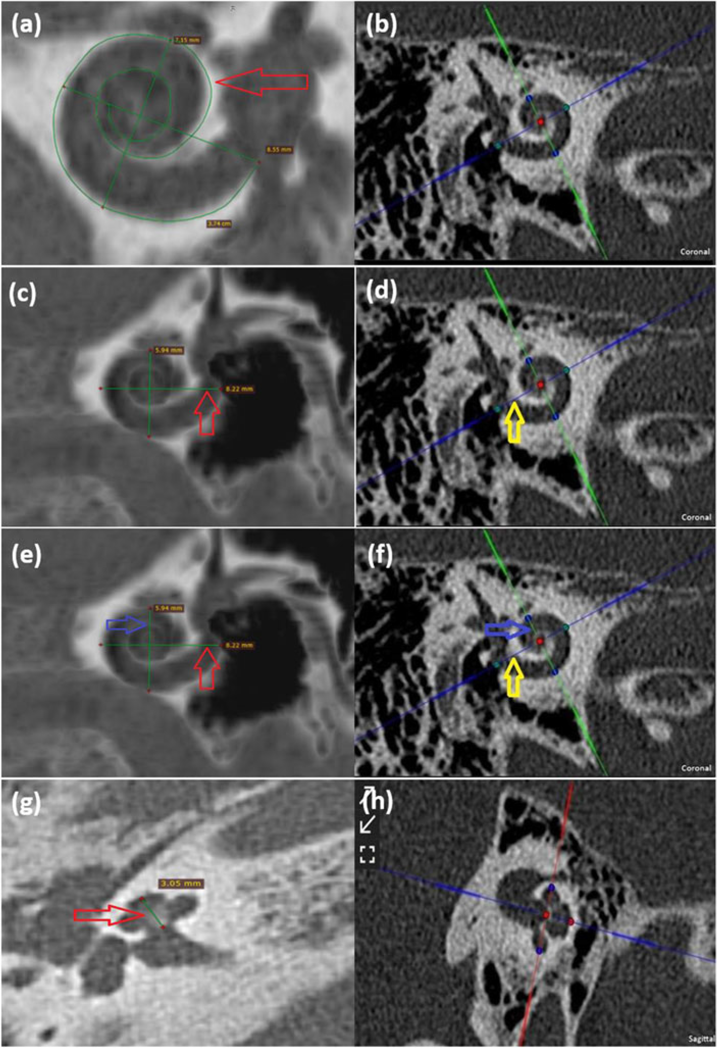
Minimum intensity projection (MiniP) was used to visualise the cochlear structures in cMPR better. The basis plane using “Cochlear View (CV)” bisected the cochlea through the basal turn of the cochlea, and the other two planes bisected the cochlea through the modiolus as the oblique sagittal and oblique axial plane. The midpoint of the RW was used as the reference point for the largest diameter measurements (Figure 1). The following CDs were recorded along with the time taken by both software for analysis in each case:
(1) Largest diameter of the cochlea (A-value): CV plane, the largest distance from the midpoint of the RW through the modiolus to the lateral wall of the basal turn.
(2) Smallest minimum width of the cochlea (B-value): CV plane, the perpendicular to the line for A value crossing the modiolus to the lateral wall of the cochlea.
(3) Height of cochlea (H-value): Oblique sagittal view, distance from the centre of the modiolus of the basal turn to the superiormost point.
(4) Cochlear duct length (CDL): length of a curved line drawn from the midpoint of RW to the terminal point of apical turn fit along the outer lateral wall of the cochlea.
(a & b) Cochlear duct length (CDL) (red arrow) calculated using curved multi-planar reconstruction (cMPR) (left) and Otoplan (right). (c & d) A value calculated by cMPR (red arrow) and Otoplan – Distance between green dots (yellow arrow). (e & f) B value calculated using cMPR (left) and Otoplan (right) – Distance between blue dots. (g & h) H value of cochlea calculated using cMPR (Left) and Otoplan (right) – Distance between red dots.

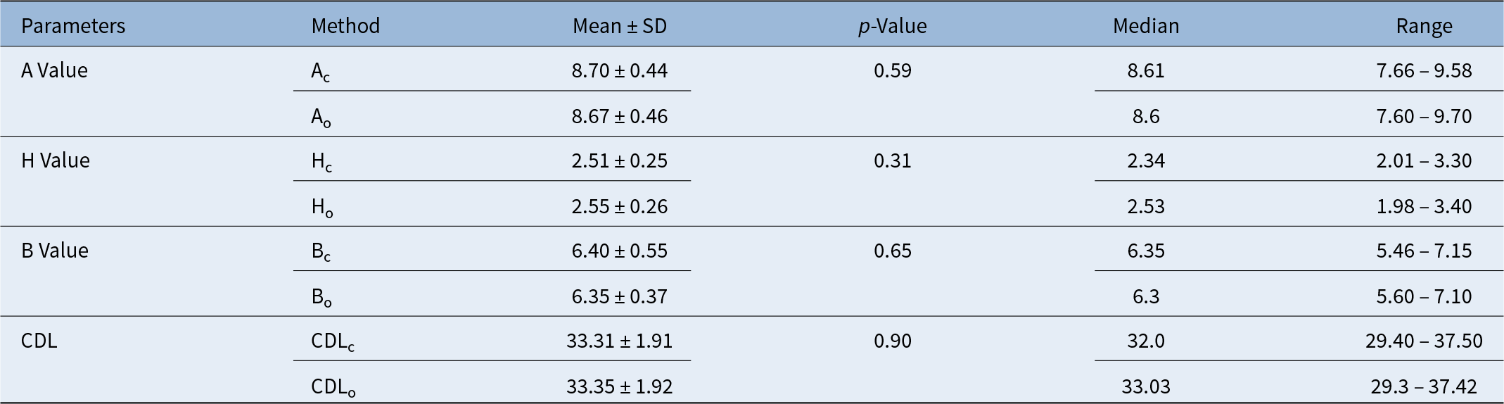
Comparison of measurements of CDs by Otoplan and cMPR

Note: Ac, Hc, Bc, CDLc: A value, B value, H value and CDL by cMPR; Ao, Ho, Bo, CDLo: A value, B value, H value and CDL by Otoplan.
CD = cochlear dimensions; CDL = cochlear duct length;
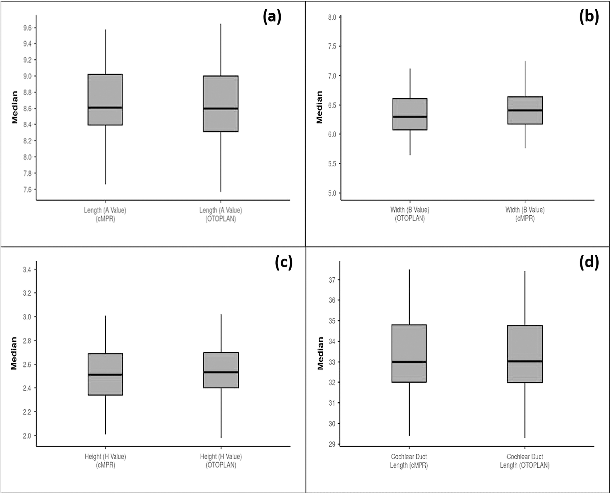
(a & b) A and B value median and range by Otoplan and curved multi-planar reconstruction (cMPR). (c & d) H and CDL value median and range by Otoplan and cMPR.

Statistical analysis was performed using Microsoft Excel 2016. A ‘Z’ test was used to compare the mean of CDs measured using cMPR and Otoplan, whereas the mean time needed for analysis was compared using the chi-squared test. A p-value of less than 0.05 was taken as significant for the relationship between CD parameters.
Results
In 55 patients (110 ears) included, the male:female ratio was 8:3. The average age was 11.5 years (range 1 to 67 years). Table 1 shows CDs measured by cMPR and Otoplan, respectively. Figure 2 shows the mean CDL calculated by cMPR was 33.31 ± 1.91 (range 29.40 to 37.50 mm), and the same by Otoplan was 33.35 ± 1.92 (range 29.3 to 37.42 mm). The mean A-value calculated by cMPR was 8.70 ± 044 (range 7.66 to 9.58 mm) and the same by Otoplan was 8.67 ± 0.46 (range 7.6 to 9.7 mm). The mean B value calculated by cMPR was 6.40 ± 0.55 (range 5.46 to 7.15 mm), and the same by Otoplan was 6.35 ± 0.37 (range 5.6 to 7.1 mm). The mean H value calculated by cMPR was 2.51 ± 0.25 (range 2.01 to 3.30 mm), and the same by Otoplan was 2.55 ± 0.26 (range 1.98 to 3.40 mm). There were no significant differences between the CDs calculated by cMPR and Otoplan; however, the time taken for analysis of a single ear, shown in Table 2, was significantly lower for Otoplan than cMPR (p < 0.01; chi-squared test).
Comparison of proportions of subjects according to time taken to measure parameters by Otoplan and cMPR

cMPR = curved multi-planar reconstruction.
* = p-value.
Discussion
The variability of the human cochlea has been studied for a century. For a long time, dissecting the cochlea and directly measuring the parameters was the only available method to study the intricacies of the cochlear anatomy.Reference Koch, Ladak, Elfarnawany and Agrawal6 With the development of newer technologies, the study of the cochlear dimensions has taken a giant leap with the use of HRCT, MRI and Otoplan. The advances in pre-operative assessment have also been paired with the advancements in CI surgery, emphasising “soft surgery” making atraumatic electrode insertion imperative. There has been growing evidence that the “one size fits all” theory for electrode arrays does not achieve the optimal hearing outcome.Reference Dutt, Gaur, Vadlamani and Nandikur9 The focus on electrode design has led to the development of a variety of CI with variable lengths and appropriate diameters that can be chosen according to the dimensions of the recipient cochlea. Deep insertion is associated with better hearing outcomes; however, the risk of traumatic insertion also increases with complications, including tip-fold-over and injury to the spiral ganglion. This creates a need for an accurate measurement of the CDL in the patient undergoing CI for optimal post-operative results. Multiple studies have shown that although the cochlea attains its adult size by the 16th to 20th weeks of gestation, a significant variation occurs between subjects, and the CDL may have a variation.Reference Meng, Li, Zhang, Li and Qin1 The variation in the CDs further depends on people from different racial backgrounds. People from the Indian subcontinent have smaller skulls, which may impact CD differences. Therefore, CD measurements should be an essential step in the pre-operative analysis of CI implant recipients.Reference Grover, Sharma, Singh, Kataria, Lakhawat and Sharma10
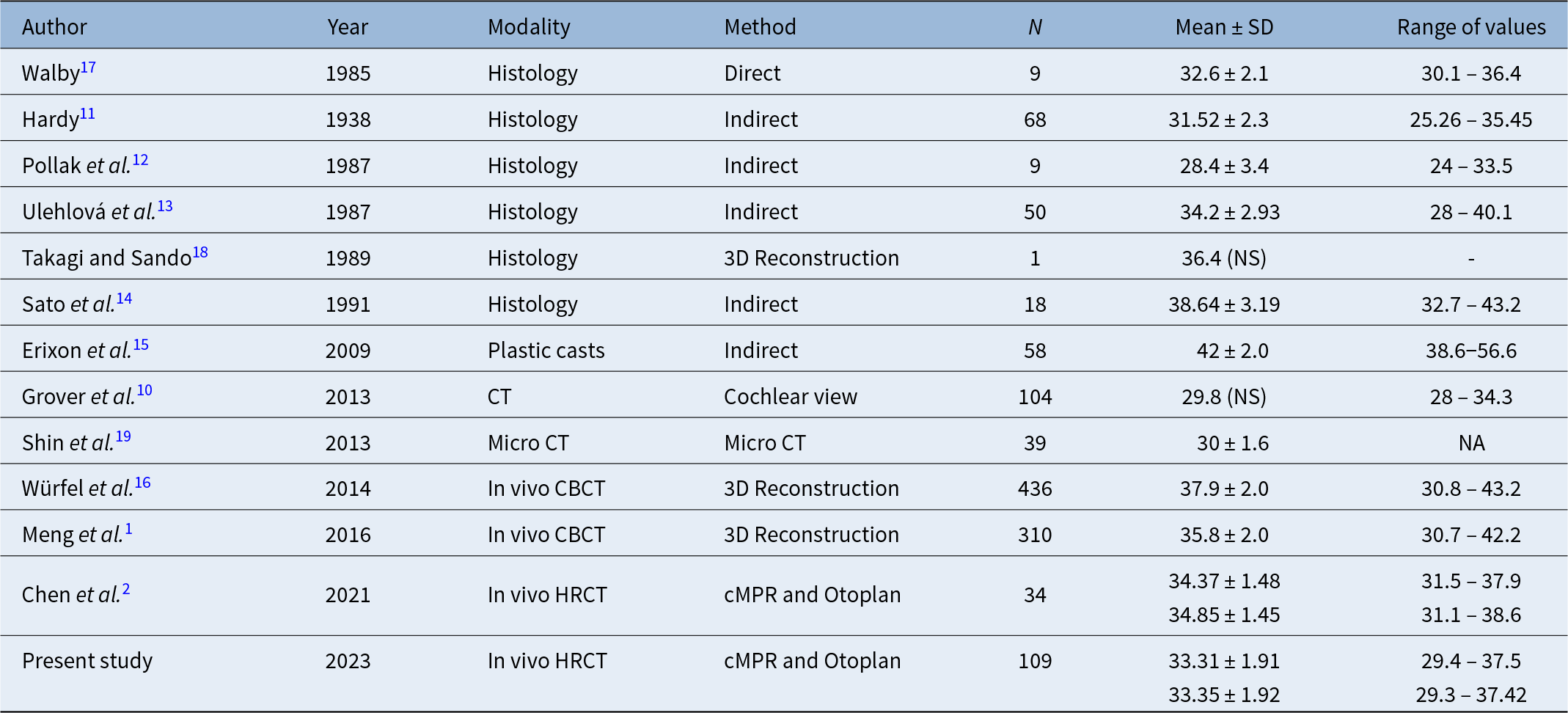
Table 3 reviews the studies that have attempted to calculate the CDs using various methods. The earliest studies were done using histological methods with dissected temporal bones. Although the studies were substantial then, the need for a dissected temporal bone greatly limited their use to a living person. Numerous studies have also reported on the density and quantity of spiral ganglion cells; however, none of the imaging techniques available today, including ours, can replicate these findings, except histology.Reference Hardy11–Reference Sato, Sando and Takahashi14 The study done by Erixon et al. (2013) used HRCT-reconstructed images of cochlear moulds/plastic casts, which is a milestone. However, the study still needed dissected temporal bones to construct the moulds/casts and was unlikely to help the individual. Furthermore, the CDL calculated in the study was done using a formula based on A and B values, whereas the CDL is calculated as an independent variable in cMPR.Reference Erixon and Rask-Andersen15 Würfel et al. (2014) conducted a study to determine CDs using cone beam computed tomography (CBCT) and analysed the images using Osirix MD software and concluded that determination of CDL is crucial for implant recipients so as to provide them with the best-suited implant from anatomical as well as audiological standpoint.Reference Würfel, Lanfermann, Lenarz and Majdani16 The study conducted by Meng et al., (2016) examined the CDs and their possible impacts on CI. Using the Osirix software, 3D-MPR MiniP was used to analyse CDS, and the findings were in line with those of earlier investigations. The slightly shorter CDL is attributed to variations in methodology, materials and racial makeup.Reference Meng, Li, Zhang, Li and Qin1 One disadvantage of the Osirix MD software is that it is only compatible with Apple operating systems and is not accessible on Windows operating systems.
Comparing the CDL measurements made by previous studies

CBCT = cone beam computed tomography; cMPR = curved multi-planar reconstruction; CT = computed tomography; HRCT = high-resolution computerised tomography; NS = not specified.
The study done by Chen et al. (2021) compared the results of Otoplan to cMPR and concluded that Otoplan offers a quicker and more accurate way to measure CDs than cMPR. Moreover, it can be used on a laptop computer or tablet and minimises the impact of subjective factors. The study was conducted on the Chinese population on a very small sample size (n = 34). Our study’s results differ from Chen et al. (2021). It shows smaller averages of CDs in the Indian cohort as compared to the Chinese.Reference Chen, Chen, Tan, Jiang, Wu and Zhang2 To our knowledge, this study is the only one conducted in the Indian subcontinent within the current paradigm.
Another major advantage of this study is that HRCT was performed on living subjects and included a wide age range. Furthermore, we found that analysis of HRCT scans is a viable option and Otoplan is easier to operate and minimises the impact of subjective factors contingent on the availability of software. The limitation of the current study is that the patient’s physical measurements, such as height or head circumference, were not taken into account and may provide a niche for further research.
• Cochlear duct length (CDL) is crucial for cochlear implant selection and placement.
• This study compared curved multi-planar reconstruction (cMPR) and Otoplan software for CDL measurement in an Indian population.
• Otoplan was significantly faster than cMPR for CDL analysis, with comparable accuracy.
• Otoplan supports efficient and accurate pre-surgical planning.
Conclusion
This study establishes that both cMPR and Otoplan software provide comparable accuracy in CD measurement, a critical factor in pre-operative cochlear implant planning. However, Otoplan significantly outperforms cMPR in terms of operational efficiency, requiring less time and less radiological expertise. Given the increasingly widespread indications for cochlear implantation and the need for patient-specific electrode selection, tools like Otoplan offer a pragmatic solution that can be employed even in resource-constrained settings. Furthermore, this study contributes uniquely to the Indian population dataset, where CD variability may influence electrode choice. Future studies correlating anatomical dimensions with audiological outcomes and incorporating anthropometric variables may further refine individualised cochlear implant strategies.
Funding
The authors declare that they have no conflict of interest and have not received any funding for the research.
Competing interests
None declared.
Ethical approval
All procedures involving human participants in the study followed the ethical standards of the Institutional and /or National Research Committee and the 1964 Helsinki Declaration and its later amendments or comparable ethical standards.
Informed consent
Informed consent was obtained from all the individual participants included in this study.