Influenza continues to cause significant mortality in children. Bacterial co-infection is present in a third of all critically ill children with influenza, and in half of all children who have influenza-associated mortality. 1 We report a child with influenza B and community-acquired methicillin-resistant Staphylococcus aureus co-infection who developed purulent pericarditis. Purulent pericarditis is very rare, and has not previously been reported as a complication of methicillin-resistant S. aureus co-infection with influenza. Influenza affects a variety of host defence mechanisms predisposing to Staphylococcal co-infection, leading to severe and fatal complications in previously healthy children.
Case
A 4-year-old boy presented with high fever and right-sided chest pain of 2 days duration. He had mild intermittent asthma but was otherwise healthy, and had received inactivated influenza vaccine about 5 months ago.
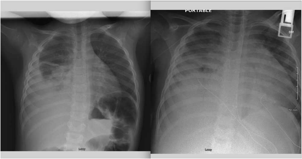
Upon admission, his body temperature was 103.7°F, heart rate was 160/minute, respiratory rate was 32/minute, blood pressure was 95/54 mmHg, and oxygen saturation was 95% on room air. He was alert, interactive, in moderate respiratory distress with subcostal retractions, and no air entry on the right side. Nasopharyngeal swab was positive for influenza B. Chest X-ray showed right lower lobe pneumonia with pleural effusion and a normal-sized cardiac silhouette (Fig 1). He was placed on high-flow nasal cannula, and intravenous ceftriaxone and oral oseltamivir were initiated. A right pleural pigtail catheter was inserted and drained 130 ml of serosanguineous fluid.
X-ray at admission and on day 6 of hospitalisation. Description: Left – X-ray at admission showing right-sided lobar pneumonia and empyema with normal cardiac silhouette. Right – X-ray on day 6 of hospitalisation showing enlarged cardiac silhouette.

CT scan showed a moderate to large loculated right-sided pleural effusion with complete consolidation of the right lung. The child underwent video-assisted thoracoscopic surgery with pleural decortication and adhesiolysis. Pleural fluid culture grew methicillin-resistant S. aureus, and vancomycin and rifampin were added to the antibiotic regimen.
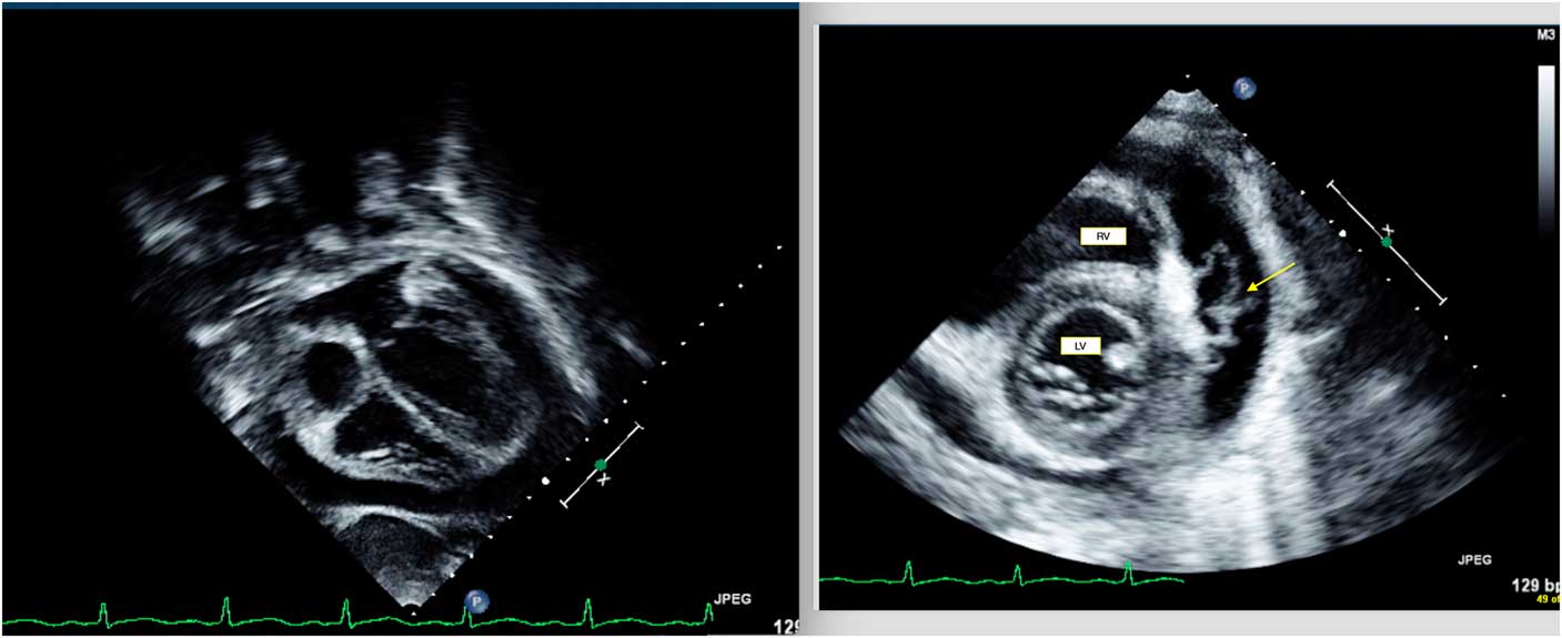
Clinical improvement did not occur as expected after video-assisted thoracoscopic surgery, with persistence of high-grade fevers and general malaise. He developed generalised oedema, diffuse abdominal pain, and feeding intolerance. Plain X-ray of the abdomen showed multiple dilated loops of small bowel suggestive of subacute obstruction. Abdominal ultrasound revealed small amount of ascites and oedema surrounding the gall bladder. Persistent tachycardia and increase in respiratory distress warranted escalation of non-invasive respiratory support. A chest X-ray on day 6 of hospitalisation (Fig 1) showed enlarged cardiac silhouette, and echocardiogram showed a moderate circumferential pericardial effusion with stranding (Fig 2). Interventricular bounce was present, suggesting haemodynamically significant effects on diastolic function. Pericardial window surgery with drainage of effusion yielded 250 ml of yellow, cloudy fluid. The pericardial fluid did not grow any organism in culture. Multiple blood cultures were also negative. There was significant clinical improvement, and the patient was discharged home after receiving 4 weeks of intravenous vancomycin and rifampin. He was doing well on follow-up.
Echocardiographic images before pericardial window drainage. Description: Left – Four-chamber view showing moderate, circumferential effusion. Right – Subcostal view showing the presence of thick fibrin stranding (arrow). LV=left ventricle; RV=right ventricle.

Discussion
Purulent pericarditis is exceedingly rare, and has not yet been reported as a complication of influenza and methicillin-resistant S. aureus co-infection. Pericardial involvement occurs most often as a result of direct extension of infection from involved lung and pleura. Pulmonary infections may spread to the pericardium through the bronchial circulation. Haematogenous dissemination from a remote site is also possible. Classic presenting features are precordial pain, pericardial friction rub, and muffled heart sounds. In rapidly progressive cases, signs of tamponade may be the presenting feature. Pericarditis may rarely present with abdominal pain, sometimes mimicking acute abdomen.Reference Donnelly, Kimball and Barr 2 Our patient had persistent abdominal pain and distension, with mild to moderate ascites and gall bladder wall oedema on ultrasound, which have been reported in association with purulent pericarditis.Reference Donnelly, Kimball and Barr 2 It is likely that diastolic dysfunction resulting from reduced ventricular compliance owing to fixed pericardial volume in our patient contributed to these abdominal symptoms as a result of increasing right-sided cardiac pressures. Fever, tachypnoea, and tachycardia – all of which are signs of pericarditis – are often attributed to underlying pulmonary or systemic infection when present. An enlarged cardiac silhouette is sometimes the only clue to the presence of pericardial effusion.
Purulent pericarditis can be fatal if undiagnosed and untreated. The rate of accumulation of pericardial fluid is vital, as gradual expansion of the parietal pericardium can accommodate relatively large quantities of fluid. Rapid accumulation of fluid can markedly increase intrapericardial pressure, and volumes as small as 100 ml may be enough to cause severe tamponade in a child. Interventricular or septal bounce, as seen in our patient, is a sign of restricted ventricular filling and occurs owing to a fixed pericardial volume. As there is rapid increase in ventricular filling during early diastole, there is a paradoxical leftward shift of the interventricular septum as right ventricular filling occurs before left ventricular filling. This is accentuated during inspiration and seen echocardiographically as a “bouncing” motion of the septum.Reference Walker, Chung and Reddy 3 This is a useful tool for ultrasonographic diagnosis of haemodynamically significant effusion before development of florid tamponade.
Preceding infection with influenza facilitates secondary bacterial infection through multiple immunological mechanisms. Influenza virus damages the epithelial layer of the tracheobronchial tree and enhances adherence of Staphylococcus. Influenza depletes resident alveolar macrophagesReference Robinson, Kolls and Alcorn 4 and suppresses nicotinamide adenine dinucleotide phosphate oxidase-dependent bacterial clearance, affecting phagocytic function of both neutrophils and macrophages.Reference Sun and Metzger 5 The virus alters the immune response of T cells and NK cells when challenged with secondary bacterial infection. Influenza and Panton Valentine leucocidin-producing S. aureus act together to induce inflammation and tissue damage in the respiratory epithelium by extensive neutrophil lysis and massive release of granule proteases, causing haemorrhage and tissue damage leading to necrotising pneumonias.Reference Niemann, Ehrhardt and Medina 6 There has been a resurgence of community-acquired methicillin-resistant S. aureus in children in the past two decades, and S. aureus co-infection with influenza has been increasing at a rapid pace.Reference Finelli, Fiore and Dhara 7 Randolph et alReference Randolph, Vaughn and Sullivan 8 reported that methicillin-resistant S. aureus co-infection was present in 44% of all previously healthy children critically ill with influenza, and led to an eightfold increase in mortality. Despite the frequent occurrence of S. aureus pneumonia, purulent pericarditis remains a rare occurrence. In a study involving 117 children with S. aureus pneumonia, only one child had a pericardial effusion.Reference Carrillo-Marquez, Hulten and Hammerman 9
It is vital that physicians caring for acutely ill children have a high index of suspicion for development of purulent pericarditis in patients with empyema, as clinical presentation may be subtle. Timely drainage is of essence to prevent complications related to ongoing pericardial inflammation.
Acknowledgements
None
Financial Support
This research received no specific grant from any funding agency or from commercial or not-for-profit sectors.
Conflicts of Interest
None