Introduction
Helminth parasites are multicellular organisms which collectively are estimated to infect over 1.2 billion people worldwide (GBD 2017 Disease and Injury Incidence and Prevalence Collaborators., 2018). These parasites cause debilitating diseases and syndromes in humans, companion animals, livestock and wildlife, leading to great economic losses and high morbidity rates. In both humans and animals, the diseases they cause are chronic and there is still a lack of effective treatments to eradicate them (McSorley and Maizels, Reference McSorley and Maizels2012). This is partly due to the scarce in vitro models available to study basic aspects of the biology of these pathogens, including factors involved in parasite multiplication and survival.
Helminth phyla relevant to human health comprise nematodes and platyhelminths (commonly known as flatworms), the latter composed of tapeworms (cestodes) and trematodes. Cestode parasites are passively transmitted between hosts and parasitize almost all vertebrate species. Among the diseases they cause is cysticercosis, produced by Taenia solium (Family Taeniidae), which is classified by the WHO within the prioritized neglected tropical diseases to be controlled by 2030 (World Health Organization, 2020). This zoonosis is the cause of severe health and economic losses in Latin America, Africa and Asia (Coral-Almeida et al., Reference Coral-Almeida, Gabriël, Abatih, Praet, Benitez and Dorny2015) and is produced by the establishment of the metacestode larval stage (cysticercus) in different loci, such as subcutaneous tissue, muscle, viscera or eyes. The life cycle of T. solium is indirect and perpetuates between humans and pigs, where the former act as both natural definitive and accidental intermediate hosts. The most serious manifestation of the disease is neurocysticercosis, which occurs when cysticerci establish in the central nervous system after ingesting infectious eggs. Currently, human neurocysticercosis is considered the most common neurological helminthic disease (Del Brutto, Reference Del Brutto2012).
To date, successful long-term in vitro models that allow cestode larvae multiplication have been established only for Echinococcus multilocularis (Hemphill and Gottstein, Reference Hemphill and Gottstein1995; Jura et al., Reference Jura, Bader, Hartmann, Maschek and Frosch1996; Spiliotis et al., Reference Spiliotis, Tappe, Sesterhenn and Brehm2004). Experimental evidence shows that the larval stage (metacestode) of this parasite requires fetal bovine serum (FBS) and factors secreted by the host to grow and multiply in vitro, indicating that host cells provide signals that promote and regulate the development of metacestodes (Hemphill and Gottstein, Reference Hemphill and Gottstein1995; Jura et al., Reference Jura, Bader, Hartmann, Maschek and Frosch1996; Spiliotis et al., Reference Spiliotis, Tappe, Sesterhenn and Brehm2004). To achieve this, parasites were grown in co-culture with primary hepatocytes or mammalian cell lines, or in the presence of media containing the corresponding host excretion/secretion (E/S) products, and best results were obtained with rat hepatoma cells (Spiliotis et al., Reference Spiliotis, Tappe, Sesterhenn and Brehm2004). In addition, the metacestode of Echinococcus spp. produces an outer acellular layer (laminated layer) that prevents the tegument of the parasite to be in contact with the feeder cells. This allowed the set-up of a unique in vitro system that mimics the growth of the parasite from primary cell cultures to the asexual multiplication of the metacestode stage (Spiliotis et al., Reference Spiliotis, Lechner, Tappe, Scheller, Krohne and Brehm2008; Spiliotis and Brehm, Reference Spiliotis and Brehm2009).
Taenia crassiceps is used as an experimental model cestode for the study of cysticercosis (Willms and Zurabian, Reference Willms and Zurabian2010) and similarly to E. multilocularis, its larvae multiply by exogenous budding. This characteristic allows the perpetuation of these parasites in experimental animals by the periodical intraperitoneal passage of larvae and, in the case of E. multilocularis, the in vitro multiplication of metacestodes. Even though some reports show that T. crassiceps cysticerci multiply asexually in vitro (Escobedo et al., Reference Escobedo, Larralde, Chavarria, Cerbón and Morales-Montor2004, Reference Escobedo, Romano and Morales-Montor2009, Reference Escobedo, Soldevila, Ortega-Pierres, Chávez-Ríos, Nava, Fonseca-Liñán, López-Griego, Hallal-Calleros, Ostoa-Saloma and Morales-Montor2010; Díaz-Orea et al., Reference Díaz-Orea, de Aluja, de L'Erosa, Gomez-Conde, Castellanos Sánchez, Willms, Sciutto and Fragoso2007; Ostoa-Saloma et al., Reference Ostoa-Saloma, Ostoa-Jacobo, Esquivel-Velázquez, Bazúa and Larralde2010; Esquivel Velázquez et al., Reference Esquivel Velázquez, Hernández, Larralde and Ostoa Saloma2014; Adalid-Peralta et al., Reference Adalid-Peralta, Rosas, Arce-Sillas, Bobes, Cárdenas, Hernández, Trejo, Meneses, Hernández, Estrada, Fleury, Laclette, Larralde, Sciutto and Fragoso2017), long-term experiments are still lacking. Here, it was aimed to assess the conditions necessary to allow T. crassiceps survival, growth and multiplication to eventually establish a long-term culture system to obtain in vitro grown cysticerci.
Materials and methods
Parasites
Taenia crassiceps cysticerci (strain HYG) were collected from experimentally infected adult female CF1 mice (6–8 weeks old). For this, mice were inoculated intraperitoneally with 25 cysticerci each and parasites were collected 4–5 months post inoculation. Animals were reared at the animal facilities from IMPaM in polycarbonate cages under controlled conditions that involved a 12 light/12 dark cycle, constant room temperature at ~ 21°C and food and water ad libitum. After euthanizing the animals by cervical dislocation, parasites were collected under sterile conditions using a 1 mL micropipette with the tips cut in order to increase the diameter of the orifice to avoid cysticerci disruption. After thorough washing with sterile phosphate-buffered saline (PBS), only floating cysticerci were recovered and parasite vitality was assessed by trypan blue staining. Briefly, 25 cysticerci per well were incubated in 5 mL of PBS and trypan blue (final concentration 0.02–0.05%) in a 6-well plate. After incubating for 10 min at room temperature, cysticerci were washed twice or thrice with 5 mL of PBS until no traces of trypan blue remained and stained (non-viable) parasites were counted. Only batches with ⩾ 90% vitality were used.
Parasite in vitro culture
Short-term culture: the aim of this experiment was to identify the minimal requirements that promote parasite survival and multiplication in order to establish a long-term in vitro system that allows cysticerci production. For this, parasites were cultured in 12-well plates for 15 days in 3 technical replicates per each biological replicate in the presence of high glucose (4.5 gr L−1) Dulbecco's modified Eagle medium (DMEM) without sodium pyruvate supplemented with sodium bicarbonate (3.7 g L−1) and antibiotics (penicillin–streptomycin 100 U mL−1) at 37°C and 5% CO2. Two parasite densities were tested: 1 cysticercus (1C) and 10 cysticerci (10C) per well. To assess parasite stimuli, cysticerci were incubated in the presence of (i) DMEM and (ii) DMEM that was daily changed (DMEM DC), without FBS in both cases. To assess host stimuli, cysticerci were incubated in the presence of: (i) DMEM + 10% FBS (DMEM FBS), (ii) filtered DMEM + 10% FBS (f-DMEM FBS), (iii) DMEM + 10% FBS + E/S products from the American Type Culture Collection (ATCC) CRL-1600 rat hepatoma cell line (cDMEM RH) and (iv) DMEM + 10% FBS + E/S products from the A549 human lung carcinoma cell line (cDMEM A549).
As previously reported, multiple mammalian cell lines were able to support in vitro the growth and asexual multiplication of the related taeniid E. multilocularis (Spiliotis et al., Reference Spiliotis, Tappe, Sesterhenn and Brehm2004). Consequently, the rat hepatoma cell line was chosen since it produced the best results in E. multilocularis. The A549 cells were used for comparative purposes. To obtain f-DMEM FBS, the DMEM FBS was passed through a 0.2 μm filter. To collect media with E/S products (cDMEM RH and cDMEM A549), each cell line was cultured in a T75 flask with 30 mL of DMEM FBS at 37°C and 5% CO2. After 7 days without changing medium, the cDMEM was collected, passed through a 0.2 μm filter and used immediately (Spiliotis et al., Reference Spiliotis, Tappe, Sesterhenn and Brehm2004).
To evaluate cysticerci growth, cultures were photographed and the major axis of each cysticercus (diameter) was measured using a reference scale (pixel cm−1). Diameter measurements were then normalized with respect to day 0. For this, the value of each well from day 0 was subtracted to the corresponding value from days 0 and 15. In the case of 10C cultures, the mean value of a given well at day 0 was subtracted to the diameter of each cysticercus in the same well at each time point. Images were processed with ImageJ 1.53 K. To evaluate parasite multiplication, the number of buds per cysticercus was counted by observation in a stereoscopic microscope. In 10C cultures, the total number of buds per well was divided by the total number of cysticerci. Parasite vitality was assessed by trypan blue staining and morphology analysis (collapsed cysticerci were excluded from the analyses).
Long-term culture: the aim of this experiment was to determine if the conditions identified in the short-term cultures were able to support parasite proliferation at the long term in order to establish a system that provides in vitro grown cysticerci. For this, 5–15 mL of cysticerci were incubated for 5 weeks in T75 flasks with DMEM FBS (control) or with DMEM FBS in co-culture with either the hepatoma cell line or the lung carcinoma cell line (feeder cells) at 37°C, 5% CO2. In the latter, freshly trypsinized cells from the corresponding cell line were added weekly. Medium change was performed once a week. The volumetric growth of parasites was measured weekly by transferring parasites to graduated conical tubes and allowing them to sediment. Then, the fold-change was calculated with respect to week 0.
Statistical analysis
For short-term culture results, the Mann–Whitney U test was performed, and for the long-term cultures the Kruskal–Wallis and Dunn's comparative tests were used. The software used for statistical analysis was GraphPad Prism version 5.01.
Results
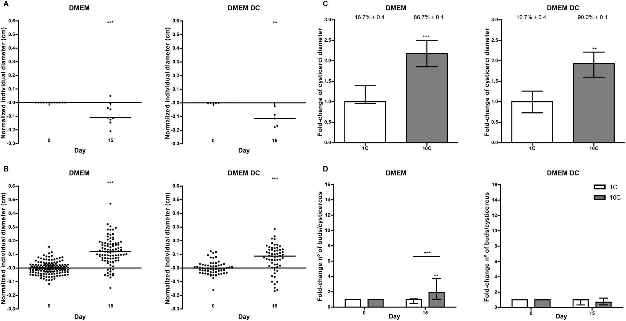
Since parasites depend on multiple factors from both pathogens and hosts, different conditions to identify those with greater potential to sustain parasite development in vitro were evaluated. Hence, the relevance of only parasite stimuli was assessed first. For this, different densities of cysticerci were incubated in the presence of DMEM and DMEM that was daily changed (DMEM DC). As a result, in 1C cultures incubated in both DMEM and DMEM DC there was a significant decrease in diameter after 15 days (P < 0.001 and P < 0.01, respectively) (Fig. 1A). The opposite was observed in 10C cultures (Fig. 1B), which showed a significant diameter increase with respect to day 0 (P < 0.001 for both media) and also a significant 2-fold change with respect to 1C cultures (P < 0.001 for DMEM and P < 0.01 for DMEM DC) (Fig. 1C). With respect to vitality, 1C cultures showed low values (16.7% ± 0.4) at day 15 in both media (Fig. 1C). Interestingly, in the presence of DMEM, 10C cultures showed a significant increase in the number of buds produced with respect to day 0 (P < 0.01) and compared to 1C cultures (P < 0.001). This effect was abolished in DMEM DC, i.e. with daily removal of E/S products from the parasites (Fig. 1D).
Assessment of short-term survival, growth and multiplication of Taenia crassiceps cysticerci in vitro at different parasite densities in the absence of host stimuli. Dots represent normalized diameter values with respect to day 0 in cultures of 1 cysticercus (A) and 10 cysticerci (B) per well. The lines indicate median values. (C) Bars represent fold-change of diameter and percentages correspond to vitality values at day 15. Median values with interquartiles are plotted. Biological replicates: DMEM = 5, DMEM DC = 2. Technical replicates = 3. (D) Bars indicate fold-change in the number of buds per cisticercus with respect to day 0. Median values with interquartiles are plotted. 1C: 1 cysticercus/well, 10C: 10 cysticerci/well. Biological replicates: DMEM = 6, DMEM DC = 3. Technical replicates = 3. The Mann–Whitney test was performed. *P < 0.05, **P < 0.01, ***P < 0.001.

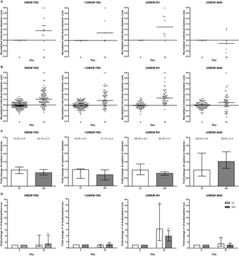
Then, we evaluated the effect of varied host stimuli such as FBS (DMEM FBS), FBS depleted in molecules/particles > 200 nm (f-DMEM FBS) and FBS together with the E/S products from rat hepatoma (cDMEM RH) and lung carcinoma (cDMEM A549) cell lines also depleted in particles > 200 nm. As shown in Fig. 2A, in 1C cultures, all media except cDMEM A549 produced a significant increase in parasite diameter (P < 0.01), while in 10C cultures a significant increase was observed in the presence of the 4 media tested (P < 0.01) (Fig. 2B). No significant differences between both parasite densities were observed (Fig. 2C). Regarding parasite vitality, the lowest value was observed in 1C cultures incubated in f-DMEM FBS (25.0% ± 0.5) while the highest values corresponded to 1C cultures in the presence of cDMEM RH and cDMEM A549 (100.0% ± 0.0). With respect to asexual multiplication, the greatest increase in the number of buds was obtained at both parasite densities in the presence of cDMEM RH, where significant 6.3-fold (2.5–14.5) and 3.9-fold (2.3–5.8) changes were observed for 1C and 10C cultures, respectively (Fig. 2D). Interestingly, parasites incubated in DMEM FBS showed a slight but significant 0.4-fold increase in the number of buds in 10 C cultures (P < 0.01), which was not observed in f-DMEM FBS. Finally, incubation in cDMEM A549 did not produce any effect on the asexual multiplication of either 1C or 10C cultures.
Assessment of short-term survival, growth and multiplication of Taenia crassiceps cysticerci in vitro at different parasite densities in the presence of host stimuli. Dots represent normalized diameter values with respect to day 0 in cultures of 1 cysticercus (A) and 10 cysticerci (B) per well. The lines indicate median values. (C) Bars represent fold-change of diameter and percentages correspond to vitality values at day 15. Median values with interquartiles are plotted. Biological replicates: DMEM FBS = 5, f-DMEM FBS = 3, cDMEM RH = 2, cDMEM A549 = 3. Technical replicates = 3. (D) Bars indicate fold-change in the number of buds per cisticercus with respect to day 0. 1C: 1 cysticercus/well, 10C: 10 cysticerci/well. Median values with interquartiles are plotted. Biological replicates: DMEM FBS = 6, f-DMEM FBS = 3, cDMEM RH = 3, cDMEM A549 = 3. Technical replicates = 3. The Mann–Whitney test was performed. *P < 0.05, **P < 0.01, ***P < 0.001.

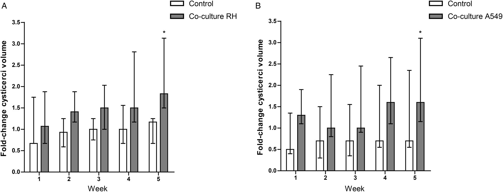
Considering that the presence of E/S products from feeder cells promotes parasite survival, and in the case of RH also growth and multiplication at the short-term, a long-term (5 weeks) incubation of cysticerci in co-culture with each cell line was evaluated. As shown in Fig. 3, co-culture with the hepatoma and lung carcinoma cell lines induced a significant increase in total parasite volume of 0.8- and 0.6-fold, respectively (P < 0.05) after 5 weeks of incubation, while axenic cultures did not show any statistically significant change with respect to week 0.
Long-term in vitro culture of Taenia crassiceps cysticerci in co-culture with rat hepatoma (A) or human lung carcinoma (B) cell lines. Bars indicate the fold change of the total volume of cysticerci with respect to week 0. Median values with interquartile ranges are plotted. The Kruskal–Wallis and Dunn's comparative tests were performed. *P < 0.05. Biological replicates: co-culture rat hepatoma (RH) and control = 3, co-culture A549 and control = 5.

Discussion
The study of parasite biology directly affects our capacity to control and detect pathogens by providing clues on therapeutic and diagnostic candidates. However, there is scarce information about the factors involved in the survival and development of cestode parasites. In this sense, in vitro systems that allow parasite culture and propagation are essential to study in detail such processes under a more controlled and simple setting than experimental or natural infections. Here, it was shown the essential role of the E/S products from both parasite and host to regulate T. crassiceps physiology. Our results demonstrate that: (i) parasite density is key to induce growth, survival and asexual multiplication when no host stimulus is present; (ii) asexual multiplication is mediated in part by parasitic secreted factors; (iii) different host stimuli show varied effects on parasite growth with non-filtered DMEM FBS and cDMEM RH being the most beneficial at both parasite densities during a short-time incubation; (iv) FBS contains factors of > 200 nm that promote survival especially at low parasite density (1C) and multiplication at high parasite density (10C); (v) E/S products from the hepatoma cell line contain factors that promote parasite multiplication independently of parasite density; and (vi) these factors are most probably < 200 nm in size since the conditioned medium was filtered before use.
The data presented here are in agreement with previous reports (Ostoa-Saloma et al., Reference Ostoa-Saloma, Ostoa-Jacobo, Esquivel-Velázquez, Bazúa and Larralde2010; Esquivel Velázquez et al., Reference Esquivel Velázquez, Hernández, Larralde and Ostoa Saloma2014), which indicated that parasite budding rate is dependent on cysticerci density and mediated by the secretion of 1 or more factors. Furthermore, other authors showed that parasite contact is also relevant for budding (Esquivel Velázquez et al., Reference Esquivel Velázquez, Hernández, Larralde and Ostoa Saloma2014). The identity of such secreted factor/s is still undetermined; however, it is known that cellular E/S products are composed of both soluble molecules and extracellular vesicles (EVs). In particular, EV secretion has been shown to constitute an active pathway for intercellular transfer of proteins, nucleic acids and lipids (Colombo et al., Reference Colombo, Raposo and Théry2014). The term EV refers to different types of vesicles among which the best characterized are microvesicles and exosomes. EVs are secreted by prokaryotic and eukaryotic cells in vitro and in vivo (van Niel et al., Reference van Niel, D'Angelo and Raposo2018) and thus, can be found in body fluids, such as the FBS used for cell culture (Théry et al., Reference Théry, Amigorena, Raposo and Clayton2006). In addition, the secretion of EVs has been described in the 3 main groups of helminths (Sotillo et al., Reference Sotillo, Robinson, Kimber, Cucher, Ancarola, Nejsum, Marcilla, Eichenberger and Tritten2020) including Taenia spp. (Galán-Puchades et al., Reference Galán-Puchades, Yang, Marcilla, Choe, Park, Osuna and Eom2016; Ancarola et al., Reference Ancarola, Marcilla, Herz, Macchiaroli, Pérez, Asurmendi, Brehm, Poncini, Rosenzvit and Cucher2017; Liang et al., Reference Liang, Mao, Zhang, Guo, Liu, Wang, Hou, Zheng and Luo2019; Wang et al., Reference Wang, Liu, Liang, Zhang, Li, Li, Liu, Mao and Luo2020). Parasite EVs have been shown to exert phenotypical alterations on host recipient cells (Sánchez-López et al., Reference Sánchez-López, Trelis, Bernal and Marcilla2021); however, the effect of host EVs on parasites or between parasites has not been reported yet. In any case, our results show that 1 parasite only is not able to sustain its own survival and growth and that bulk-incubated cysticerci require the continuous secretion, or the accumulation of a certain concentration, of E/S products to positively influence survival and development.
Regarding the host soluble factor/s, at the long term both cell lines either secrete it or secrete multiple factors with similar effects on total parasite volume. This was also observed by Spiliotis et al. (Reference Spiliotis, Tappe, Sesterhenn and Brehm2004), who concluded that these factors could either be common to mammalian cells or, alternatively, to particular immortal cell lines. Additionally, it was demonstrated that primary hepatocyte cultures secrete soluble factors of 3.5–14 kDa which support E. multilocularis growth and multiplication (Jura et al., Reference Jura, Bader, Hartmann, Maschek and Frosch1996). This suggests that at least part of the soluble factors is not associated with EVs, but secreted as soluble proteins or lipoproteins. Finally, FBS also contains components that promote parasite multiplication, which may be associated with large EVs or nanoparticles due to the estimated size of >200 nm. However, their sole presence is not enough in the long term to induce an effect similar to the one caused by feeder cells, indicating that cysticerci also uptake factors actively secreted by host cells which are absent in the FBS or present at an insufficient concentration.
Our results set the basis for the long-term in vitro culture of T. crassiceps that will allow a steady provision of parasites independently of experimental animals and consequently, with significantly less contamination with host cells. Furthermore, it was shown that growth, asexual multiplication and survival of T. crassiceps in vitro depend on a complex network where both host and parasite signalling molecules are necessary.
Author's contributions
L. C. A. G., M. G. P., M. E. A., M. C. R. and M. A. C. conceived and designed the study. L. C. A. G., M. G. P., M. E. A. and M. A. C. performed the experiments. L. C. A. G. and M. A. C. performed statistical analyses. L. C. A. G. and M. A. C. wrote the article.
Financial support
This work was supported by Agencia Nacional de Promoción Científica y Tecnológica (ANPCyT), Argentina, project PICT 2017 No 2062; Pérez Guerrero Trust Fund for South-South Cooperation (PGTF) and Consejo Nacional de Investigaciones Científicas y Técnicas (CONICET).
Conflict of interest
The authors declare there are no conflicts of interest.
Ethical standards
Procedures involving the use of experimental animals were approved by the Institutional Committee for the Care and Correct Treatment of Laboratory Animals, School of Medicine of University of Buenos Aires, Argentina (protocol number CD No 1229/2015).