Introduction
Fish of the Rivulidae family are distributed across southern, North, Central and South America, and comprise about 450 valid species (Fricke et al., Reference Fricke, Eschmeyer and Van der Laan2018). They are sexually dimorphic freshwater fish, with precocious sexual maturation (Errea and Danulat, Reference Errea and Danulat2001). Popularly known as killifish, the rivulid species are small bodied animals (usually between 50 to 80 mm total length) presenting a great variety of colour patterns, and making them desirable for the aquarium fish trade (Costa, Reference Costa, Reis, Kullander and Ferraris2003).
Many rivulids are also known as annual fish, because they inhabit temporary shallow puddles that are formed only during the rainy season in the summer but that become dry for a longer periods during other seasons. Therefore, these fish reproduce and die over a short period of time (Costa, Reference Costa2002) and eggs survive during this dry season due to the mechanism of diapause. Embryos arrest and hatch only in the next rainy season (Wourms, Reference Wourms2011). Because of this particular reproductive characteristic, many rivulids could become extinct following a short rainy season or an extended dry season, in addition to loss of habitat caused by anthropic actions such as urbanization, deforestation and drainage, which has increased the vulnerability of these species (Rosa and Lima, Reference Rosa and Lima2008). Just one genus of the Rivulidae family, Hypsolebias, represents at least 32 endangered species (ICMBio, 2018).
In this context, much research effort has been made to preserve rivulids, as knowledge in the fields of biology and ecology is necessary to establish protocols for species conservation and environmental reconstitution action (Volcan et al., Reference Volcan, Lanés and Gonçalves2010, Reference Volcan, Gonçalves and Lanés2011). In this sense, studies on genetic characterization (Nascimento et al., Reference Nascimento, Bezerra, Lima-Filho, Yamamoto, Chellapa and Molina2014; Kim et al., Reference Kim, Lee, Lee, Rhee and Lee2016), ecology (Abilhoa et al., Reference Abilhoa, Vitule and Bornatowski2010; Keppeler et al., Reference Keppeler, Lanés, Rolon, Stenert and Maltchik2013), taxonomy (Costa et al., Reference Costa, Amorim and Mattos2012, Costa and Amorim, Reference Costa and Amorim2014) and reproduction (García et al., Reference García, Loureiro and Tassino2008; Cassel et al., Reference Cassel, Mehanna, Mateus and Ferreira2013; Passos et al., Reference Passos, Tassino, Reyes and Rosenthal2014; Schalk et al., Reference Schalk, Montaña and Lisbon2014) have already been performed for some Rivulidae species. They present a huge diversity of reproductive strategies that include external (Jara et al., Reference Jara, Soto and Palma1995; Volcan et al., Reference Volcan, Gonçalves and Lanés2011) and internal fertilization (Costa et al., Reference Costa, Amorim and Mattos2016), in addition to the self-fertilization (Avise and Tatarenkov, Reference Avise and Tatarenkov2015), a uniquely reported case among vertebrates.
The aim of this study was to describe aspects of Hypsolebias sertanejo biology, such as ploidy status, reproductive behaviour and performance. Moreover, we also performed experiments to determine the best conditions for incubation and hatching of eggs obtained from fish acclimated in the laboratory. The choice of H. sertanejo for such a study was motivated by its absence from the lists of threatened species. For ethical reasons, we avoided using endangered species, but the knowledge acquired here may be used for reproduction and conservation of other Rivulidae species, mainly those of the same genera (Hypsolebias, which total at least 32 endangered species (ICMBio, 2018)), as phylogenetic proximity facilitates the application of the protocols developed here.
Materials and methods
Field sampling and maintenance
Adult fish were caught in temporary ponds from Itacarambi city, Minas Gerais State, Brazil (15°4′56.4′′S, 44°5′17.9′′W) (licence number 48541) during March 2014 and March 2015. Sampling was performed using dip-nets (3 mm mesh size) and the sampled fish were distributed into plastic containers (2 litres), kept at 26°C, and taken to the laboratory where the experiments were performed. Fish were maintained in 60-litre glass aquaria in a recirculation system with the temperature set at 27°C, and photoperiod of 12 h of light. They were fed twice a day with earthworms and Artemia salina nauplii.
Ploidy analyses
Before the start of the behavioural and biotechnological experiments, to better understand the basic biology of the species, the ploidy status of the fish was checked using flow cytometry and a standard protocol for nuclear staining with 4′,6-diamidino-2-phenylindole (DAPI). A piece of the caudal fin from each animal was sampled (Xavier et al., Reference Xavier, Senhorini, Pereira-Santos, Fujimoto, Shimoda, Silva, Dos Santos and Yasui2017) and analyzed using a Partec CyFlowPloidy Analyzer (Partec GmbH, Münster, Germany). Ploidy status was determined by comparing relative DNA content with a standard haploid DNA content (C) obtained from the sperm of Astyanax altiparanae fish species.
Experiment 1. Description of the reproductive behaviour
In this experiment, three couples of diploid animals were placed in three 60-litre glass aquaria containing a 1-litre transparent plastic recipient filled with sand, which was used as substrate for reproduction. The sand was washed and selected using a soil analysis sieve (0.42 mm diameter) to standardize the particle size. Aiming to better describe the reproductive behaviour of the species, video sequences using a DCR-DVD 308 camera (Sony, Japan) were recorded and digital images were obtained from them.
Experiment 2: Evaluation of reproduction
Diploid and mosaic fish were used to perform the reproduction experiment. They were divided into two groups for different treatments: Treatment 1: one diploid male was placed with three mosaic females; Treatment 2: one mosaic male was placed with three diploid females. The experiments took place in 60-litre glass aquaria.
Additionally, histological analysis of testes and ovaries from diploid and mosaic adult fish was performed. Briefly, gonads were removed, cut into transverse and longitudinal sections and fixed in Bouin’s solution for 24 h. Samples were dehydrated in increasing concentrations of alcohol solution, embedded in Paraplast (Sigma-Aldrich, P3558), sectioned at 5.0-µm thickness on a microtome equipped with steel blade (LEICA RM 2235), and stained with haematoxylin and eosin. The material was examined using an optical microscope (Nikon, Eclipse Ci-L) and digital images were captured using NIS-Ar Elements software (Nikon, Tokyo, Japan) using a charge coupled device (CCD) camera (Nikon, DS-Fi1).
Experiment 3: Optimizing conditions for hatching
Treatment 1: Hatching eggs under dry conditions
Seven couples of H. sertanejo were allocated for reproduction as described in Experiment 2. The resulting eggs (96 samples) were collected from March to June 2014 and transferred to 6-well microplates (35 mm diameter) filled with three different semi-humid substrates (1, coconut fibre; 2, felt made of polyester fibre wool and; 3, dishwasher sponge, mainly made of polyurethane) and kept in a biological oxygen demand (BOD) incubator in the dark at 25°C. After 30 days under those conditions, eggs were collected and counted to determine recovery rate. Eggs were immersed in distilled water, and hatching rate was measured.
Treatment 2: Hatching eggs at wet conditions
Seven males and 21 females were distributed into aquaria (1 male and three females in each aquarium) as previously described (see above). Eggs collected from May to July 2015 (26 samples) were transferred to 6-well microplates (35 mm diameter) filled with 15 ml ultrapure water, and kept in BOD incubator in the dark at 25°C. The microplates were checked daily to remove dead eggs and change the water. Hatching rates were measured for each egg batch.
Results
Ploidy analyses
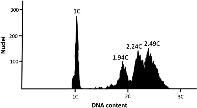
Ploidy analysis indicated that 85.71% of the fish were diploid and 14.29% (one male and three females) showed chromosome mosaicism. In these animals, some of the analyzed somatic cells presented a DNA content of 2N and some showed a DNA content between diploidy and triploidy (Fig. 1).
Relative DNA content of H. sertanejo compared against a standard haploid DNA content (1C) obtained from A. altiparanae sperm and against a standard diploid DNA content (1.94C) obtained from H. sertanejo. H. sertanejo tail cells had different DNA contents (2.24C and 2.49C).

Description of reproductive behaviour (Experiment 1)
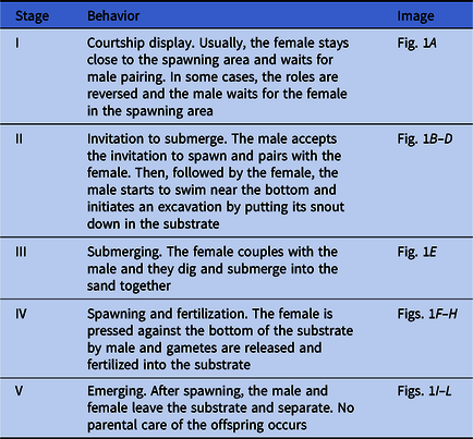
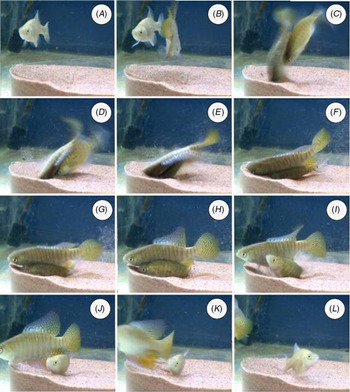
Reproductive behaviour occurred spontaneously and was divided into five stages based on the description by Belote and Costa (Reference Costa2002) (Table 1, Fig. 2).
Stages of the reproductive behavior in H. seertanejo

Spawning behavior of H. sertanejo divided in five stages. (A) Courtship displays. (B, C) Invitation to submerge. (D) Excavation. (E) Submerging. (F–H) Spawning and fertilization. (I–L) Emerging.

Evaluation of reproduction (Experiment 2)
When mosaic females were put together with a diploid male, spawning and fertilization occurred. Conversely, the mosaic male did not breed with diploid females and no reproductive behaviour was observed.
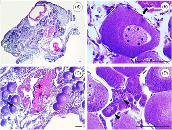
Histologically, ovaries and testes from diploid and mosaic animals did not show differences. In ovaries, most of the oocytes were perinucleolar oocytes, which are in primary growth (Fig. 3A). They had a central nucleus with an irregular border and many peripheral spherical nucleoli, basophilic cytoplasm, surrounded by zona radiata and follicle cells (Fig. 3B). oogonia (Fig. 3D) and the post-ovulatory follicle complex (POC) (Fig. 3C), which is formed by hypertrophied follicular cells; basement membrane and theca protruding towards the lumen after ovulation were also observed
Morphology of H. sertanejo ovaries. (A) Overview. (B) Pre-vitellogenic oocyte in primary growth; arrow: follicular cell. (C) Asterisk: post-ovulatory follicle complex; arrowhead: oogonia. (D) Arrowheads: oogonia. Scale bars represent 50 µm.

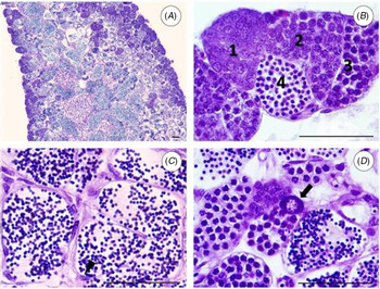
Testes were lobular with spermatogonia restricted to the lobule periphery and spermatogenesis was cystic (Fig. 4A–C). A primary growth oocyte was found in the testicle of one male (Fig. 4D).
Morphology of H. sertanejo testes. (A) Overview. (B) 1. Spermatogonia. 2. Spermatocytes in pachytene phase. 3. Spermatocytes in diplotene phase. 4. Spermatids. (C) Spermatozoa into the lumen, arrowhead: Sertoli cell. (D) Arrow: oocyte in primary growth. Scale bars represent 50 µm.

Optimizing conditions for hatching (Experiment 3)
Treatment 1: Hatching eggs at dry conditions
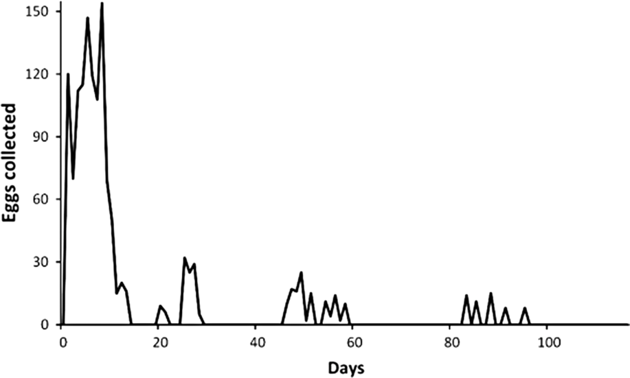
During the experimental period 1403 eggs were collected, and females presented spawning peaks (Fig. 5). Eggs stored in felt and sponge substrates were not able to be recovered, whereas 18% of the eggs kept in coconut fibre were recovered. In total, 5.04% of the eggs hatched when immersed in distilled water.
Number of H. sertanejo eggs collected daily during the experimental period.

Treatment 2: Hatching eggs at wet conditions
During the experimental period, 2137 eggs were collected and hatching rate was 3.79% (81 hatchings). Average time of incubation before hatching was 75.84 ± 27.23 days, varying from 29 to 128 days.
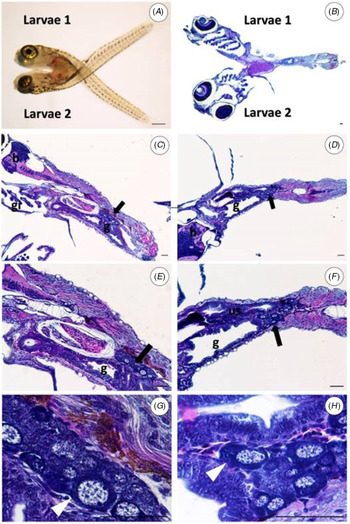
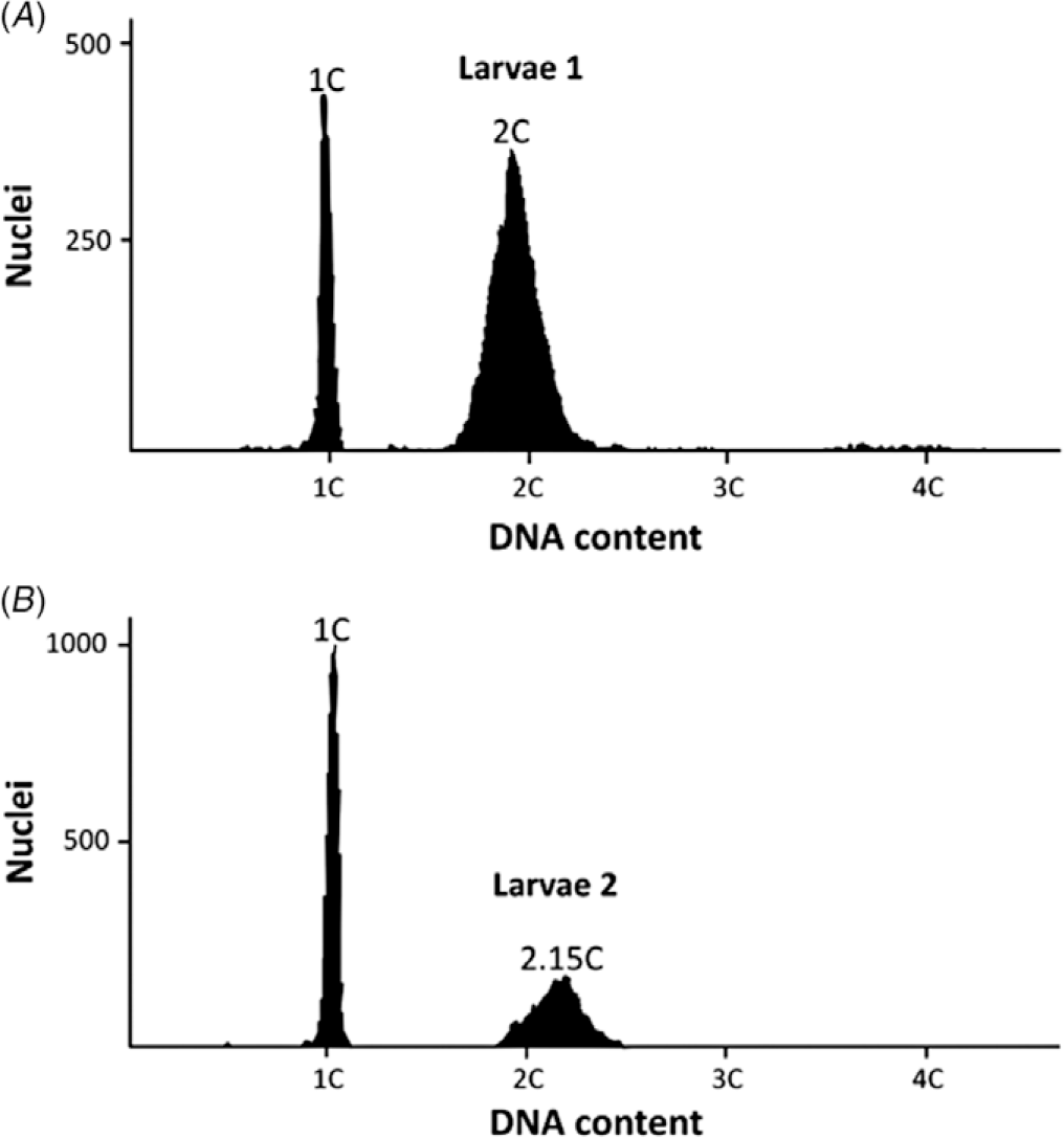
The occurrence of conjoined embryo was verified (Fig. 6A, B). Histological analysis showed that organs such as gills, guts, urinary and reproductive system were independent in each larvae (Fig. 6C, D), which were joined by connective tissue. Both larvae had gonads already differentiated in ovaries with pre-vitellogenic oocytes (Fig. 6E, F). Flow cytometric analysis from tails of the larvae showed that relative DNA content from cells of each tail was different (Fig. 7). Larvae 1 had DNA content as diploid (2C), whereas larvae 2 showed higher relative DNA content (2.15C).
Hypsolebias sertanejo larvae connected. (A, B) Larvae 1 (l1) and 2 (l2). (C) Larvae 1, b: brain, g: gut, gi: gill. (D) Larvae 2, b: brain, g: gut. (E, F) us: urinary system. Arrows: ovary. (G, H) Arrowheads: pre-vitellogenic oocytes. Scale bars represent 50 µm.

Relative DNA content of connected H. sertanejo larvae compared against a standard haploid DNA content (1C) obtained from A. altiparanae sperm. Larvae 1 (A) shows the DNA content as diploid (2C), in comparison larvae 2 (B) has a relatively higher DNA content (2.15C).

Discussion
The breeding protocol used here has proven to be successful for H. sertanejo and may be used as a model for other related species, including threatened ones. Although the reproductive behaviour of H. sertanejo presented similarities to other species of related genera, such as Austrolebias reicherti (García et al., Reference García, Loureiro and Tassino2008.), Cynolebias albipunctatus (Belote and Costa, Reference Belote and Costa2002) and Simpsonichthys boitonei (Shibatta, Reference Shibatta2006), many peculiarities inherent to its reproductive biology could be observed.
Our data suggested that several chromosome abnormalities occur in this species such as mosaicism, polyploids and morphological abnormalities, including conjoined fish presenting different ploidy status. Mosaicism did not affect males regarding maturation of germ cells (sterility), as our results showed, but suggested infertility. The origin of this alteration is still unknown but naturally occurring polyploidy may arise from hybridization and production of unreduced gametes (Fujimoto et al., Reference Fujimoto, Yasui, Yoshikawa, Yamaha and Arai2008; Yasui et al., Reference Yasui, Arias-Rodriguez, Fujimoto and Arai2009), thermal treatments after fertilization (Adamov et al., Reference Adamov, Nascimento, Maciel, Pereira-Santos, Senhorini, Calado, Evangelista, Nakaghi, Guerrero, Fujimoto and Yasui2017; Garcia-Abiado et al., Reference Garcia-Abiado, Lynch, Dabrowski and Hartman2001; Kalbassi et al., Reference Kalbassi, Dorafshan, Pourkazemi and Amiri2009; Xavier et al., Reference Xavier, Senhorini, Pereira-Santos, Fujimoto, Shimoda, Silva, Dos Santos and Yasui2017) and delayed egg fertilization (Aegerter and Jalabert, Reference Aegerter and Jalabert2004; Flajšhans et al., Reference Flajšhans, Kohlmann and Rab2007; Samarin et al., Reference Samarin, Policar and Lahnsteiner2015, Reference Samarin, Blecha, Uzhytchak, Bytyutskyy, Zarski, Flajšhans and Policar2016). The occurrence of such chromosomal abnormalities can lead to formation of aneuploidy gametes (Hamasaki et al., Reference Hamasaki, Takeuchi, Miyaki and Yoshizaki2013; Linhart et al., Reference Linhart, Rodina, Flajšhans, Mavrodiev, Nebesarova, Gela and Kocour2006), which results in inviable eggs and may be related to the low hatching rates.
Hypsolebias sertanejo inhabits a limiting and selective environment that accelerates the speciation process. Such a phenomenon may explain a wide number of species even if the fish are morphologically similar and separated by small distances, as is the case for H. sertanejo and other species of the H. flavicaudatus complex such as H. janaubensis, as described by Costa and colleagues (Costa et al., Reference Costa, Amorim and Mattos2012). Such similarity suggested that hybridization may occur especially regarding interpopulation hybridization because, during the rainy season, ponds could be connected and then generate massive introgression. High temperatures may also affect the ploidy status in this species because fertilized eggs naturally enter diapause and embryos can remain for long periods in small wetlands at extreme temperatures (Markofsky and Matias, Reference Markofsky and Matias1977). According to Costa and colleagues (Costa et al., Reference Costa, Amorim and Mattos2012), species of this genera are commonly found in temporary ponds that are more exposed to sunlight.
The conjoined embryo found in this study was of the same sex, but had a different ploidy status. Sex may be explained by the monozygotic origin that suggested a common genetic sex determination. Additionally, both embryos were maintained together under the same conditions and environmental sex determination was the same. The explanation for different ploidy status conversely is still unknown, but asychronic development within the blastoderm was observed in some embryos. This development may be related to partitioning of the blastoderm, generating these morphological and chromosomal differences in the conjoined embryos.
In conclusion, several reproductive, chromosomal and morphological problems arose in our study species. In addition, a very low percentage of the collected eggs survived as far as the hatching stage, suggesting that these deleterious effects may reduce the long-term survival of H. sertanejo under natural conditions, which limits the perpetuation of this species and emphasizes the need for more preservation efforts, including artificial propagation and gene banking for the killifish species.
Acknowledgements
The authors acknowledge CEPTA/ICMBio for gently providing experimental fish and the facilities.
Financial support
AES Tietê/ANEEL project 4690000174 and São Paulo Research Foundation (FAPESP, 2010/17429-1 and 2011/11664-1) for financial support.
Conflicts of interest
The authors declare no conflict of interest
Ethical standards
The authors assert that all procedures contributing to this work complied with the ethical standards of the relevant national and institutional guides on the care and use of laboratory animals.