INTRODUCTION
The tropical melongenid Melongena melongena is commonly found buried in the sediment of muddy bottoms of coastal lagoons and of muddy bottoms of open waters. The species is distributed from Mexico to Surinam including the Netherlands Antilles, except Puerto Rico (Work, Reference Work1969). In Venezuela, M. melongena has been reported in several areas along the coast, from east to west, mostly in coastal lagoons and seagrass beds, as well as in off-shore islands (Rehder, Reference Rehder1962; Work Reference Work1969; Prince, Reference Prince1973; Flores, Reference Flores1978; Carvajal & Capelo, Reference Carvajal and Capelo1992; Ramos & Robaima, Reference Ramos and Robaina1994; Capelo & Buitriago, Reference Capelo and Buitriago1998).
Bandel (Reference Bandel1976), Flores (Reference Flores1983) and Penchaszadeh (Reference Penchaszadeh1981) have described the egg capsule morphology, and reported the number of embryos per capsule as well as the hatching mode for M. melongena on the south coast of Venezuela and Colombia. The egg capsules of M. melongena are anchored in the mud or sand and follow a structural pattern which is similar to that reported for other species of the family Melongenidae, that is, capsules aligned in parallel and joined together by a collar-shaped cord. A few egg capsules of M. melongena act as anchorage in the substratum for the rest of the spawn. The capsules are relatively large (about 1 cm in diameter), flat, white-yellowish, semitranslucid, with an ovoid-shaped exit plug on the upper edge, and increase in size the farther they are from the substrate. Embryos and larvae of M. melongena are embedded in a gelatinous matrix within the egg capsules. Such a gelatinous matrix has been reported to be for some species a nutritional source for the developing embryos (De Mahieu et al., Reference De Mahieu, Penchaszadeh and Casal1974; Penchaszadeh & Miloslavich, Reference Penchaszadeh and Miloslavich2001). The changes in the biochemical composition of the embryos and intracapsular fluid during intracapsular development are well known for a few gastropod species (De Mahieu et al., Reference De Mahieu, Penchaszadeh and Casal1974; Stöckmann & Althoff, Reference Stöckmann and Althoff1989; Miloslavich & Dufresne, Reference Miloslavich and Dufresne1994; Penchaszadeh & Rincón, Reference Penchaszadeh and Rincón1996; Calvo, Reference Calvo1999; Miloslavich, Reference Miloslavich1999; Penchaszadeh & Miloslavich, Reference Penchaszadeh and Miloslavich2001), but of these, the variations in the protein content of the embryos in relation to changes in the intracapsular fluid has only been reported for Voluta musica (Penchaszadeh & Miloslavich, Reference Penchaszadeh and Miloslavich2001). In V. musica, the embryos feed on proteins and carbohydrates contained in the intracapsular liquid at an early intracapsular veliger stage. In this study, we will quantify the protein content present in the embryos and intracapsular fluid at several stages of development, as well as determine if the embryos feed on extraembryonic food sources during intracapsular development.
MATERIALS AND METHODS
Specimens
Egg capsules of M. melongena were collected in February 2002, April 2002 and February 2003 at a coastal lagoon within the Golfete de Cuare, which is part of the Cuare Wildlife Reserve, Falcon State, Venezuela (10°48′N 68°14′W). The egg capsules were found between 1 and 2 m depth, anchored to the muddy substrate and covered by the seagrass Syringodium filiforme.
Development and protein content
We studied the size of the egg capsules, the number and size of the eggs and developing embryos within the egg capsules at the different stages of development and the volume of the intracapsular liquid.
The egg capsule content consisted of two fractions: embryos and intracapsular liquid. Embryos were carefully separated from the intracapsular fluid, placed in 1.5 ml Eppendorf tubes in groups of approximately 40 embryos each, and frozen at –20°C. The volume of the intracapsular liquid was measured by extracting it from the capsule with a 20 µl Pipetmann micropipette (precision ±1 µl). The liquid was placed in a 1.5 ml Eppendorf tube and frozen at –20°C. The protein content present in the eggs, embryos and intracapsular fluid was determined following the Bio-Rad protein assay procedure based on the Bradford method (Bradford, Reference Bradford1976). Bovine serum albumin (BSA) was used as a standard. Samples were left overnight in 500 µl of NaOH 0.5 N and thoroughly homogenized. The protein concentration was measured with a spectrophotometer at 595 nm.
In order to determine whether there existed any differences in the protein content per embryo between the different stages of development, a one-way ANOVA was applied followed by Tukey multiple comparisons.
RESULTS
Development
The buried capsules of M. melongena contained no eggs or embryos, and showed an average width and height of 1.56 cm and 1.26 cm respectively. The capsules found in the water column above the substratum contained either eggs, or embryos or larvae, and showed an average width and height of 2.70 cm and 2.08 cm respectively.
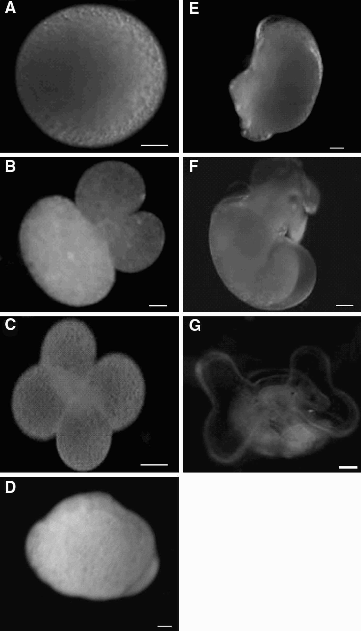
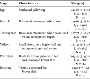
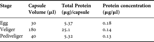
We found egg-capsules at several stages of embryonic development, however, within the same spawn, all embryos developed synchronously. The embryonic stages were easily distinguishable through the capsule walls. At early development, the capsule content is white-yellowish due to the yellow colour of the eggs and the white colour of the intracapsular fluid. At the final stages of development, the capsule interior looks brownish-red due to shell coloration of the larvae and the transparency of the intracapsular liquid. Egg-capsules contained between 308 and 675 eggs each, 95% of these eggs developed and hatched as pediveligers. The eggs measured between 352 and 480 µm in diameter. The 4 cell stage was characterized by 4 blastomeres of approximately the same size. The gastrula was round shaped, vitellum-rich and with a rotating movement, their length varied between 332 and 561 µm. The trochophore was whitish with two lobes (from which the velum develops), and measured between 434 and 663 µm in length. The early veliger was characterized by a large cephalic region, a very small velum and a fragile shell. These larvae show signs of movement due to small cilia in the velum. The fully developed veliger had a brown coiled shell, the velum was larger and ciliated and the eyes were distinguishable at this stage. During veliger development, packed yolk could be observed on the inside of the larva. The shell-length of these veligers varied between 306 and 485 µm, and the velum measured on average 338 µm in width and 144 µm in height (Table 1; Figure 1).
Embryonic development of Melongena melongena. (A) Uncleaved egg; (B) three blastomeres; (C) four blastomeres; (D) gastrula; (E) trochophore; (F) veliger; (G) pediveliger. Scale bar 100 µm.
Characteristics of the embryos during intracapsular development. Values represent mean±SD, numbers in parentheses indicate range. N, number of observations.

The pediveliger measured between 510 and 689 µm in shell length and was characterized by a bilobed velum which measured about 639 µm in width and 407 µm in height. The foot was very transparent and showed some purple pigmentation. At the hatching stage, the shell measured between 714 and 740 µm, it had a well developed purple foot and the shell was dark brown (Table 1; Figure 1). During development, no adelphophagy of nurse eggs, or cannibalism among sibling embryos was observed.
Protein content
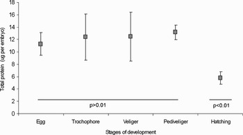
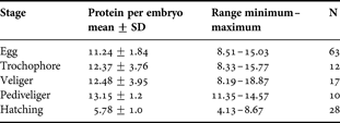
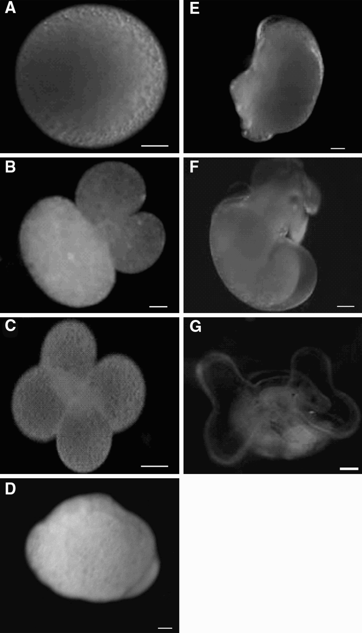
During the embryonic development of M. melongena there was a slight increase, however, not significant in the protein content from the egg stage (11.24 µg/egg) to the trochophore (12.37 µg/larva), veliger (12.48 µg/larva) and pediveliger (13.15 µg/larva) stages. From this stage, the protein values decreased significantly to the hatching stage (5.78 µg/juvenile) (one-way ANOVA, P < 0.01, Table 2; Figure 2). Tukeýs multiple comparisons showed no differences in the intracapsular protein content for the egg, veliger and pediveliger stage (P > 0.05) but a significant difference to the protein content at the hatching stage (P < 0.05).
Total protein present in the different stages of development. The vertical bar indicates the standard deviation. The point indicates the average values of the proteins and the horizontal bars indicate different non-significative stages.

Average protein content per embryo (µg)±SD for the different developmental stages of Melongena melongena, along with minimum and maximum values and number of observations (N).
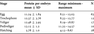
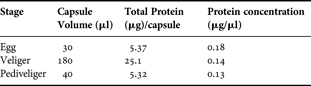
Table 3 reports the volume of intracapsular fluid, total protein content in this fluid and protein concentration in this fluid for the various developmental stages. A slight decrease was observed in the protein concentration of the intracapsular fluid as the embryo developed. In the egg stage, the protein concentration in the intracapsular fluid was 0.18 µg/µl, while in the pediveliger stage the concentration was 0.13 µg/µl. However, no significant differences were found (P > 0.05) in the intracapsular fluid protein concentration as the organisms developed.
Capsule volume, total protein/capsule and protein concentration (µg/µl) in the intracapsular fluid for the different stages of development de Melongena melongena.
DISCUSSION
Development
Melongena melongena egg capsules follow a structure pattern similar to that reported for other species of the family Melongenidae. This pattern consists of egg capsules attached to a common string and disposed in parallel as in a collar. D'Asaro (Reference D'Asaro1970) pointed out that egg capsules of M. melongena are similar to those of M. patula, however, the egg capsules of M. patula are much larger and higher (5.3 cm × 5.0 cm). D'Asaro (Reference D'Asaro1970) has related the egg capsules of the genus Melongena with those of the family Xancidae, while Bandel (Reference Bandel1975) related them to those of the genus Busycon emphasizing the modality of capsules aligned in parallel and bound by a cord.
The egg capsules of M. melongena collected in the Golfete de Cuare are characterized by presenting between 30 and 37 capsules per egg capsules with an average of 503 eggs/capsule, which is similar to the number reported by Bandel (Reference Bandel1976) who found in the Colombian Caribbean egg capsules of M. melongena with 41, 43 and 51 capsules an average of 445 eggs/capsules. On the other hand, Penchaszadeh (Reference Penchaszadeh1981) found between 15 and 50 capsules with an average of 465 eggs/capsule at Cayo Animas, Morrocoy National Park. Miloslavich (Reference Miloslavich2005) found capsules containing 436 eggs also at Morrocoy National Park, and Flores (Reference Flores1983) reported between 26 and 42 capsules for this species on the eastern coast of the Venezuelan Caribbean, with an average of 1500 eggs/capsule. We found that during the embryonic development of M. melongena, 5% of the eggs in the capsules did not develop, while those that did develop, presented the typical developmental stages of the caenogastropods, hatching as a pediveliger larva. We also observed that the embryos develop in a synchronized way, which could indicate an absence of competition among siblings for extraembryonic food sources such as nurse eggs or the intracapsular liquid. Development and hatching herein reported for M. melongena is similar to other species of the family Melongenidae. Hathaway (Reference Hathaway1958) and Woodbury (Reference Woodbury1986) found that M. corona hatches as a pediveliger, with a hatching size of 700–900 µm. Hemifusus tuba and Hemifusus ternatanus also hatch as pediveligers with hatching sizes of 554 µm and 600 µm respectively (Hahn, Reference Hahn1993). Castagna & Kraeuter (Reference Castagna and Kraeuter1994) and Power et al. (Reference Power, Covington, Recicar, Walker and Eller2002) reported that Busycon carica hatches as juveniles measuring between 4.0 and 5.6 mm. Other studies in M. melongena, report that this species hatches as a veliger larva (Bandel, Reference Bandel1976; Flores, Reference Flores1983; Miloslavich, Reference Miloslavich2005). The fact that the larva of M. melongena might hatch as veligers or as a more developed pediveliger, may be a consequence of environmental conditions, which particularly at the Golfete de Cuare are very stable (temperature, salinity and sediments), allowing for the larva to remain inside the egg capsule for a longer time than in other more variable localities.
Protein content
No significant changes in the protein content of the embryos between the egg and the early pediveliger stages was observed, however, at the hatching stage, this amount of protein decreases significantly probably due to the fact that the pediveligers have exhausted at this point all the protein reserves of their own eggs, which in this species, seems to be the major protein source during embryonic development. In some caenogastropod species (Buccinum undatum and Serpulorbis arenaria), a decrease in the protein content has been found between the last stage of the intracapsular development and the hatching stage (Miloslavich & Dufresne, Reference Miloslavich and Dufresne1994; Calvo, Reference Calvo1999 respectively).
In M. Melongena, no significant consumption of the protein in the intracapsular liquid by the embryos was observed, indicating that despite representing an additional source of food for the embryos, the intracapsular liquid does not represent a primary food source for the developing embryos. This statement can be justified by the small variation in the protein concentration found in the intracapsular fluid as the embryos developed, by the low protein concentration present in the fluid and by the fact that no significant differences were found in the protein concentration present between the egg stage and the intracapsular pediveliger stage. Miloslavich & Penchaszadeh (Reference Miloslavich and Penchaszadeh2001) points out that egg size is an important factor because it determines the amount of albumin available for the embryo's development when other sources of extraembryonic food, such as nutritional eggs, cannibalism among siblings or intracapsular fluid are not available. Penchaszadeh & Rincón (Reference Penchaszadeh and Rincón1996) and Calvo (Reference Calvo1999) have reported the importance of the egg's own albumin as a nutritional source during embryonic development.
The protein concentration present in M. melongena's intracapsular fluid is very low as compared to the concentration reported for other species of the same family, as is the case with Busicon carica and B. canaliculatum, where 3.8 and 8.7 mg/ml of protein content were found, respectively (Harasewych, Reference Harasewych1978). However, despite the fact that no significant differences were found in the intracapsular fluid's protein content as the embryos developed, a slight increase in protein concentration at the last stages of intracapsular development could only be attributed to the consumption of the proteins available in the intracapsular fluid. This stage corresponds with the development of structures which have been reported to be used in the uptake of extraembryonic food from the intracapsular liquid, such as the ciliated velum (Penchaszadeh & Miloslavich, Reference Penchaszadeh and Miloslavich2001). Many works on gastropod embryonic development have shown the importance of the intracapsular fluid and its nutritional value for some species (Bayne, Reference Bayne1968; De Mahieu et al., Reference De Mahieu, Penchaszadeh and Casal1974; Stöckmann & Althoff, Reference Stöckmann and Althoff1989; Calvo, Reference Calvo1999; Penchaszadeh & Miloslavich, Reference Penchaszadeh and Miloslavich2001), but not for others (Miloslavich & Dufresne, Reference Miloslavich and Dufresne1994).
Finally, despite the fact that between 2 and 5% of the eggs do not develop, these are not used as nurse eggs by the developing embryos, since they were observed intact at the hatching stage. Melongena melongena is a species that inhabits coastal lagoons which have important salinity variations throughout the year. Changes in salinity are known to produce variations in the number of egg capsules (Kirkegaard, Reference Kirkegaard2006 for Theodoxus fluviatilis). In the genus Neritina, there are several examples of species that live in brackish waters that have marine larvae (Bandel & Kowalke, Reference Bandel and Kowalke1999; Kano & Kase, Reference Kano and Kase2003; Strong et al., Reference Strong, Gargominy, Ponder and Bouchet2008). In these changing environments, inputs of either marine or freshwater may induce or delay hatching of the larvae from the egg capsules. As mentioned earlier, M. melongena has reported to hatch as veliger larvae in some marine coastal environments, so these food sources, nurse eggs and the intracapsular liquid represent two potential sources of protein for the embryos if these have to remain longer in the capsule due to environmental variations.
ACKNOWLEDGEMENTS
We wish to thank the Houston Museum of Natural Sciences for their support through the ‘Connie Boone Grant to Malacology’ given to Nicida Noriega. This work was also supported by the Decanato de Estudios de Postgrado and Decanato de Investigación y Desarrollo (DID-USB), Universidad Simón Bolívar. We also wish to acknowledge the Limnology Laboratory and the Protein Biochemistry Laboratory, Universidad Simón Bolívar for technical support, as well as to Lic Samuel Narciso and MSc Carlos del Mónaco for their help in the fieldwork.