




No CrossRef data available.
Article contents
Cellular infiltration at skin lesions and draining lymph nodes of sheep infested with adult Hyalomma anatolicum anatolicum ticks
Published online by Cambridge University Press: 15 July 2005
Abstract
Immunohistochemical analysis of skin and draining lymph nodes of sheep repeatedly infested with the ixodid tick Hyalomma anatolicum anatolicum were studied for different antigen-presenting cells and lymphocyte subpopulations. Infiltration of neutrophils, macrophages and lymphocytes adjacent to the tick bite site were observed. Skin biopsies showed significant increases in dermal infiltration of CD8+ and γδ+ T cells at 72 h and 8 days after both primary and secondary infestation. Infiltrations of MHC-II DR/DQ decreased at 72 h after tick infestation, whereas significant increases were recorded for 8-day skin biopsies. CD1+ cellular infiltrations were observed during secondary infestations at the dermis. Decreased ratios of CD4[ratio ]CD8 T cells and MHC-II[ratio ]CD1 antigen-presenting cells were observed in both infestations compared to healthy skin biopsies. Ratios of αβ[ratio ]γδ T cells increased gradually during infestation compared to uninfested skin. The regional lymph nodes from tick-infested sheep showed an increased CD8+, γδ+ T and CD1+ cellular infiltration compared to control lymph nodes. CD4+ T cells were decreased. There were no significant changes in CD45R+ cellular infiltration either at skin lesions or regional lymph nodes.
Keywords
Information
- Type
- Research Article
- Information
- Copyright
- © 2005 Cambridge University Press
INTRODUCTION
Blood-feeding arthropods modulate host immune responses that could impair their ability to obtain a bloodmeal (Brossard and Wikel, 2004). The days required for an ixodid tick to obtain a bloodmeal provide an ample opportunity to interact with host immune defences. Cutaneous immune interactions at the tick–host interface are being characterized at the cellular and molecular levels (Wikel, 1996; Nuttall and Labuda, 2003; Brossard and Wikel, 2004). Interactions of feeding ticks with the host immune system at the cutaneous interface are central to acquisition and expression of acquired resistance and an important determinant in pathogen transmission (Schoeler and Wikel, 2001).
Laboratory animal–tick associations are generally characterized by more intense expression of acquired resistance than naturally occurring tick–host relationships (Ribeiro, 1989). Acquired resistance is expressed as reduced engorgement weight, longer duration of feeding, reduced numbers of ova, blocked moulting and death of ticks (Willadsen, 1980; Wikel, 1996). Elements of the host immune system implicated in acquisition and expression of acquired resistance include: antigen-presenting (dendritic) cells, T- and B-lymphocytes, immunoglobulins, complement, granulocytes, monocytes/macrophages, cytokines and numerous other biologically active molecules (Wikel, 1996; Brossard and Wikel, 2004). Specific elements involved depend upon both the species of tick and the host. Some ticks, such as Ixodes scapularis, do not induce acquired resistance during the course of repeated infestations in BALB/c and C3H/HeN mice (Schoeler, Manweiler and Wikel, 1999).
Guinea-pigs resistant to Demacentor andersoni (Allen, 1973) and Bos taurus breed cattle previously exposed to Ixodes holocyclus (Allen, Doube and Kemp, 1977) both developed intense accumulations of basophils, suggesting a cutaneous basophil hypersensitivity (CBH) response at tick attachment sites on tick-sensitized animals. Kinetics of mast cell, basophil and eosinophil accumulation at Hyalomma anatolicum feeding sites on cattle was studied by Gill (1986). Rabbits repeatedly infested with Ixodes ricinus ticks showed degranulation of mast cells and basophils at tick attachment sites (Brossard and Fivaz, 1982). Riek (1962) observed differences in the type and intensity of cellular infiltration in the skin of resistant and susceptible cattle infested with Boophilus microplus.
The cell types observed at the tick–host interface are constituents of the skin-associated lymphoid tissues (SALT), which are central to the regulation of cutaneous innate and specific acquired immune responses and immunity against ticks (Bos and Kapsenberg, 1993; Wikel and Bergman, 1997). Tick antigens injected into the skin through tick saliva are taken up by Langerhans cells and presented to T-lymphocytes in lymph nodes draining the bite site (Allen, Khalil and Wikel, 1979). Langerhans cells leave the epidermis and migrate to the draining lymph node via lymphatic vessels (Silberberg-Sinakin et al. 1976). Presentation of antigen to naïve T cells occurs within the lymph nodes. Interactions between the T lymphocytes and antigen-bearing, antigen-presenting cells, such as Langerhans cells, initiate intracellular signalling cascades leading to T lymphocyte proliferation and clonal expansion within the lymph node. These are essential steps in the acquired immune response to antigens introduced into the skin (Meeusen, 1998).
Cutaneous immune responses of BALB/c mice infested with nymphal I. ricinus were examined immunohistochemically by use of cryostat sections of skin biopsies obtained 72 h after attachment, and it was observed that CD4+ outnumbered CD8+ T lymphocytes (Mbow, Rutti and Brossard, 1994). Immunohistological examination of a human skin lesion, marked by an itch response of the patient after I. ricinus tick bite, revealed infiltration of CD1+ Langerhans cells and CD8+ T lymphocytes (Beaudouin et al. 1997).
Our previous study examined changes induced in circulating ovine lymphocytes during the course of infestation with Haemaphysalis bispinosa and H. a. anatolicum, either individually or together (Boppana et al. 2004). Hyalomma a. anatolicum showed significant depletions of in vivo circulating ovine total T-lymphocyte (CD5+), gamma/delta (γδ+) and CD8+ T-cell numbers as evaluated by flow cytometry at different stages of primary and secondary infestation (Boppana et al. 2004).
The present immunohistochemical study examines antigen presenting cells and lymphocyte sub-populations in skin and draining regional lymph nodes at selected times during the course of repeated infestations of sheep with adult H. a. anatolicum ticks.
MATERIALS AND METHODS
Sheep
Twelve Madras red breed sheep of either sex and aged approximately 1 year, with no previous history of tick infestation, were procured from Tamil Nadu Veterinary and Animal Sciences University Livestock Research Station. All the animals were housed in tick-proof sheds and given similar access to uniform conditions of food and water ad libitum.
Ticks
Pathogen-free colonies of Hyalomma anatolicum anatolicum were maintained in the Department of Veterinary Parasitology, Madras Veterinary College, India. Different life-cycle stages were fed on tick naïve New Zealand white rabbits, and all life-cycle stages were held in 10 ml glass vials kept in a desiccator (Tarsons Products, India) over a saturated solution of potassium chloride (KCl) in a refrigerated incubator (Forma Scientific, USA) at 85% relative humidity and 25 °C±2 °C until they moulted to the next instar (Palmer, Grewal and Dhillon, 1996).
Infestations
Sheep were randomly divided into 2 groups (Group I and Group II). Lambs in Group I (n=4) were treated as the tick-free control group. Lambs in Group II (n=8) were infested with 25 pairs of unfed adult H. a. anatolicum ticks contained in a cotton bag secured with adhesive tape to either the right or left ear. Four of the sheep in Group II were euthanized on the eighth day after primary infestation and the remaining sheep were infested again (secondary infestation) with a similar number and species of ticks 35 days after initiation of primary infestation on the same ear. Skin biopsies were collected at 72 h and 8 days after initiation of tick infestation (primary and secondary) from both left and right sides of the ear from Group II lambs. Skin biopsies and parotid lymph nodes were also collected from tick-free control (Group I) sheep. The tick-infested ear and opposite ear (uninfested) of the same (Group II) lambs were designated as infested and control skin biopsies and lymph nodes respectively. Skin biopsies and parotid lymph nodes collected from Group I animals were treated as tick-free control group.
Monoclonal antibodies
Monoclonal antibodies (mAb) reactive with antigen-presenting cell and lymphocyte–cell surface antigens were obtained from the Center for Animal Biotechnology, University of Melbourne, Australia. Details of the mAbs are provided in Table 1. All the mAbs were received as freeze-dried tissue culture supernatants and each vial was reconstituted with distilled water as per manufacturer's recommendations.

Skin biopsy
Ovine skin biopsies from tick-infected and uninfested ears were collected adjacent to the tick mouthparts from the infested ear and equivalent area on the opposite (control) ear using 0·5 cm diameter sterile biopsy punch forceps. Skin samples were collected at 72 h and 8 days after initiation of primary and secondary infestations and placed into ice-cold phosphate buffered saline, pH 7·2, and immediately snap frozen and stored at −70 °C.
Lymph nodes
Lambs in Groups I and II were euthanized and 8 days after primary (Group I, n=2 and Group II, n=4) and secondary (Group I, n=2 and Group II, n=4) infestations. Parotid lymph nodes draining tick infested and uninfested ears were collected from Group II animals and lymph nodes were collected from tick-free animals and placed into ice-cold PBS and immediately transferred to the laboratory. Gross lymph node weights (mg) were recorded and tissue samples of 0·1 to 0·2 mm thickness (including lymph node surface capsule and upto deep medulla) were collected. Skin biopsies and lymph node tissue specimens were immediately embedded in Tissue-Tek® O C T compound and snap frozen in liquid nitrogen, then wrapped in aluminum foil and stored at −70 °C until further processing.
Cryostat sections
Serial 5 μm thick cryostat sections were prepared using a Leica cryostat and mounted onto clean glass slides. One in every 4 slides was fixed in 10% formaldehyde for 10 min at room temperature and stained with haematoxylin and eosin (H&E). Remaining sections were dried in a cold chamber and fixed with ice-cold absolute ethanol for 8 min. Slides were then wrapped in aluminum foil and stored at −70 °C until processed for immunohistochemistry.
Immunohistochemical staining
Immunohistochemical staining was done according to the method described by Jorundsson, Press and Landsverk (2000). Tissue sections were incubated with 10% normal goat serum for 30 min at room temperature and then reacted with 50 μl of the respective monoclonal antibodies (Table 1) diluted (1[ratio ]20) in PBS, containing 1% bovine serum albumin (BSA). Sections were incubated in a moist chamber overnight at 4 °C. Sections were rinsed with PBST (2% Tween-20 in PBS) at room temperature and allowed to react with goat anti-mouse IgG-biotin conjugate (Sigma – diluted with PBS at 1[ratio ]200) for 1 h at 37 °C.
Endogenous peroxidase activity was quenched by incubating sections in 0·3% hydrogen peroxide (H2O2) in methanol for 30 min. Following washing, sections were allowed to react with streptavidin–horseradish peroxidase complex (Sigma – diluted with PBS at 1[ratio ]200) for 30 min at room temperature. Tissue sections were reacted with 3,3′-diaminobenzidine tetrahydrochloride (DAB) liquid substrate system (Sigma). Sections were post-fixed with 2% glutaraldehyde and rinsed with Milli Q equivalent purified water. Sections were counter-stained with methyl green for 5 min and dehydrated with acetone, cleared with xylene and mounted in DPX medium.
Enumeration of cellular response
Cells showing a positive brown colour indicating reactivity with the mAb were counted according to the method described by Bos et al. (1989), which was modified for ovine skin. Skin sections were examined in the context of epidermis and dermis for enumeration of cells. Cells were counted at a magnification of 400× for a minimum of 10 non-overlapping microscopic fields for each individual section stained with specific mAb.
Results were expressed as mean±S.E of cells in the epidermis and dermis reacting with each specific mAb. Positive immunohistological reactions in lymph node sections were scored as ± (below 25 cells), + (about 25 cells), ++ (25 to 50 cells),+++ (50 to 75 cells) and ++++ (more than 75 cells) per 400× microscopic field for each specific mAb.
Statistical analysis
The mean number of positive cells in infested and control skin for epidermis and dermis were compared using Student's t-test according to the method of Snedecor and Cochran (1980) for each mAb.
RESULTS
Histological examination of haematoxylin and eosin stained sections of ovine skin infested with adult H. a. anatolicum at 8 days after primary infestation showed deposition of attachment cement and infiltration of inflammatory cells around mouthparts (Fig. 1A). Mouthparts penetrated deep into the dermis and inflammatory infiltrate extended to the cartilage of the pinna. Infiltrate consisted of degranulated neutrophils, eosinophils, a few macrophages, epithelioid cells and lymphocytes. Epidermis adjacent to the bite site was infiltrated with neutrophils, macrophages, lymphocytes and spongiocytes. Epidermal keratinocytes were altered in appearance with vacuolation, and parakeratosis (Fig. 1B). There was a marked subepidermal collection of leukocytes in tissues adjacent to the bite site. Histological sections collected at 72 h of primary infestation showed similar types of inflammatory cells with a lesser degree of infiltration.

Fig. 1. (A) Mouthparts of tick penetrated into the skin: mp, tick mouthparts; C, cement; D, dermis. (B) Epidermis showing parakeratosis, cellular infiltration and vacuolations: Ep, epidermis. (C) Focal infiltration during secondary infestation. (D) Parotid lymph nodes: Inf, draining lymph node from tick-infested ear side; C, uninfested side. (E) Control lymph node section showing paracortical and cortical zones, distinct germinal follicles. (F) Tick-infested draining lymph node showing infiltration of newly recruited cells in paracortical zone. (G) Tick-infested lymph node showing indistinct follicle surrounded by dense population of newly recruited cells in cortical zone. (H) Showing damage to the lining of endothelium of lymphatic vessel and desquamation of endothelium.
Repeated infestation at 35 days after initiation of the primary infestation revealed multifocal infiltrations characteristic of a hypersensitivity reaction. Inflammatory cells observed included abundant eosinophils, neutrophils and lymphocytes with mild serofibrinous exudate in the dermis at the eighth day of a secondary infestation (Fig. 1C). Whereas 72 h after initiation of secondary infestation showed less intense cellular infiltration around the mouthparts consisted mainly of degranulating neutrophils and macrophages.
Parotid lymph nodes draining the infested ear that were collected after 8 days of primary infestation were enlarged when compared to control lymph nodes (Fig. 1D). The weight (mean±S.E) of lymph nodes draining infestation sites (primary; 1·43 ±0·15 and secondary; 1·28±0·18) were significantly (P[les ]0·05) increased compared to control (0·55±0·04) lymph nodes. Haematoxylin and eosin stained sections of infested lymph nodes revealed infiltrations of lymphoid cells in the cortical region along with large macrophages (Fig. 1F). Cortical follicles of infested lymph nodes were indistinct and often surrounded by dense populations of darkly-stained lymphoid cells and scattered eosinophils (Fig. 1G). Sections of lymph nodes draining the uninfested tick-free side showed distinct cortical follicles with normal architecture (Fig. 1E). Secondary infestation with H. a. anatolicum resulted in slight damage to the endothelial lining of lymphatic vessels and desquamation of the endothelium as evidenced by cells entering into the lymph (Fig. 1H).
Immunohistochemical studies
Skin–primary infestation. Skin biopsies obtained from sheep infested with H. a. anatolicum ticks showed significantly (P[les ]0·01) increased infiltration of CD8+ and γδ+ T-lymphocytes in both the epidermis and dermis at 72 h after the start of primary infestation. By contrast, significantly lower numbers of MHC II+ cells were observed in the dermis of infested skin sites when compared to uninfested controls (Table 2). At 8 days after initiation of primary infestation immunohistochemical analysis of skin T-lymphocyte subpopulations (CD4+, CD8+ and γδ+ T-cells) and antigen-presenting cells (MHC II+ and CD1+) revealed a significant increase in mean number of positive cells (P[les ]0·01) when compared to control skin sites. Ovine skin stained with different mAbs, showed positive reactions and those distribution patterns are depicted in Fig. 2A–E. Control and infected epidermal layers did not react with the CD45R+ mAb, whereas the dermis showed an insignificant increase in CD45R+ cells during the course of infestation.

Fig. 2. (A) Infiltration of CD4+ lymphocytes in tick-infested dermis. (B) CD8+ lymphocyte distribution pattern in skin at 72 h after initiation of tick infestation. (C) CD8+ lymphocytes at 8 days of tick infestation. (D) γδ+ T-cell infiltration at dermis during 8 days of tick infestation. (E) MHC II+ antigen-presenting cell infiltration at epidermis of skin infested with ticks. (F) CD1+ cellular infiltration in dermis of skin infested with ticks.

Ratios of CD4+[ratio ]CD8+ T-lymphocytes (Fig. 4A) and MHC II+[ratio ]CD1+ APC's (Fig. 4C) were reduced at different levels during early (72 h) and later (8 days) periods of tick infestation. Ratios of αβ (total CD4+ and CD8+ cells)[ratio ]γδ T-lymphocytes were increased at both 72 h and 8 days of primary infestation (Fig. 4B).

Fig. 3. (A) CD4+ lymphocytes in paracortical and cortical zones of control lymph node. (B) CD4+ lymphocyte depletion at cortical zone of infested draining lymph node at 8 days of infestation. (C) CD8+ lymphocytes in cortical zone of control lymph node. (D) Excessive infiltration of CD8+ lymphocytes in paracortical, cortical and subcapsular zone of draining lymph node at 8 days of tick infestation. (E) Distribution of γδ T cells around germinal follicles in cortical zone (control). (F) Excessive infiltration of γδ+ T-lymphocytes in cortical zone of lymph node infested with ticks. (G) Lymph node section CD45R+ (B-cell) distribution in germinal follicles of paracortical zone. (H) Scattered distribution of CD1+ antigen-presenting cells in control lymph node section. (I) Cortex of infested draining lymph node section showing excessive infiltration of CD1+ cells.

Fig. 4. (A) CD4+[ratio ]CD8+ ratio in ovine skin infested with Hyalomma anatolicum anatolicum. (B) αβ[ratio ]γδ T-cell ratio in ovine skin infested with H. a. anatolicum. (C) MHC-II[ratio ]CD1 cell ratio in ovine skin infested with H. a. anatolicum.
Skin–secondary infestation
Infiltration rates of T-lymphocytes and antigen-presenting cells during tick infestations at 72 h and 8 day after initiation of secondary infestations are provided in Table 3. Secondary infestations resulted in significant increases in CD45R+ B-lymphocytes at both 72 h and 8 days post-infestation when compared to control skin sites. An intense infiltration of lymphocyte subpopulations and antigen-presenting cells was observed in 8-day post-infested skin biopsies compared to tissues collected at 72 h of tick feeding. Ratios of CD4+[ratio ]CD8+ T-lymphocytes (Fig. 4A) were 0·24, 0·19 during 72 h and 8 days of initiation of secondary infestation, respectively, when compared to control skin (0·91). The MHC II+[ratio ]CD1+ antigen-presenting cell ratios were also reduced compared to control skin (Fig. 4C). The αβ+ (total CD4+ and CD8+ cells): γδ+ T-lymphocytes ratios were 0·52 and 1·05 at 72 h and 8 days, respectively when compared to 0·47 in control skin (Fig. 4B).

Lymph nodes
Immunohistochemical analysis of lymph node sections at 8 days after initiation of primary and secondary infestations are reported in Table 4. CD8+ T-lymphocytes were more extensively infiltrated at cortical zones of parotid lymph nodes during both primary and secondary infestations when compared to lymph nodes of respective control groups. Lower numbers of CD4+ T-lymphocytes infiltrated in lymph nodes draining infested sites, when compared to controls. Distribution patterns of different populations of T-lymphocytes and APC's are depicted in Fig. 3A–I. The number of positive CD45R+ B-cells and MHC II+ cells were not altered during different infestations when compared to their respective controls. Tick infestation resulted in increased numbers of γδ+ T-cells and CD1+ cells in lymph node sections during both primary and secondary infestations.

DISCUSSION
Histological examination of tick attachment sites and infiltration of predominantly inflammatory cell types varied with respect to tick species, host and host immune status (Wikel, 1982). Riek (1962) observed eosinophils and Schleger and Lincoln (1976) described eosinophils and degranulated mast cells at skin sites of bovines infested with B. microplus. Cattle infested with 30 pairs of adult H. a. anatolicum tick showed predominantly neutrophils and mononuclear cells during primary infestation and basophils and eosinophils during tertiary infestation (Gill, 1986). Mice repeatedly infested with Rhipicephalus sanguineus exhibited several-fold increase in dermal eosinophils and neutrophils compared to non-infested animals (Ferreira et al. 2003). The present study characterized the distribution of lymphocyte and antigen-presenting cell populations adjacent to tick attachment sites and in regional draining lymph nodes at different times during the course of tick infestations.
Neutrophils and other mononuclear cells were the predominant cell types at bite sites of H. a. anatolicum, an observation in agreement with the report by Gill (1986). Degranulation of basophils, tissue mast cells and eosinophils beneath the mouthparts and beside the attachment cement was a consistent observation during that prior study with H. a. anatolicum infestation on cattle (Gill, 1986). Predominance of neutrophils at tick bite site was noticed either in non-resistant (Ferreira et al. 2003) or animals which did not show acquired resistance to that tick species (Gill, 1986). Increased infiltration of neutrophils may be due to chemotactic molecules as previously reported in Dermacentor variabilis saliva (Berenberg, Ward and Sonenshine, 1972) or more likely due to chemokines generated as a result of the inflammatory response at the bite site (Mackay, 2001).
Several studies examined the topic of cellular infiltrations at the bite sites of tick feeding on susceptible hosts or animals with varying degrees of resistance to tick infestation (Gill and Walker, 1984; Wikel and Bergman, 1997; Ferreira et al. 2003). Sheep repeatedly infested with H. a. anatolicum and H. bispinosa showed partial development of acquired resistance as evidenced by a non-significant reduction in engorgement weight and prolonged feeding period (Boppana et al. 2004).
Findings of this study emphasize that tick infestation stimulates either infiltration and/or upregulation of antigen-presenting cells CD1+, MHC-II+ and T-lymphocyte subpopulations CD8+, γδ+ T cells at skin and draining lymph nodes during different stages of tick feeding. Our previous experiments to examine the changes induced during the course of infestation with two different ixodid ticks, H. bispinosa and H. a. anatolicum, on ovine circulating blood lymphocytes. H. a. anatolicum showed significant depletions of in vivo circulating ovine total T-lymphocytes (CD5+), gamma/delta (γδ+) and CD8+ T-cell numbers as evaluated by flow cytometry at different stages of feeding during primary and secondary infestation (Boppana et al. 2004). These changes might be due to the selective cellular infiltrations at tick feeding sites and draining lymph nodes.
Characterization of different immune effector/regulatory cells in tick-infested skin and regional draining lymph node is central to the understanding of the innate and specific acquired immune responses to infestation and tick-induced immunomodulation of the host. Immunohistochemical analysis of tick attachment sites on the eighth day after initiation of both primary and secondary infestations showed significant increases in the infiltration of CD1+ and MHC II+ cells into the dermis. Mean number ±S.E of positive cells per microscope field (400×) with CD1+ outnumbered over MHC II+ at the eighth day of infestation. The CD1+ antigen-presenting cells were also significantly increased in draining lymph node sections collected during primary and secondary infestations. Upregulation in the level of expression of CD1+ on Langerhans cells at the epidermis and dendritic cells at the epidermis in tick-infested skin biopsy suggests that CD1+ cells play important roles in the response to H. a. anatolicum tick salivary antigens.
Increased numbers of CD1+ and γδ+ T-cells in regional lymph nodes in response to exposure to tick feeding is intriguing. In humans, a major subset of γδ T-cells recognizes CD1c molecules (Spada et al. 2000). Although the relationship between ovine CD1+ molecules and γδ+ T-cells have yet to be studied, an intriguing question for future study is the role of CD1+ and/or γδ+ T cells in innate or acquired responses in the skin as well as the draining lymph nodes.
The mean number ±S.E of CD4+ T cells increased significantly in tick-infested dermis on the eighth day after the start of primary and secondary infestations. However, lower numbers of these cells were observed in lymph nodes draining tick infestation sites compared to controls. CD8+ and γδ+ T cells infiltrated the dermis at tick attachment sites and in draining lymph nodes, which correlated with our previous observations that circulating ovine CD8+ and γδ+ T cells were significantly (P[les ]0·01) depleted during the course of H. a. anatolicum feeding (Boppana et al. 2004). These results suggest selective recruitment of these cells into the tick bite site and subsequently the draining lymph node. In contrast to the present observations, immunohistochemical analysis of BALB/c mice skin at 72 h post-attachment of I. ricinus revealed that CD4+ T cells outnumbered CD8+ T cells in all infestations (Mbow et al. 1994). Differences between that study and the present one could be due to different host and tick species, with variations in the nature of salivary antigens.
Secondary infestation showed increased numbers of CD8+ T cells in efferent draining lymph nodes. Significant reduction was observed in the CD4+/CD8+ T-cell ratio in tick-infested skin compared to controls. The mild infiltration of CD4+ T-cells in the dermis of sheep infested with H. a. anatolicum is combined with a more intense influx of CD8+ T-cells. Tick saliva contains multiple activities that can influence CD4+ T cells and other cell type activation and effector functions (Brossard and Wikel, 2004).
Significant accumulation of γδ+ T cells at skin and draining lymph nodes were consistent with previous studies of Nash et al. (1996). γδ+ T cells also accumulate in large numbers at sites of inflammation and they function as a link between early innate immune mechanisms and subsequent αβ+ T cell-mediated immune responses (Jutila, 1996). Saliva from I. scapularis negates the in vivo functions of γδ T cells, as evidenced by the production of a similar antibody isotype in both wild type and γδ+ T cell-deficient BALB/c mice against Borrelia burgdorferi infection (Bockenstedt et al. 2003). T-helper cell bias could possibly be attributed to the action of tick saliva on epidermal γδ+ T cells. The γδ+ T-cells may contribute to the local cytokine milieu involved in maturing dendritic cells in the skin and thus influence priming of α/β T cell responses in the lymph nodes. A number of studies have reported that bovine γδ T cells mimic natural killer (NK) cells as well as CTL activity during viral and parasitic infestations. Non-MHC restricted NK-like cytotoxicity has been shown by the WC1+ γδ T cell line against Babesia bovis (Brown et al. 1994) and Theileria parva-infected target cells (Daubenberger et al. 1999). Functional integrity of those cells depends on the effective cellular cross-talk mediated through the cytokine milieu.
Based on the present study immune effector cells CD1, MHC-II, CD8 and γ/δ T cells are infiltrating in response to saliva deposited at the host cutaneous interface. Several studies of tick–laboratory host models, established that tick-mediated immunomodulation acts in part through changes in cytokines (Mbow et al. 1994; Bergman et al. 2000; Gillespie et al. 2001). In conclusion H. a. anatolicum infestation modulates cellular infiltrations at the tick–host cutaneous interface and in draining regional lymph nodes of sheep. The effector functions of these cells depend at least in part on the appropriate cytokine milieu. Tick-induced immune modulation plays an important role not only at the bite site but systemically (Schoeler and Wikel, 2001).
The authors would like to thank the Dean, Madras Veterinary College for the facilities provided during the present research.
References
REFERENCES
Allen, J. R. (
1973).
Tick resistance: basophils in skin reactions of resistant guineapigs.
International Journal for Parasitology
3,
195–200.CrossRefGoogle Scholar
Allen, J. R.,
Doube, B. M. and
Kemp, D. H. (
1977).
Histology of bovine skin reactions to Ixodes holocyclus, Neuman.
Canadian Journal of Comparative Medicine
41,
26–35.Google Scholar
Allen, J. R.,
Khalil, H. M. and
Wikel, S. K. (
1979).
Langerhans cells trap tick salivary gland antigens in tick-resistant guinea-pigs.
Journal of Immunology
122,
563–565.Google Scholar
Beaudouin, E.,
Kanny, G.,
Guerin, B.,
Guerin, L.,
Plenat, F. and
Moneret-Vautrin, D. A. (
1997).
Unusual manifestations of hypersensitivity after a tick bite: report of two cases.
Annals of Allergy, Asthma and Immunology
79,
43–46.CrossRefGoogle Scholar
Berenberg, J. L.,
Ward, P. A. and
Sonenshine, D. E. (
1972).
Tick-bite injury: mediation by complement-derived chemotactic factor.
Journal of Immunology
109,
451–456.Google Scholar
Bergman, D. K.,
Palmer, M. J.,
Caimano, M. J.,
Rodolf, J. D. and
Wikel, S. K. (
2000).
Isolation and molecular cloning of a secreted immunosuppressant protein from Dermacentor andersoni salivary gland.
Journal of Parasitology
86,
516–525.CrossRefGoogle Scholar
Bockenstedt, L. K.,
Shanafelt, M. C.,
Belperron, A.,
Mao, J. and
Barthold, S. W. (
2003).
Humoral immunity reflects altered T helper cell bias in Borrelia burgdorferi infected γδ T-cell-deficit mice.
Infection and Immunity
71,
2938–2940.CrossRefGoogle Scholar
Boppana, D. K. V.,
Dhinakar Raj, G.,
John Lalitha.,
Wikel, S. K.,
Latha, B. R. and
Gomathinayagam, S. (
2004).
In vivo immunomodulatory effects of ixodid ticks on ovine circulating T- and B-lymphocytes.
Parasite Immunology
26,
83–93.CrossRefGoogle Scholar
Bos, J. D.,
Hagenaars, C.,
Das, P. K.,
Krieg, S. R.,
Vroom, W. J. and
Kapsenberg, M. L. (
1989).
Predominance of ‘memory’ T cells (CD4+ CDw29+) over ‘naïve’ T cells (CD4+ CD45R+) in both normal and diseased human skin.
Archives of Dermatology Research
281,
24–30.CrossRefGoogle Scholar
Bos, J. D. and
Kapsenberg, M. L. (
1993).
The skin immune system: progress in cutaneous biology.
Immunology Today
14,
75–78.CrossRefGoogle Scholar
Brossard, M. and
Fivaz, V. (
1982).
Ixodes ricinus L: mast cells, basophils and eosinophils in the sequence of cellular events in the skin of infested or reinfested rabbits.
Parasitology
85,
583–592.CrossRefGoogle Scholar
Brossard, M. and
Wikel, S. K. (
2004).
Tick immunobiology.
Parasitology
129,
S161–S167.CrossRefGoogle Scholar
Brown, W. C.,
Davis, W. C.,
Choi, S. H.,
Dobbelaere, D. A. E. and
Splitter, G. A. (
1994).
Functional and phenotypic characterization of WCI+ γδ+ T cells isolated from Babesia bovis – stimulated T cell lines.
Cellular Immunology
153,
9–27.CrossRefGoogle Scholar
Daubenberger, C. A.,
Taracha, E. L.,
Gaideelis, L.,
Davis, W. C. and
Mc Keever, D. J. (
1999).
Bovine γδ+ T cell responses to the intracellular protozoan parasite.
Infection and Immunity
67,
2241–2249.Google Scholar
Ferreira, B. R.,
Szabo, M. J. P.,
Cavassani, K. A.,
Bechara, G. H. and
Silva, J. S. (
2003).
Antigens from Rhipicephalus sanguineus ticks elicit potent cell-mediated immune responses in resistant but not in susceptible animals.
Veterinary Parasitology
115,
35–48.CrossRefGoogle Scholar
Gill, H. S. (
1986).
Kinetics of mast cell, basophil and eosinophils populations at Hyalomma anatolicum feeding sites on cattle and the acquisition of resistance.
Journal of Parasitology
93,
305–315.CrossRefGoogle Scholar
Gill, H. S. and
Walker, A. R., (
1984).
Histological analysis of tick feeding sites during acquisition of resistance of Hyalomma anatolicum anatolicum.
Proceedings of the British Society for Parasitology, Spring meeting, 1984. Parasitology
89,
IXXiV.Google Scholar
Gillespie, R. D.,
Dolan, M. C.,
Piesman, J. and
Titus, R. G. (
2001).
Identification of an IL-2 binding protein in the saliva of the Lyme disease vector tick, Ixodes scapularis.
Journal of Immunology
166,
4319–4326.CrossRefGoogle Scholar
Jorundsson, E.,
Press, C. MCL. and
Landsverk, T. (
2000).
Distribution of MHC-II and CD1 molecules in the skin of lambs and changes during experimentally-induced contact hypersensitivity.
Veterinary Immunology and Immunopathology
74,
87–101.CrossRefGoogle Scholar
Jutila, M. A. (
1996),
γ/δ T cell/endothelial cell interactions.
Veterinary Immunology and Immunopathology
54,
105–110.CrossRefGoogle Scholar
Mackay, C. R.,
Maddox, J. F.,
Gogolin-Ewens, K. J. and
Brandon, M. R. (
1985),
Characterization of two sheep lymphocyte differentiation antigens, SBU-T1 and SBU-T6.
Immunology
55,
729–737.Google Scholar
Mackay, C. R.,
Maddox, J. F. and
Brandon, M. R. (
1987).
A monoclonal antibody to the P220 component of sheep LCA identifies B cells and a unique lymphocyte subset.
Cellular Immunology
110,
46–55.CrossRefGoogle Scholar
Mackay, C. R.,
Maddox, J. F.,
Wijffels, G. L.,
Mackay, I. R. and
Waker, I. D. (
1988).
Characterization of a 95,000 molecule on sheep leucocyte homologous to murine pgp-1 and human CD44.
Immunology
65,
93–99.Google Scholar
Mackay, C. R. (
2001).
Chemokines: immunology's high impact factors.
Nature, Immunology
2,
95–99.CrossRefGoogle Scholar
Maddox, J. F.,
Mackay, C. R. and
Brandon, M. R. (
1985).
Surface antigens, SBU-T4 and SBU-T8 of sheep T lymphocyte subsets defined by monoclonal antibodies.
Immunology
55,
739–748.Google Scholar
Mbow, M. L.,
Rutti, B. and
Brossard, M. (
1994).
Infiltration of CD4+, CD8+ T cells, and expression of ICAM-1, Ia antigens, IL-1α and TNF-α in the skin lesion of BALB/c mice undergoing repeated infestations with nymphal Ixodes ricinus ticks.
Immunology
82,
596–602.Google Scholar
Meeusen Els, N. T. (
1998).
Differential migration of Th1 and Th2 cells-implications for vaccine and infection studies.
Veterinary Immunology and Immunopathology
63,
157–166.CrossRefGoogle Scholar
Nash, A. D.,
Egan, P. J.,
Kimpton, W.,
Elhay, M. J. and
Bowels, V. M. (
1996).
Local cell traffic and cytokine production associated with ectoparasite infection.
Veterinary Immunology and Immunopathology
54,
269–279.CrossRefGoogle Scholar
Nuttall, P. A. and
Labuda, M. (
2003).
Dynamics of infection in tick vectors and at the tick-host interface,
Advances in Virus Research
60,
233–272.Google Scholar
Palmer, A.,
Grewal, A. S. and
Dhillon, P. (
1996).
Immunological cross-reactivity between salivary gland proteins of Hyalomma anatolicum anatolicum and Boophilus microplus ticks.
Veterinary Immunology and Immunopathology
51,
345–352.Google Scholar
Puri, N. K.,
Gogolin-Ewens, K. J. and
Brandon, M. R. (
1987).
Monoclonal antibodies to sheep MHC class I and class II molecules: biochemical characterization of three class I gene products and four distinct subpopulations of class II molecules.
Veterinary Immunology and Immunopathology
15,
59–86.CrossRefGoogle Scholar
Ribeiro, J. M. C. (
1989).
Role of saliva in tick/host interactions.
Experimental and Applied Acarology
7,
15–20.CrossRefGoogle Scholar
Riek, R. F. (
1962).
Studies on the reactions of animals to infestation with ticks. VI. Resistance of cattle to infestation with the tick Boophilus microplus.
Australian Journal of Agricultural Research
13,
532–550.Google Scholar
Scheger, A. V. and
Lincoln, D. T. (
1976).
Boophilus microplus: characterization of enzymes introduced into the host.
Australian Journal of Biological Sciences
29,
482–497.Google Scholar
Schoeler, G. B.,
Manweiler, S. A. and
Wikel, S. K. (
1999).
Ixodes scapularis: effects of repeated infestations with pathogen-free nymphs on macrophage and T lymphocyte cytokine responses of Balb/c and C3H/HeN mice.
Experimental Parasitology
92,
239–248.CrossRefGoogle Scholar
Schoeler, G. B. and
Wikel, S. K. (
2001).
Modulation of host immunity by haematophagous arthropods.
Annals of Tropical Medical and Parasitology
95,
755–771.CrossRefGoogle Scholar
Silberberg-Sinakin, I.,
Thorbecke, G. J.,
Baer, R. L., Rosenthal, S. A. and
Berezowksy, V. (
1976).
Antigen-bearing Langerhans cells skin, dermal lymphatics and in lymph nodes.
Cellular Immunology
25,
137.CrossRefGoogle Scholar
Snedecor, G. W. and
Cochran, W. G. (
1980).
Stastical Methods.
Iowa State University Press, Ames, Iowa, USA.
Spada, F. M.,
Grant, E. P.,
Peters, P. J.,
Sugita, M.,
Melian, A.,
Leslie, D. S.,
Lee, H. K.,
Van Donselaar, E.,
Hanson, D. A.,
Krensky, A. M.,
Majdic, O.,
Porcelli, S. A.,
Morita, C. T. and
Brenner, M. B. (
2000).
Self-recognition of CD1 by gamma/delta T cells: implications for innate immunity.
Journal of Experimental Medicine
191,
937–948.CrossRefGoogle Scholar
Wikel, S. K. (
1982).
Immune responses to arthropods and their products.
Annual Review of Entomology
27,
21–48.CrossRefGoogle Scholar
Wikel, S. K. (
1996).
Host immunity to ticks.
Annual Review of Entomology
41,
1–22.CrossRefGoogle Scholar
Wikel, S. K. and
Bergman, D. (
1997).
Tick-host immunology: significant advances and challenges opportunities.
Parasitology Today
13,
383–389.CrossRefGoogle Scholar
Willadsen, P. (
1980).
Immunity to ticks.
Advances in Parasitology
18,
293–313.CrossRefGoogle Scholar
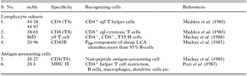
Table 1. Monoclonal antibodies used against antigen-presenting cells and lymphocyte subsets
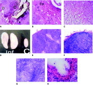
Fig. 1. (A) Mouthparts of tick penetrated into the skin: mp, tick mouthparts; C, cement; D, dermis. (B) Epidermis showing parakeratosis, cellular infiltration and vacuolations: Ep, epidermis. (C) Focal infiltration during secondary infestation. (D) Parotid lymph nodes: Inf, draining lymph node from tick-infested ear side; C, uninfested side. (E) Control lymph node section showing paracortical and cortical zones, distinct germinal follicles. (F) Tick-infested draining lymph node showing infiltration of newly recruited cells in paracortical zone. (G) Tick-infested lymph node showing indistinct follicle surrounded by dense population of newly recruited cells in cortical zone. (H) Showing damage to the lining of endothelium of lymphatic vessel and desquamation of endothelium.
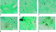
Fig. 2. (A) Infiltration of CD4+ lymphocytes in tick-infested dermis. (B) CD8+ lymphocyte distribution pattern in skin at 72 h after initiation of tick infestation. (C) CD8+ lymphocytes at 8 days of tick infestation. (D) γδ+ T-cell infiltration at dermis during 8 days of tick infestation. (E) MHC II+ antigen-presenting cell infiltration at epidermis of skin infested with ticks. (F) CD1+ cellular infiltration in dermis of skin infested with ticks.
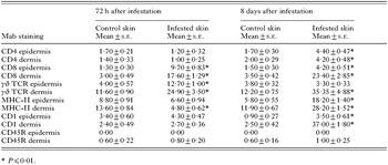
Table 2. Immunohistochemical analysis of lymphocytes and antigen-presenting cell infiltration in skin lesions during primary infestation with adult H. a. anatolicum
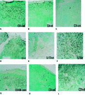
Fig. 3. (A) CD4+ lymphocytes in paracortical and cortical zones of control lymph node. (B) CD4+ lymphocyte depletion at cortical zone of infested draining lymph node at 8 days of infestation. (C) CD8+ lymphocytes in cortical zone of control lymph node. (D) Excessive infiltration of CD8+ lymphocytes in paracortical, cortical and subcapsular zone of draining lymph node at 8 days of tick infestation. (E) Distribution of γδ T cells around germinal follicles in cortical zone (control). (F) Excessive infiltration of γδ+ T-lymphocytes in cortical zone of lymph node infested with ticks. (G) Lymph node section CD45R+ (B-cell) distribution in germinal follicles of paracortical zone. (H) Scattered distribution of CD1+ antigen-presenting cells in control lymph node section. (I) Cortex of infested draining lymph node section showing excessive infiltration of CD1+ cells.

Fig. 4. (A) CD4+[ratio ]CD8+ ratio in ovine skin infested with Hyalomma anatolicum anatolicum. (B) αβ[ratio ]γδ T-cell ratio in ovine skin infested with H. a. anatolicum. (C) MHC-II[ratio ]CD1 cell ratio in ovine skin infested with H. a. anatolicum.
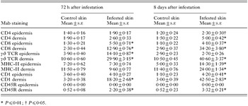
Table 3. Immunohistochemical analysis of lymphocytes and antigen-presenting cell infiltration in skin lesions during secondary infestation with adult H. a. anatolicum

Table 4. Immunohistochemical analysis of lymphocytes and antigen-presenting cell infiltration in draining lymph nodes during primary and secondary infestations with adult H. a. anatolicum
 You have
Access
- 11
- Cited by