Clostridioides difficile is an anaerobic, gram-positive bacterium that causes ~453,000 infections and ~29,300 attributable deaths per year in the United States, making it the most common hospital-acquired infection in the country. Reference Lessa, Winston and McDonald1,2 C. difficile infection (CDI) represents a spectrum of disease ranging from mild diarrhea to sepsis, toxic megacolon, and even death. Solid-organ transplant (SOT) recipients are at an increased risk for developing CDI, likely due to higher rates of hospitalization and exposure to immunosuppressants and antimicrobials. Reference Riddle and Dubberke3,Reference Boutros, Al Shaibi and Chan4 The incidence of CDI in SOT patients ranges from 1.5% to 31% depending on the organ transplanted; the highest incidence is seen after lung transplantation. Reference Riddle and Dubberke3,Reference Boutros, Al Shaibi and Chan4
Generally, C. difficile colonization refers to the presence of C. difficile without symptoms of infection (eg, diarrhea). Rates of C. difficile colonization among inpatients at acute-care facilities range from 3% to 21%, depending on the specific population and testing method. Reference Crobach, Vernon and Loo5 Many of the risk factors linked to CDI have also been implicated in C. difficile colonization, including gastric acid suppressant use, exposure to immunosuppressants, and recent hospitalization. Reference Eyre, Griffiths and Vaughan6–Reference Kong, Dendukuri and Schiller8
Although colonization is thought to be a necessary step in the development of CDI, Reference Martin, Monaghan and Wilcox9 not all C. difficile carriers go on to develop active infection. Reference Longtin, Paquet-Bolduc and Gilca10 Thus, underlying host factors, high-risk exposures (ie, antibiotics, acid suppressants, immunosuppressants) and/or characteristics of bacterial strains are thought to affect disease progression. Recent studies have explored the link between C. difficile colonization and CDI in hematopoietic stem cell transplant patients Reference Bruminhent, Wang and Hu11,Reference Jain, Croswell and Urday-Cornejo12 ; however, this has not yet been examined in the SOT population. Reference Revolinski and Munoz-Price13 We explored the association between C. difficile colonization and CDI in SOT patients.
Methods
This retrospective cohort study was performed at a 600-bed academic medical center in Milwaukee, Wisconsin. All SOT patients admitted to hospital units with high incidences of CDI underwent C. difficile screening within 72 hours of admission according to the hospital’s infection control policy. Screening was then performed weekly for the duration of a hospitalization as well as subsequent readmissions until the patient developed CDI, died, or was lost to follow-up. All consecutive SOT patients with a screening test between May 2017 and April 2018 were included. Screening tests performed within 3 days of a positive clinical test were excluded to ensure that patients were asymptomatic at the time of screening. Patients whose only screening test occurred within 72 hours of a positive clinical test were excluded.
Stool samples for C. difficile screening were collected by nursing staff according to standard unit orders. Units sent stool samples in collection cups or used rectal swabs. Although screening test results were available to providers, the process for ordering a clinical test did not change on the basis of screening results. Clinical tests were collected according to unit protocol and were tested using the 2-step algorithm outlined below.
A C. difficile colonized patient was defined as any patient with a positive screening test in the absence of a concomitant positive clinical test. Patients with incongruent screening tests (ie, not all positive or all negative) were defined as colonized or noncolonized based on the last screening test result prior to CDI, death, or loss to follow-up. Clinical tests were defined as C. difficile tests ordered by providers due to concern for CDI. Clinical tests were considered positive based on C. difficile nucleic acid amplification test (NAAT) results, regardless of toxin enzyme immunoassay (EIA) results. Diarrhea was defined as 3 or more loose bowel movements in 24 hours. CDI was defined as diarrhea in the absence of laxatives, a positive C. difficile clinical test, and the use of C. difficile-directed antimicrobial therapy as ordered by managing clinicians. Hospital-onset CDI was defined as CDI diagnosed > 48 hours after hospital admission, and community-onset CDI was defined as CDI occurring within 48 hours of admission or >12 weeks after discharge. Severe CDI was defined as CDI with a white blood cell (WBC) count ≥15,000 cells/mL and/or a serum creatinine >1.5 mg/dL. Patients with a history of CDI < 3 months prior to first screening test were excluded. Patients who were treated with a new laxative in the 24 hours preceding CDI diagnosis were also excluded.
All patients included in the study underwent chart review to identify demographic variables, SOT date and type, dates of admission and discharge, relevant laboratory data, and clinical outcomes. We used medication administration records to assess relevant exposures: acid suppressants, antimicrobials, and immunosuppressants. All data points were evaluated from the time of first screening until CDI, death, or final discharge.
Microbiology
The C. difficile NAAT screening tests were conducted using the Xpert C. difficile Assay (Cepheid, Sunnyvale, CA). This test targets a conserved region of the cytotoxin B (tcdB) gene required for virulence. The test was internally validated for off-label use with rectal swab specimens and formed stools. Formed stools were tested by inserting the swab into or by rolling the swab on the surface of the stool specimen until the swab was visibly coated. The swab was then eluted into Xpert C. difficile test buffer (provided) and tested in accordance with the product insert.
The C. difficile NAAT clinical tests were conducted using the Xpert C. difficile assay in accordance with the manufacturer’s product insert (Cepheid, Sunnyvale, CA). Unformed stool samples collected by healthcare providers were submitted to the laboratory. A flocked swab (provided) was used to sample the submitted stool and the swab was then eluted into Xpert C. difficile test buffer. The eluted specimen was vortexed and tested in accordance with the product insert. All stool specimens testing positive for C. difficile by the Xpert test were then automatically reflexed for TcdA and TcdB toxin enzyme immunoassay (EIA) testing. Toxin testing was conducted using the C. diff Quik Chek Complete lateral flow test (Alere, Waltham, MA). This test provides a result for glutamate dehydrogenase (GDH) and a combined result for TcdA/TcdB.
Statistical analysis
The data analysis was conducted using SAS 9.4 software (SAS Institute, Cary, NC). The χ2 or Fisher exact tests were used to compare categorical variables. Student t tests or Wilcoxon’s rank sum tests were used to compare continuous variables. The same statistical methods were employed for bivariate analysis between CDI and non-CDI patients. Using multiple logistic regression with a backwards selection method, multivariate analysis was performed for the odds of CDI. Only variables with a P value <.15 in bivariate analysis were included as candidates. All P values were 2-sided, and P < .05 was considered statistically significant.
Results
Study patients
In total, 364 SOT patients underwent C. difficile screening between May 2017 and April 2018. We excluded 10 patients due to history of CDI within the 3 months preceding the patient’s first screening test. Six additional patients were excluded because their only C. difficile screening test occurred within the 72 hours prior to CDI diagnosis.
Colonization
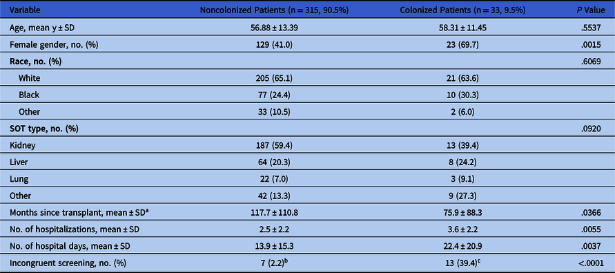
Of the 348 SOT patients included in our study, 33 (9.5%) were colonized with C. difficile while the remaining 315 (90.5%) were not colonized. Table 1 shows the characteristics of these 2 patient populations. Compared to their noncolonized counterparts, patients colonized with C. difficile were more likely to be female (P = .0015) and had more recent transplant surgery (P = .0366). Colonized patients also had higher number of hospitalizations (P = .0055) and hospital days (P = .0037). Of the 33 patients who were colonized at the time of study completion, 13 (39.4%) had a preceding negative screening test. Only 7 (2.2%) of the patients who were non-colonized at study completion had a preceding positive screening test (P < .0001).
Characteristics of Solid-Organ Transplant Patients With and Without Clostridioides difficile Colonization

Note. SD, standard deviation; SOT, solid-organ transplant.
a Measured as months from SOT to last colonization test.
b Refers to patients who were noncolonized at the time of study completion but had previously been colonized during the course of the study. Mean time between incongruent tests was 39.2 days.
c Refers to patients who were colonized at the time of study completion but had previously been noncolonized during the course of the study. Mean time between incongruent tests was 37.1 days.
CDI
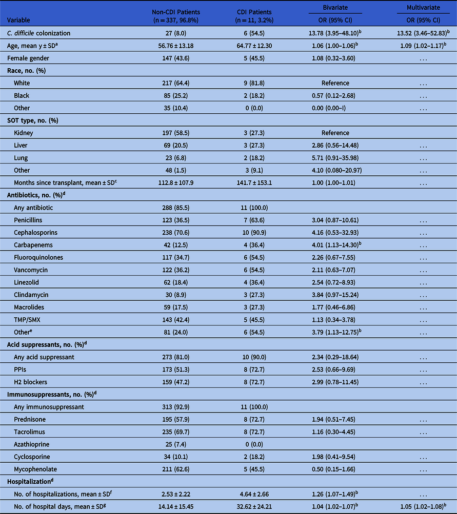
The incidence of CDI in our study was 3.2% (11 of the 348 patients). As shown in Table 2, 6 of these 11 patients (55%) were colonized with C. difficile while the remaining 5 were not (odds ratio [OR], 13.78; 95% confidence interval [CI], 3.95 to 48.10; P < .0001). Patients with CDI were significantly older (P = .0483) and were more likely to have been exposed to carbapenems (OR, 4.01; CI, 1.13 to 14.30; P = .0320) and “other” antibiotics (OR, 3.79; CI, 1.13 to 12.75; P = .0312). CDI patients also had a higher number of unique hospitalizations (P = .0063) and hospital days (P = .0011). In a multivariate analysis for odds of CDI, only C. difficile colonization (OR, 13.52; CI, 3.46–52.83; P = .0002), age (OR, 1.09 per additional year; CI, 1.02–1.17; P = .0135), and number of hospital days (OR, 1.05 per additional day; CI, 1.02–1.08; P = .0017) were shown to be independently associated with CDI (Table 2). In an additional multivariate analysis excluding patients who met criteria for CDI but had a negative EIA, only C. difficile colonization (OR, 12.62; 95% CI, 2.31–69.01; P = .0034) was independently associated with CDI (Supplementary Table 1, online).
Analysis of Solid-Organ Transplant Patients With and Without Clostridioides difficile Infection
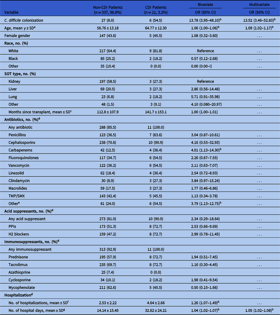
Note. CDI, Clostridioides difficile infection; OR, odds ratio; CI, confidence interval; SD, standard deviation; SOT, solid-organ transplant; TMP/SMX, trimethoprim/sulfamethoxazole; PPI, proton pump inhibitor; H2 blocker, histamine 2 receptor blocker.
a Odds ratio calculated for 1 year increase in age.
b Denotes a P value <.05.
c Measured as months from SOT to last colonization test.
d Assessed from first screening test to CDI, death or loss to follow-up.
e Includes aztreonam, dapsone, daptomycin, metronidazole, rifaximin and tetracyclines.
f Odds ratio calculated for 1 additional hospitalization.
g Odds ratio calculated for 1 additional hospital day.
Clinical outcomes
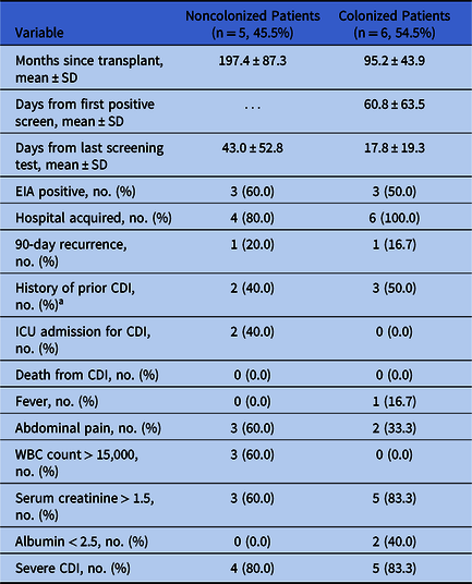
Table 3 shows the characteristics and clinical data points for the 11 patients who developed CDI. Time from transplant to CDI was greater in noncolonized patients than in colonized patients (197.4 months vs 95.2 months). Among colonized patients, the mean time from first positive screening test to CDI was 60.8 days. None of the colonized patients required intensive care unit (ICU) admission for CDI, while 2 of the 5 noncolonized patients (40%) required ICU care. No patient died as a result of CDI.
Characteristics and Clinical Data for Patients With Clostridioides difficile Infection
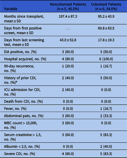
Note. SD, standard deviation; EIA, enzyme immunoassay; CDI, Clostridioides difficile infection; ICU, intensive care unit; WBC, white blood cell.
a Occurring >90 d prior to current CDI episode.
Discussion
In this study, colonization with toxigenic C. difficile was common in the SOT population; it should be considered as a clinically significant risk factor for development of CDI. Although SOT patients colonized with C. difficile were much more likely to develop CDI, most colonized patients did not go on to develop CDI.
Previous studies have shown rates of colonization among inpatients ranging from 3% to 21%, depending on the specific patient population. Reference Crobach, Vernon and Loo14 The overall rate of colonization with toxigenic C. difficile in our study was 9.5%. Although this was the first study to our knowledge that specifically assesses the burden of C. difficile colonization in SOT patients, other recent work has examined toxigenic colonization in patients undergoing hematopoietic stem cell transplantation (HSCT). Reference Bruminhent, Wang and Hu11,Reference Jain, Croswell and Urday-Cornejo12 In these studies, C. difficile colonization was detected in 9.3% to 12% of HSCT patients and was a statistically significant risk factor for CDI. Reference Bruminhent, Wang and Hu11–Reference Revolinski and Munoz-Price13 The association of female gender with C. difficile colonization replicates the finding of Jain et al, Reference Jain, Croswell and Urday-Cornejo12 which showed a similar gender difference in colonization among HSCT patients. Further work will be required to elucidate whether this finding is the result of differences in the gut microbiome or innate immunity of female patients.
Among patients who developed CDI, colonization with C. difficile proved to be the strongest predictor by far. The high utilization of acid suppressants (>80%), antimicrobials (~100%), and immunosuppressants (>90%) in our cohort makes identifying independent risk factors challenging. Regardless, the higher incidence of CDI in patients colonized with toxigenic C. difficile points to the importance of infection control efforts related to the spread of C. difficile. Although isolation of asymptomatic carriers remains controversial, data do support this practice. Reference Longtin, Paquet-Bolduc and Gilca10,Reference Grigoras, Zervou and Zacharioudakis15,Reference Lanzas and Dubberke16 Several studies have shown the implementation of antimicrobial stewardship programs to be effective in reducing rates of CDI Reference Feazel, Malhotra and Perencevich17,Reference Wenzler, Mulugeta and Danziger18 ; however, data on the utility of stewardship efforts targeted specifically towards C. difficile colonized patients are limited.
This study has several limitations. It was performed at a single center, and these results may not be generalizable to all other institutions. This study was also subject to the usual shortcomings of retrospective chart review and was limited by the low incidence of CDI. Because medications were not examined as time-dependent variables, we did not account for the dose or duration of particular exposures. Perhaps most importantly, providers in this study had access to screening test results. Although providers may have limited high-risk exposures or may have reflexively ordered a clinical test in response to a positive screening test, a few points argue against this. Based on our unpublished experience, attempts to de-escalate antibiotic exposures among C. difficile colonized transplant patients were often unsuccessful, even with focused outreach by an antimicrobial stewardship team. Additionally, the average time from last screening test to CDI diagnosis was 17.8 days in colonized patients. Thus, clinical testing does not appear to have been ordered in direct response to a screening result, although we cannot rule out its effect entirely.
In summary, in this study, nearly one-tenth of SOT patients were colonized with toxigenic C. difficile, which demonstrates that colonization is the single most significant risk factor for development of CDI. Because the use of acid suppressants, immunosuppressants, and antimicrobials is so widespread among this patient population, large-scale studies are required to further assess the effect of specific medication exposures. Additionally, future work will be needed to examine different strategies for utilizing colonization data in clinical care with the ultimate goal of preventing CDI in this high-risk population.
Acknowledgments
Financial support
No financial support was provided relevant to this article.
Conflicts of interest
L.S.M.P. received a grant from Cepheid. N.A.L. has served as a consultant for Luminex and Cepheid; he has also received grants from Cepheid, Quidel, Luminex, and First Light Diagnostics. All other authors have nothing to disclose.
Supplementary material
To view supplementary material for this article, please visit https://doi.org/10.1017/ice.2020.431