Ageing of the human population presents probably the most urgent biomedical challenge facing developed nations today. Although it is possible to age in relatively good health, most people face not just an increased risk of death but also an elevated likelihood of developing a plethora of degenerative conditions and functional impairments. This burden of morbidity is probably the most distressing aspect of old age and it has negative effects at both the level of populations (incurring significant costs on the healthcare system) and individuals (morbidity is a thoroughly miserable experience) (Refs Reference Khaw1, 2, Reference Seshamani and Gray3).
Faced with this problem, one valid clinical approach has been to focus efforts on the development and evaluation of interventions designed to reduce either the extent of functional impairments or the duration of the morbid period (Refs Reference Fries4, Reference Fries5). This approach is commonly known as ‘compression of morbidity’ and has met with some success. This article does not discuss classical compression of morbidity interventions but instead focuses on research efforts to increase our understanding of how the mechanisms driving human ageing operate. This approach is related to but distinct from compression of morbidity activities and is particularly attractive at the current time because it is evident that, in model organisms, single ageing mechanisms cause multiple morbidities. Thus, it is possible that interventions that simultaneously postpone multiple causes of morbidity could be developed and delivered.
Although ageing is sometimes painted in positive terms, the truth of the situation is that morbidity means that growing older is bad news for most people, most of the time (Refs Reference Bowling and Dieppe6, Reference Strawbridge, Wallhagen and Cohen7). If a proper understanding of the cellular and molecular mechanisms underlying this process can be obtained then the potential healthcare benefits will be exceptionally large. This leads us to pose two rather obvious questions: what is this thing we call ‘ageing’ and why do we do it at all?
Background to ageing
What is ageing?
The study of the biology of ageing is roughly akin to the study of the consequences of wear and tear or the mechanical properties of worn vehicle parts. As such, a precise and concise definition of ageing is quite difficult to achieve. The best generally accepted one is probably that of Strehler and Mildvan (Ref. Reference Strehler and Mildvan8), who defined ageing as a process that is: universal (i.e. all members of a population of organisms will show it, which distinguishes ageing from infectious disease); progressive (the process is continual and incremental rather than sudden as in the case of ‘programmed’ organismal death or suicidal reproduction); intrinsic (this distinguishes ageing from death due to outside events); and degenerative (this captures the idea that ageing is associated with both increasing chances of mortality but also an increasing level of morbidity).
Although these criteria are a good start in distinguishing ageing from other pathological processes, they tend to underplay the increased physiological frailty of old organisms compared with their young counterparts. Put simply, aged organisms often die as a result of stresses that young organisms are able to survive. Saccharomyces cerevisiae, for example, reproduces asexually but shows an ageing process marked by the eventual cessation of reproductive capacity in older mother cells (Ref. Reference Powell, Quain and Smart9). If young and old cultures of yeast are exposed to a stimulus that induces physiological stress (such as ultraviolet radiation), old yeast cells are markedly more prone to die (Ref. Reference Kale and Jazwinski10). Elevated deaths from cold among elderly humans compared with their young counterparts might be an interesting ‘real world’ example of this type of physiological frailty (Refs Reference Meyer11, Reference Mercer12).
Why do organisms age? The evolutionary rationale
When considering ageing it is important to distinguish between life expectancy and maximum lifespan. The patterns of ageing and mortality seen in human populations illustrate this distinction well. Estimated average life expectancies at birth for members of human populations living under prehistoric conditions are typically of the order of 25–35 years (Refs Reference Bocquet-Appel and Bacro13, Reference Pietrusewsky14). By contrast, life expectancy at birth in the USA in 2001 was 77.2 years and is predicted to rise. However, there is no evidence that the maximum lifespan of Homo sapiens (which appears to be ~120–125 years) has increased over historical time. What has changed is the chance of a given individual reaching an advanced age.
Many other species in the biosphere show similar population dynamics. In the wild, aged organisms are rare because they are rapidly culled by predators or killed by disease. However, when transferred to protected environments ageing changes can be readily observed in a wide variety of species including plants, animals and fungi. What is driving this process?
A compelling explanation for the evolution of ageing is based on the observation that the force of natural selection declines with age. This means that, even in a population of nonageing organisms, there are always far fewer chronologically ‘old’ ones than ‘young’ ones around (because the longer any individual has been around the more likely it is to have been killed or eaten). Thus, any mutation that favours early life fecundity will be selected for even if it results in deleterious effects later on in the lifetime (a type of gene action termed ‘antagonistic pleiotropy’) (Ref. Reference Williams15). This view of ageing argues against the operation of a ‘programme’ controlling the ageing of individuals (i.e. the existence of genes that have the sole function of causing ageing). Rather it suggests that ageing will result from the accumulation of a wide range of unrepaired faults that are too energetically costly to repair under normal conditions. ‘Disposable soma theory’, developed in the 1970s by Kirkwood and Holliday (Ref. Reference Kirkwood and Holliday16), draws essentially the same conclusions as antagonistic pleiotropy but discusses the evolution of ageing in terms of resource allocation between reproduction and somatic maintenance.
Mechanisms of ageing: whom to study and what to look for?
Since ageing is nothing more than the unprogrammed result of selection for early reproductive success, it presents a ‘soft’ phenotype by comparison to those seen when single-gene inheritance or discrete clinical entities are studied. There is limited consensus on what ‘normal ageing’ looks like, which gives rise to two conceptual problems for gerontologists. These may be summarised as: (1) which individuals within a population should be studied to try and uncover ageing mechanisms and (2) what would an ageing mechanism look like if you found it? Before discussing mechanisms that we see as plausible, we offer some initial words of explanation to allow readers to follow our chain of conceptual reasoning in what remains a confused and contested area.
The first problem (‘Whom do you study?’) has proved divisive for biogerontologists. One approach is to treat ageing as though it is a clinical entity in its own right. Thus, some researchers have attempted to identify a ‘pure case of ageing’ by screening individuals on the basis of health status prior to study and excluding those who are unhealthy. The result is that the individuals recruited are atypical for the general aged population and show minimal changes in the parameter being studied. The SENIEUR protocol for the study of immunosenescence provides a good example of this type of thinking. Set up around the idea that the study of ageing necessitated study of the healthy aged, it sets strict admission criteria that exclude those who show disease or are on medication. The result has been that many immune deficits that are found in the general elderly population are simply absent in those who meet SENIEUR criteria (Refs Reference Ligthart17, Reference Ershler18). Similar problems surrounded attempts to gauge the likelihood that cellular senescence (see below) operates as an ageing mechanism. Lacking markers that could identify senescent cells in vivo, large-scale studies of the in vitro divisional capacity of fibroblast cultures from donors of different ages were undertaken. These show that a small decline occurs with chronological age in unselected populations, which becomes statistically insignificant if cultures from healthy donors are used (Refs Reference Martin, Sprague and Epstein19, Reference Goldstein20, Reference Cristofalo21). These findings seem surprising but are consequences of the design of the studies. If an individual showed an age-related change in factor ‘x’ but remained healthy, that would make ‘x’ a good means of estimating chronological age but a poor causal mechanism of age-linked decline. The annual cement lines laid down in human teeth are a good example of this type of physiologically passive but strongly age-linked process. This debate is more visible within the literature on human ageing than it is on that concerning ageing in yeast or Caenorhabditis elegans (given the difficulty of assessing health status in these species), but it is still present and needs to be factored in when thinking critically about the area. None of the above should be surprising but it continues to cause widespread confusion – possibly because there is confusion within the field concerning the properties an ‘ageing mechanism’ is expected to possess.
With regard to this latter question, we consider that any candidate ageing mechanism must meet the following criteria: (1) it must be present in vivo; (2) it must be capable of exerting degenerative effects; and (3) altering the rate of the process should alter the rate of organismal ageing. These criteria are easy to write down but often extremely difficult to test experimentally. However, there are examples within the literature that demonstrate what can be achieved.
The oxidative damage hypothesis of ageing is well known within the scientific community and, in a vulgarised form, has helped to sell beauty products. It is less well known that the hypothesis, as usually stated, is probably wrong. In essence the hypothesis postulates that the degenerative changes seen in ageing are the result of damage arising from free radical reactions (Refs Reference Houthoofd and Vanfleteren22, Reference Harman23). This is, in itself, entirely plausible. Reactive oxygen species are generated in vivo and can clearly produce degenerative effects on cells and proteins. However, attempts to alter rates of ageing by altering antioxidant defences have failed to yield convincing results. Two series of studies are particularly pertinent to this question. Richardson and co-workers (Ref. Reference Pérez24) have undertaken a series of 18 distinct genetic manipulations in transgenic mice designed to either compromise or enhance antioxidant defences. Of these, only deletion of the sod1 gene showed any effect on organismal lifespan (reducing mean survival from ~900 to ~730 days). Using a similar strategy, Gems and co-workers (Ref. Reference Doonan25) investigated the role of the sod genes in C. elegans and saw no major effects on lifespan. These data are not consistent with the hypothesis that the major constraint on organismal survival is the capability to resist oxidative stress.
By contrast, the accumulation of extrachromosomal rDNA circles (ERCs) in S. cerevisiae does appear to be a plausible ageing mechanism (Ref. Reference Kaberlein and Powers26). ERCs are formed by homologous recombination between adjacent rDNA repeats in the yeast genome. Such ERCs are asymmetrically inherited by mother cells at cell division and are capable of autonomous replication, increasing ERC levels with age. ERCs are clearly present in yeast, and mutations that increase ERC formation shorten lifespan. Conversely, mutations that reduce ERC formation rates increase lifespan. Although the direct link to degenerative effects is complex (Refs Reference Falcon and Aris27, Reference Jazwinski28), it is clear that ERC formation is probably a cause of ageing in yeast. The only problem is that it doesn't seem to be a cause of ageing in anything else!
Cell and molecular mechanisms of ageing
The ‘public’ and ‘private’ divide
There are clearly mechanisms of ageing that are species or lineage specific (sometimes referred to as ‘private’ ageing mechanisms). Aside from ERC accumulation, perhaps the best example is the toxic effect on female Drosophila of seminal fluid components secreted from the accessory gland of the male fruit fly (Ref. Reference Barnes29). Seminal fluid toxicity is a major cause of ageing and death in female fruit flies but is not a serious contender as a human ageing mechanism. However, there are a small class of mechanisms that do appear to be truly multispecies in scope and thus probably evolutionarily conserved (sometimes known as ‘public’ ageing mechanisms). These form the focus of the sections below.
Insulin/IGF signalling and dietary restriction
Mutants in the insulin/insulin-like growth factor (IGF) signalling pathway (IIS mutants) display significantly extended lifespans compared with wild-type controls in a wide variety of species. These include the daf-2 mutant in C. elegans (a null mutation in the insulin/IGF-1 receptor), chico in Drosophila (an insulin receptor substrate protein mutant) and a variety of mutations in mice that affect the insulin receptor in selected tissues or global circulating insulin levels and insulin receptor substrates. In addition to being long-lived, IIS mutants show retarded presentation of multiple markers of ageing, including the development of ulcerative dermatitis, bone thinning, increased memory T cell number, and reduced motor coordination (Ref. Reference Selman30). Thus, it appears that interference in IIS pathways can delay age-associated pathology and morbidity (Ref. Reference Bartke31).
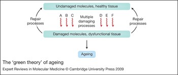
Interestingly, although altered IIS signalling is clearly a conserved longevity assurance mechanism, a cross-species study has shown little evolutionary conservation of the individual genes that are regulated by it (Ref. Reference Piper32). Instead, it seems that there is conservation of processes. The primary mechanisms conserved between these mutants appear to be altered protein biosynthesis (long-lived mutants show a downregulation of genes involved in mRNA translation) and enhanced phase I/phase II xenobiotic metabolism (there is a marked upregulation of cytochrome p450 genes, short-chain dehydrogenase reductases and glutathione S-transferases). These observations, together with the increasingly unsatisfactory nature of the oxidative damage theory of ageing (Ref. Reference McElwee33), have led Gems and co-workers to propose a new hypothesis based on broad-spectrum detoxification. This theory, nicknamed the ‘green theory’ of ageing (Refs Reference Gems and McElwee34, Reference Gems, Faragher, Thornally and Foyer35), proposes that ageing is the result of macromolecular damage that has accumulated as a result of the action of toxic metabolic byproducts (including, but not restricted to, damage caused by reactive oxygen species). A primary determinant of lifespan is thus the efficiency with which potentially damaging species are either removed or the products of damage are repaired (Fig. 1). The green theory is compatible with antagonistic pleiotropy because phase I/phase II metabolism is an energetically costly process to carry out. Thus, there is likely to have been selective pressure to render it no more efficient than necessary in wild-type organisms.
The ‘green theory’ of ageing. The organism can be visualised as initially composed of undamaged molecular and cellular components. Levels of damage increase in this system as a result of multiple damaging processes including (but not restricted to) oxidative stress. Phase I/phase II xenobiotic metabolism functions as a repair process but is energetically costly.

There is also good evidence (Refs Reference Chou36, Reference Cho37, Reference Spindler and Dhahbi38, Reference Tóth39, Reference Tsuchiya40) for significant overlap between the pathways controlled by IIS signalling and those activated by a regime of calorie or dietary restriction (DR). Animals enter a DR state when they consume significantly fewer calories without malnutrition, a process that results in a significant extension of lifespan. DR appears to be a general anti-ageing mechanism since the state can be triggered in a wide variety of experimental organisms including Drosophila, laboratory rodents and primates (Ref. Reference Mattison41). Until recently, it was considered likely that DR mediated its beneficial effects primarily by altering either the production of reactive oxygen species or by enhancing antioxidant defences (Ref. Reference Houthoofd and Vanfleteren22). However, since oxidative damage appears increasingly unlikely to be a major ageing mechanism, it seems more plausible that the enhanced survival produced by DR results from entry of the animal into a ‘recycling’ state marked by repeated cycles of apoptosis and autophagic cell death combined with elevated protein turnover and repair (processes very similar to those invoked by green theory). Similarly, reduction in the activity of the TOR (target of rapamycin) protein by RNA interference (RNAi) or direct inhibition can extend lifespan in yeast, C. elegans and Drosophila (Refs Reference Vellai42, Reference Kapahi43). Since in both yeast and mammalian cells a primary role of TOR is essentially to couple protein synthesis to nutrient availability, inhibition of the enzyme may extend lifespan, at least in part, by mimicking starvation and shifting the emphasis of metabolism more to recycling than de novo protein synthesis.
It needs to be stressed that the entry of an organism into a DR state is not without fitness costs. It has recently been shown that aged DR mice exhibit an impaired natural killer cell function, leading to increased mortality in response to viral infection compared with age-matched controls (Refs Reference Ritz and Gardner44, Reference Ritz45).
Cellular senescence
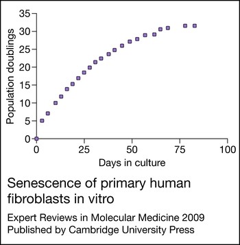
Progressive accumulation of cells that can no longer divide has also been suggested as a general ageing mechanism. This mechanism is sometimes known as the cell senescence hypothesis (or just as ‘replicative senescence’) and is relevant only to organisms that turn over somatic cells during life – not to those with a postmitotic soma (such as C. elegans). Unlike IIS or DR, replicative senescence was not discovered as the result of in vivo manipulation of lifespan through genetic or environmental means but rather as an in vitro phenomenon limiting the capacity of populations of normal human fibroblasts to continue to expand (Fig. 2). Senescence was proposed as an organismal ageing mechanism when it was first observed in the 1960s. However, only recently has sufficient in vivo work accumulated to render that proposal compelling.
Senescence of primary human fibroblasts in vitro. Growth of a population of primary human diploid fibroblasts (Strain 249.Br). The vertical axis measures cell growth in cumulative population doublings. This generic measure of growth is calculated as: population doublings = (log10 number of cells harvested − log10 number of cells seeded) ÷ log10 2. Adapted, with permission, from Ref. Reference Faragher46 (© 2008 Biochemical Society).

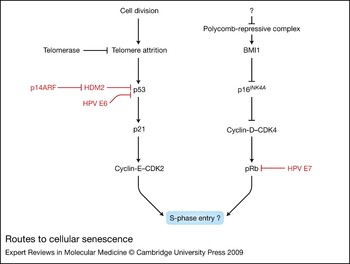
Senescence is the permanent entry of individual cells into a viable but nondividing state, usually as the result of repeated cell division. The molecular pathways that trigger this process are complex but are now relatively well understood (Fig. 3). For the purposes of this article it is sufficient to say that the controls on the number of times an individual cell may divide before entry into the senescent state occurs are tissue and species specific (Ref. Reference Faragher46). Cell senescence can be observed in vitro in cells from a wide variety of different species and has been proposed to act as an anticancer mechanism (Ref. Reference Campisi and Sedivy47). Considered in these terms, senescence appears to be an example of antagonistic pleiotropy at the process level. In the early part of the organismal lifespan, entry into the senescent state probably prevents the growth of tumours, thus contributing to organismal survival. However, cellular senescence is typically associated with the heavily upregulated secretion of pro-inflammatory factors (e.g. the interleukins IL-6, IL -7, IL-8 and the chemokine CCL8) and similar changes in phenotype (Refs Reference Faragher46, Reference Coppé48, Reference Kipling49), which have the potential to produce degenerative effects. However, for senescence to be a plausible ageing mechanism it must satisfy the criteria given above: first, senescent cells must be present in tissue; and second, those changes must produce deleterious effects. The evidence for this is summarised below.
Routes to cellular senescence. The ‘pathways’ triggering replicative senescence shown here are components of a simplified network. The left side of the diagram essentially summarises components of the network associated with telomere attrition (prevented by the specialised reverse transcriptase telomerase), the tumour suppressor protein p53 and the cyclin-dependent kinase inhibitor p21 (p21CIP1/WAF1/CDKN1A). The right side summarises growth suppression associated with the activity of the cyclin-dependent kinase inhibitor p16INK4A, which acts on complexes of cyclin D and cyclin-dependent kinase 4 (CDK4). Activity at this locus is partly controlled by the BMI1 proteins (the activity of which is enhanced by EZH2-containing polycomb complexes). EZH2 levels are reduced at senescence leading to general derepression at the INK4A locus. These pathways can be interdicted by a variety of gene products including SV40 T antigen, the adenovirus E1A and E1B proteins and the human papilloma virus E6 and E7 proteins (denoted HPV E6 and E7 in the schema). Multiple linkages exist but one of the easiest conceptually is the action of the ARF protein on p53 levels via reduction of HDM2 levels. Useful orientation in this area can be found in Refs Reference Bracken78, Reference Cánepa79 and Reference Fridman and Tainsky80. Abbreviations: p14ARF, alternate reading frame INK4A locus protein (CDKN2A); BMI1, B-lymphoma Moloney murine leukaemia virus insertion region-1 protein; EZH2, enhancer of zeste, Drosophila, homologue 2 histone methyltransferase; HDM2, human homologue of MDM2 (E3 ubiquitin ligase; p53-binding protein); pRb, retinoblastoma protein; SV40, simian virus 40.

With regard to the presence of senescent cells in tissue, several studies have now directly demonstrated that they accumulate with in vivo age in the tissues of a variety of species. The most comprehensive of these (Ref. Reference Herbig50) has shown that at least 15% of all dermal cells in very old baboons can be classed as senescent (by immunocytochemical detection of multiple makers of the senescent state). With regard to the effects that the accumulation of senescent cells may have, there is evidence that the accumulation of senescent cells plays a role in liver fibrosis (Ref. Reference Krizhanovsky51), in immune dysfunction (Ref. Reference Effros52), osteoarthritis (Ref. Reference Loeser53) and in the development of atheroma (Ref. Reference Burton54). In an interesting example of antagonistic pleiotropy, there is also good evidence that if senescent cells form ~10% of the total cell population, the pro-inflammatory factors and matrix-degrading enzymes that they secrete can stimulate nearby premalignant and malignant epithelial cells to proliferate and form tumours (Ref. Reference Krtolica55).
Although animals in a DR state show greatly reduced rates of senescent cell accumulation (see below), the best evidence that cellular senescence has the potential to play a causal role in ageing derives from humans suffering from diseases that mimic accelerated or premature ageing (Ref. Reference Cox and Faragher56). There are many of these ‘progeroid syndromes’ but the best known are probably Werner syndrome and Hutchinson–Gilford progeria. Progeria (MCK 176670, Ref. Reference Merideth57) is a sporadic autosomal dominant disorder caused by gain-of-function mutations in the gene coding for nuclear lamin A (LMNA). Mature lamin A is formed from a precursor that is first farnesylated in the C-terminal region and then has this region removed by proteolytic cleavage. In progeria, incorrect splicing of the LMNA mRNA occurs. When translated this produces a protein missing the protease cleavage site present in the wild-type protein. As a result, incorrectly processed prelamin A, termed ‘progerin’ is formed. This retains an additional 15 amino acid C-terminal sequence including the farnesylated residue and remains anchored at the nuclear membrane. At the level of light microscopy, progerin accumulation is observable in individual fibroblasts. These show highly aberrant nuclear structures and chromosomal positioning as the result of disruption of the nuclear lamina (Ref. Reference Meaburn58). Compared with normal controls, populations of such fibroblasts (Refs Reference Bridger and Kill59, Reference Paradisi60) show hypersensitivity to heat stress, elevated rates of apoptosis hyperproliferation and premature accumulation of senescent cells.
Patients with Werner syndrome (MCK 277700) carry loss-of-function mutations in a gene coding for a member of the recQ helicase family (WRN/RECQL2) and show a wide range of pathologies highly reminiscent of normal ageing. Fibroblasts from these patients show a greatly abbreviated divisional capacity in vitro as the result of accelerated exit from the cell cycle. This accelerated senescence is not observed in cells (such as T cells) from tissues that do not show evidence of clinical involvement. Similar observations have been made with Werner syndrome knockout mice (Ref. Reference Kipling61), suggesting that the accumulation of cells with a pro-inflammatory phenotype is driving the disease. The clinical implications of these findings are discussed below.
Clinical implications
Intervention in the progeroid syndromes
The mechanistic understanding of both Werner syndrome and progeria has now reached the stage when clinical interventions are feasible. Trials aimed at the treatment of progeria in particular are already at an advanced stage. These are based on the use of molecules that block the accumulation of progerin at the nuclear membrane by directly inhibiting the enzyme responsible for farnesylation of the protein. One clinical candidate farnesyltransferase inhibitor in particular, tipifarnib (R115777, Zarnestra), shows dose-dependent prevention of both the onset and progression of cardiovascular disease (the primary cause of death in progeria) when administered orally in a mouse model of the syndrome (Ref. Reference Capell62). However, since lamin A also undergoes alternative prenylation by geranylgeranyltransferase, inhibition of farnesyltransferase alone may be insufficient to prevent the build-up of progerin. If this should prove to be the case then alternative treatment routes are available. Combination treatment with statins and aminobisphosphonates has recently been shown to inhibit both farnesylation and geranylgeranylation of progerin in a mouse model of the disease (Ref. Reference Varela63). The prospects for effective treatments for progeria are brighter now than at any time since the disease was first described.
With Werner syndrome, progress has been somewhat slower. The crucial breakthrough from a treatment perspective is the observation by Kipling and co-workers (Ref. Reference Davis64) that Werner syndrome fibroblasts show activation of p38 mitogen-activated protein (MAP) kinase (probably as a downstream consequence of telomere shortening and replication stress caused by replication fork stalling). Treatment of Werner syndrome fibroblasts with compound SB203580 normalises the lifespan, growth rate and morphology of the cells. SB203580 [4-(4′-fluorophenyl)-2-(4′-methylsulphinylphenyl)-5-(4′-pyridyl) imidazole] is a cell-permeable and semiselective inhibitor of several MAP kinase homologues. There is clear potential for the effects of SB203580 or related compounds to be evaluated in rodent models of Werner syndrome but as yet these trials have not been undertaken. SB203580 itself is not suitable for human use because of issues of selectivity and safety, but p38 MAP kinase is a heavily targeted molecule and many potential alternative inhibitors are available (e.g. RO3201195 and BIRB 796), which may prove to be of clinical utility (Refs Reference Bagley65, Reference Bagley66).
Intervention in the ageing immune system
One sign of the physiological frailty that is the hallmark of ageing is increased susceptibility to infection. The ability to produce naive T cells for release into the peripheral T cell pool is dependent in mammals on the presence of an intact thymus. However, with increasing age the thymus atrophies (Ref. Reference Aspinall and Mitchell67). This leads to a dramatic decline in the production and export of T cells bearing new antigen receptor specificities, leading to shrinkage of the overall immune repertoire. Concurrently with this process, replicative senescence within the peripheral T cell pool further compromises the ability to meet an immune challenge. This senescence is driven in part by persistent, often asymptomatic, life-long infection with a variety of viruses such as cytomegalovirus and Epstein–Barr virus (Refs Reference Koch68, Reference van Baarle69). Treatment for this age-related immune dysfunction could potentially take place by a combination of therapies including simple vaccination against the major viruses in childhood (Ref. Reference Pass70), use of telomerase activators such as TAT2 to retard replicative senescence in T cell populations (Ref. Reference Fauce71), and treatment with IL-7 to enhance thymic function and naive T cell output (Ref. Reference Aspinall72).
Insulin/IGF signalling in the clinic?
There is little doubt that the insights into ageing mechanisms that are being obtained using IIS mutants have been some of the most exciting research developments of recent years. Interventions based on IIS remain a considerable way off, but it is interesting to note that mutations and particular alleles in genes involved in these pathways appear to correlate with increased healthy life expectancy in humans. It has recently been reported (Ref. Reference Willcox73) that polymorphisms in the FOXO3A gene strongly (P < 0.05) associate with human longevity. FOXO genes are transcription factors that serve as downstream targets of IIS (Ref. Reference Daitoku and Fukamizu74). Of the Japanese males studied, 75% of those homozygous for a minor allele were healthy compared with 57% of those homozygous for the major FOXO3A allele. Health was defined as the absence of several major diseases in combination with both high physical and cognitive function. Study members carrying the minor FOXO alleles also had better self-rated health than those carrying the major allele. Other studies have linked variation in IIS in humans to old age survival (Ref. Reference van Heemst75) and this will clearly be an area to watch closely in the near future as the links between IIS and healthy human longevity become better understood.
Outstanding research questions
Evolutionary biology: linking species together?
Although antagonistic pleiotropy provides a convincing explanation for the evolution of ageing, there are studies that do not fit altogether smoothly into standard versions of fecundity theory. A case in point concerns recent work conducted on ageing in the guppy (Poecilia reticulate). A series of studies have compared guppies that live in high-predation environments with those in low-predation environmental niches in the same river system in Trinidad. Classical resource allocation theory would predict that fish in low-predation environments should display relatively longer lifespan and a reproductive strategy tending more towards the prudent end of the spectrum. By contrast, guppies in high-predation environments should be driven towards prodigal strategies. However, it has been shown that guppies that evolved with predators mature at an earlier age but also show longer lifespans than their counterparts in environments with low extrinsic mortality (Ref. Reference Reznick76). This strongly suggests that our current evolutionary models are incomplete and additional work (e.g. factoring in foraging costs and kin selection) would be very worthwhile.
Extended lifespan and replicative senescence: linking mechanisms together?
There is intense interest in the mechanisms of both IIS- and DR-based lifespan extension. However, very few studies to date have attempted to link these observations to the theory that replicative senescence plays a causal role in ageing. A notable exception to this is the work of Wolf and colleagues (Ref. Reference Li77) who established (1) that increasing numbers of cells from the mitotic compartment of the rodent lens became senescent with advancing organismal age and (2) that placing the animals on a DR regime significantly decreased the rate at which senescent cells built up. Thus, at least for DR, it is possible that it exerts its ‘anti-ageing’ effects by preventing the build up of senescent cells. This is an intriguing possibility and worthy of further study.
No studies have as yet been undertaken to determine the frequency of senescent cells in key tissues in rodents carrying IIS mutations. This represents an important gap in our understanding of how alterations in IIS could lead to extended lifespan. Reduced accumulation of senescent cells in such mutants is quite plausible and could result from altered rates of apoptosis and tissue turnover, from direct effects of altered IIS signalling on pathways governing cell cycle traverse or from autophagy. A reduced rate of senescent cell accumulation would be consistent with extension of lifespan via reduced numbers of senescent cells (and would raise the prospect that increased rates of cell senescence would block healthy lifespan extension in such animals). Alternatively, extended lifespan with normal rates of senescent cell accumulation would, in essence, falsify the hypothesis that lifespan in these animals is limited by the accumulation of senescent cells but would raise the possibility that the systemic environment, rather than cell-intrinsic changes, plays the major role in the extended lifespan phenotype.
Acknowledgements and funding
The authors gratefully acknowledge the reviewers of this manuscript and the financial support of the BBSRC, the BBSRC-EPSRC, the SPARC Network and Research Into Ageing.