Implantable cardioverter defibrillators can be life-saving for children who are at increased risk of sudden arrhythmic death; however, implantable cardioverter defibrillators are primarily designed for the adult population. In children, implantable cardioverter defibrillators are associated with a higher risk of complications, including inappropriate shocks, infection, and lead problems.Reference Marron, Spirito and Ackerman 1 – Reference DeWitt, Triedman and Cecchin 3 Unfortunately, with the exception of previous cardiac arrest, risk stratification for determining who needs a cardioverter defibrillator in childhood is poor.
In clinical situations where the risk/benefit ratio of implantable cardioverter defibrillators is unclear, it may be reasonable to consider an external automated defibrillator either as a bridge to implantable cardioverter defibrillator or until further diagnostic information is available. In addition, an external automated defibrillator may be considered as one of the preventative measures in children at increased risk of arrhythmic death.Reference Pundi, Bos, Cannon and Ackerman 4
The aim of this study was to review our experience of prescribing automated external defibrillators to families with children at potential increased risk of arrhythmic sudden death.
Methods
A retrospective review was undertaken of all children who were issued an automated external defibrillator by The Scottish Paediatric Cardiac Electrophysiology Service from January, 2005 – issue of the first automated external defibrillator – to July, 2015.
In every case, the automated external defibrillator issued was a Philips HeartStart HS1 (Koninklijke Philips NV, Amsterdam, Netherlands), each costing around £1000. Paediatric pads were used for all children under 8 years of age. All parents were given resuscitation training according to the Paediatric Resuscitation guidelines, including use of the automated external defibrillator.Reference Maconochie, de Caen and Aickin 5 Training was provided either by the hospital resuscitation training service or by the St John’s Ambulance Service.
The following information was obtained from patient records:
-
∙ age at which the automated external defibrillator was issued;
-
∙ gender;
-
∙ diagnosis and results of any genetic testing;
-
∙ medications;
-
∙ symptoms after the automated external defibrillator was issued;
-
∙ any events for which the automated external defibrillator was or should have been utilised;
-
∙ documented rhythm during symptoms or events;
-
∙ outcome when the automated external defibrillator was used;
-
∙ whether the automated external defibrillator was returned when no longer deemed to be needed.
Results
Over a period of 10.5 years, 36 automated external defibrillators were issued to 36 families for 44 children, 27 (61%) of whom were male. The age of the children at the time the automated external defibrillator was issued ranged from 1 day to 15 years (mean 8.8 years). Follow-up ranged from 12 to 138 months, with a median of 50 months (4.1 years) and a mean of 75.5 months (6.2 years).
Diagnoses
The primary diagnosis was prolonged QT in 22 (50%), broad complex tachycardia of uncertain aetiology associated with syncope or cardiac arrest in six (14%), hypertrophic cardiomyopathy in five (11%), catecholaminergic polymorphic ventricular tachycardia in four (9%), Brugada in two (4%), and one each of Barth syndrome, Andersen–Tawil, anomalous coronary artery with cardiac arrest, cardiac tumour with ventricular tachycardias, and a novel SCN5A mutation, the phenotype of which has previously been described in detail in the literature.Reference Maconochie, de Caen and Aickin 6 One of the children with long QT syndrome 2 also had hypertrophic cardiomyopathy. Depending on age and clinical presentation, investigation of patients presenting with broad complex tachycardia included echocardiography, ambulatory electrocardiography, exercise testing, genetic testing for channelopathy, parental 12-lead electrocardiograms, invasive electrophysiology studies, and implantation of loop recorder.
Among all, 31 (70%) patients had one or more pathogenic gene mutations. Gene testing was not performed only in two children, one with anomalous coronary artery and the other with cardiac tumour. Genetic testing varied depending on the availability at the time of diagnosis. Individuals with suspected catecholaminergic polymorphic ventricular tachycardia had analysis of the RYR2 gene, and those with suspected long QT syndrome had analysis of the following genes: KCNQ1, KCNH2, KCNE1, KCNE2, and SCN5A. The six-gene hypertrophic cardiomyopathy panel included MYBPC3, MYH7, TNNT2, TNNI3, TPM1, and MYL2. Genetic analysis was carried out by direct sequencing of the coding regions and immediate flanking regions of the genes.
Genetic mutations
Pathogenic mutations for long QT syndrome
Of the 22 patients with prolonged QTc, 21 had a pathogenic gene mutation. The only child who tested negative for long QT genetic mutations is now thought to have neurocardiogenic syncope and not long QT syndrome, based on the documentation by implanted loop recorder of bradycardia or asystolic pause during symptoms, rather than ventricular arrhythmia. Of the 21 patients with long QT mutations, 11 (52%) had one or more mutations in the KCNQ1 gene (long QT1) and 10 (47%) had mutations in the KCNH2 gene (long QT2). Of the 11 patients with long QTS1, six (54%) had two mutations in the KCNQ1 gene – including four with compound heterozygous mutations and two with homozygous mutations with sensorineural deafness. In addition, two (20%) unrelated children of the 10 who were heterozygous for a mutation in the KCNH2 gene (long QT2) had a coexisting pathogenic mutation in the MYBPC3 gene – including one who had echocardiographic features of restrictive hypertrophic cardiomyopathy and presented with cardiac arrest.
Pathogenic mutations for ventricular tachycardia
Of the four children with a diagnosis of catecholaminergic polymorphic ventricular tachycardia, two (50%) had a pathogenic gene mutation in the RYR2 gene. In the other two patients, gene testing was negative. In addition, two other children had a mutation in the RYR2 gene of uncertain pathogenicity – the child with Andersen–Tawil who also had a pathogenic mutation in the KCNJ2 gene and a child with broad complex tachycardia and cardiac arrest in whom gene testing was otherwise negative.
Pathogenic mutations for hypertrophic cardiomyopathy
Of five children with an echocardiographic diagnosis of hypertrophic cardiomyopathy, three (66%) had a pathogenic gene mutation – one in the TPM1 gene and two in the MYPBC3 gene, one of whom also had a mutation in the KCNH2 gene. Another child, who had a mutation in the KCNH2 gene, also had a pathogenic mutation in the MYBPC3 gene, but no echocardiographic features of hypertrophic cardiomyopathy.
Anti-arrhythmics
A total of 37 (84%) children were prescribed β-blockers, including 20 of the 22 (91%) children who were issued an automated external defibrillator for long QT. Only one child with long QT stopped taking β-blockers because of reported side effects including behavioural problems. In one patient, the parents declined β-blockers. The majority, 31 (84%) of the 37 children on β-blockers, was taking nadolol; otherwise, two were taking propranolol, three atenolol, and one bisoprolol; three children were taking amiodarone, two in combination with a β-blocker and one as a single agent; and two other children were initially on amiodarone in combination with a β-blocker, but the amiodarone was stopped and the β-blocker was continued. The child with Andersen–Tawil was on flecainide in combination with β-blockers.
Reasons for issuing an automated external defibrillator
Of the 44 children, 35 (79%) were issued an automated external defibrillator on recommendation of the physician. This group included six children for whom an implantable cardioverter defibrillator had been recommended, but implant was delayed on account of small patient size (three), chronic infection (two), and parental uncertainty about proceeding with implantable cardioverter defibrillator placement (one). In one child with catecholaminergic polymorphic ventricular tachycardia, two transvenous implantable cardioverter defibrillators had been explanted for lead fracture, resulting in inappropriate shocks, and an automated external defibrillator was issued until the child was deemed to be of a suitable size for subcutaneous implantable cardioverter defibrillator implant. An automated external defibrillator was chosen over an implantable cardioverter defibrillator for two children in whom there was a high likelihood of unnecessary implantable cardioverter defibrillator shocks in response to recurrent asymptomatic self-terminating ventricular arrhythmias despite medication – one with a novel mutation in the SCN5A gene and one with Andersen–Tawil syndrome; 17 (39%) other patients were issued an automated external defibrillator as they were thought to be at possible increased risk of arrhythmia despite treatment with medication or surgery, but were not considered to be at enough known risk to justify implantation of an implantable cardioverter defibrillator. This group included eight patients with long QT syndrome who had two pathogenic gene mutations, a recognised risk factor for more severe disease.Reference Westenskow, Splawski, Timothy, Keating and Sanguinetti 7 , Reference Schwartz and Spazzolini 8
For nine (20%) patients, the automated external defibrillator was issued because of parental request and anxiety, even though not recommended by the physician.
Almost 1:5 children had the defibrillator with them at all times. For 28 (65%) children, there was an automated external defibrillator installed at school or the child was allowed to take the automated external defibrillator to school, with school staff trained in its use.
Symptoms and events after issuing the automated external defibrillator
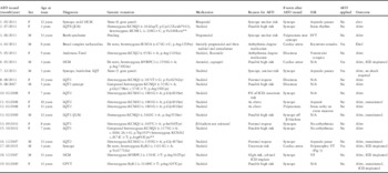
Of the 44 children, 19 (43%) had symptoms or events after the automated external defibrillator was issued (Table 1). In all, 11 children had one or more syncopal events, three had episodes of dizziness, and two had episodes of palpitations. In addition, three children (7%) had a cardiac arrest, and 11/19 patients with symptoms or events had an implantable loop recorder. Documented arrhythmias during syncopal events on an implantable loop recorder or non-invasive monitor included asystole in four patients and ventricular fibrillation in one patient – a 12-year-old boy with hypertrophic cardiomyopathy for whom an implantable cardioverter defibrillator had been recommended but his parents were unsure whether to proceed with implantation (Table 1, patient 18). The automated external defibrillator was not with him when the episode occurred, but he reverted to sinus rhythm spontaneously after 5 minutes of ventricular fibrillation as recorded on his implantable loop recorder. He had a rapid, normal neurological recovery, and an implantable cardioverter defibrillator was implanted.
Symptoms after issue of automated external defibrillator (AED).

CPVT=catecholaminergic polymorphic ventricular tachycardia; F=female; FH=family history; HCM=hypertrophic cardiomyopathy; ICD=implantable cardioverter defibrillator; ILR=implantable loop recorder; JLN=Jervell–Lange Nielsen; LQTS=long QT syndrome; N/A=not applicable; M=male; SCD=sudden cardiac death; SVT = supraventricular tachycardia; VF=ventricular fibrillation; VT=ventricular tachycardia
*Frame shift mutation
**Variant uncertain significance
Automated external defibrillator use
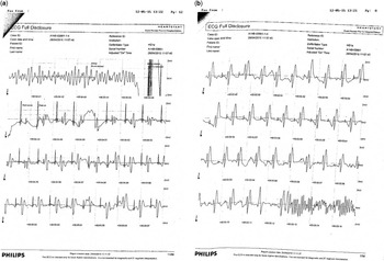
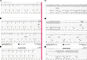
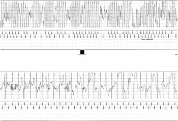
During the study period, there were four events where the automated external defibrillator was applied (Table 1). Of the four events, one child was correctly identified by the automated external defibrillator to be in a rhythm that did not require a shock – sinus rhythm as documented on his implantable loop recorder – and no shock was delivered. The automated external defibrillator correctly identified shockable rhythms in the other three events and one or more shocks were appropriately delivered in all three cases, as described in Figures 1–3. Of the three children, two survived, both with normal neurological recovery, but one died as a result of a recurrent polymorphic ventricular tachycardia, as documented on his implantable loop recorder (Fig 2b). Both survivors had implantation of an implantable cardioverter defibrillator.
( a ) (patient 6, Table 1) Recording from The Philips HeartStart automated external defibrillator, showing termination of ventricular fibrillation with a DC shock in a 5-year-old boy with hypertrophic cardiomyopathy, surgical resection of the left ventricular outflow tract 5 months previously, and dual-chamber pacemaker for postoperative heart block. The event happened while in a children’s playpark. The automated external defibrillator was applied by his mother and aunt, a police officer. ( b ) (patient 6, Table 1) Ventricular fibrillation recurs and was correctly identified as a shockable rhythm. A further DC shock was delivered, which successfully cardioverted him to his paced rhythm. He had normal neurological recovery. There was no damage to his pacemaker from the automated external defibrillator. An implantable cardioverter defibrillator was implanted.

( a ) (patient 4, Table 1) ECG recording from the implantable loop recorder (St Jude Medical Confirm; St Jude Medical Inc., Minnesota, United States of America) following two DC shocks in a 9-year-old boy with sodium channelopathy, who collapsed at home while playing on his computer. The automated external defibrillator was applied by his mother. Following the two DC shocks, the rhythm initially appears to be an atrial flutter with variable block, but is suggestive of junctional rhythm later in the recording. The automated external defibrillator correctly identified this as “non-shockable”. There were no earlier ECG recordings as the device was programmed to update and store the most recent automatic recordings. ( b ) (patient 4, Table 1) The family lived in a remote area and while waiting for emergency services the implantable loop recorder showed that the rhythm degenerated into recurrent torsades de pointes, which, although spontaneously terminated, were followed by further episodes of torsades. Sadly, he did not survive to the hospital. The automated external defibrillator was damaged during transfer of the patient, and the data from it could not be retrieved.

(patient 17, Table 1) Automatic ECG recording from the implantable loop recorder (Medtronic LINQ; Medtronic, Dublin, Ireland) in a 4-year-old with syncope, subsequently found to have a pathogenic mutation in the RyR2 gene. The implantable loop recorder shows polymorphic ventricular tachycardia. The child collapsed while running to the hayloft, and the automated external defibrillator was applied by his parents. The automated external defibrillator correctly identified a shockable rhythm and successfully cardioverted him with one DC shock. The boy had a rapid and normal neurological recovery, and an implantable cardioverter defibrillator was implanted.

Of note is that all three patients for whom the automated external defibrillator was used for cardiac arrest secondary to polymorphic ventricular tachycardia or ventricular fibrillation were male, none had a family history of the cardiac condition or of sudden death, and all had a de novo gene mutation, one in the MYBPC3 gene, one in the SCN5A, and one in the RyR2 gene. Indeed being male and having a de novo mutation for an inherited arrhythmia were 100% predictive for cardiac arrest within our small cohort of patients.
Return of the automated external defibrillator
The automated external defibrillator was returned by six patients; five of them returned the device after placement of an implantable cardioverter defibrillator. The other four children, who underwent implantable cardioverter defibrillator placement, did not return the automated external defibrillator, either because another member of the family is affected by the condition or because the family wishes to keep the automated external defibrillator as a “back-up”. The automated external defibrillator has been returned by only one of the seven families where the child was transitioned to adult services.
Discussion
The chance of surviving a cardiac arrest secondary to ventricular fibrillation is directly dependent on the time it takes to defibrillation. If a shock is delivered within 3 minutes, the chance of survival is >50%, but thereafter the chance of survival reduces by 7–10% for every minute of delay, dependent also on the presence and quality of bystander cardiopulmonary resuscitation.Reference White, Vukov and Bugliosi 9
Studies such as the Chicago Airport and Casino automated external defibrillator trials have shown that automated external defibrillators in public places save lives, especially where there are personnel trained to recognise cardiac arrest and initiate resuscitation.Reference Valenzuela, Roe and Nichol 10 – Reference Caffrey, Willoughby, Pepe and Becker 12 A study on the use of automated external defibrillators in high schools in the United States of America has shown that, combined with a response plan involving emergency services, automated external defibrillators significantly improve survival from out-of-hospital cardiac arrest in student athletes.Reference Drezner, Toresdahl, Rao, Huszti and Harmon 13 There are a small number of case reports describing the use of an automated external defibrillator in younger children with out-of-hospital cardiac arrest.Reference Gurnett and Atkins 14 – Reference Divekar and Soni 16 More recently, a study by Ackerman et al looked at the incidence of automated external defibrillator rescues in a group of 291 children treated for long QT syndrome without implantable cardioverter defibrillators, for whom an automated external defibrillator had been advised.Reference Pundi, Bos, Cannon and Ackerman 4 They reported successful use of an automated external defibrillator – not in all cases the child’s personal automated external defibrillator – in three children with cardiac arrest, recommending that an automated external defibrillator be considered as part of the preventative measures for patients treated for long QT syndrome. The risk of cardiac arrest, however, was only 1% (<1/1700 patient years) in this patient group, who had been referred to a specialist centre; therefore, it is expected to have skewed disease towards the more severe end of the spectrum. In our study, there were no cardiac arrests or episodes of ventricular tachycardia or ventricular fibrillation in the long QT group, but four (18%) of the 22 children issued an automated external defibrillator for an arrhythmic condition other than long QT had an episode of ventricular fibrillation or polymorphic ventricular tachycardia. Our study indicates that issuing an automated external defibrillator may be even more appropriate for children diagnosed with arrhythmic conditions other than treated long QT syndrome, where the risk of life-threatening arrhythmia is likely to be higher, perhaps especially in boys with de novo mutations. It is notable that two of the three patients for whom an automated external defibrillator was used in Ackerman’s group required drugs in addition to multiple shocks to control arrhythmia, showing that defibrillation alone is not always sufficient to treat ventricular arrhythmias in patients with channelopathies. This was the case for our patient with a sodium channelopathy who died with recurrent torsades de pointes despite appropriate use of the automated external defibrillator, for whom arrival of the emergency services was delayed because he lived in a remote area. It is unknown whether more rapid defibrillation together with pacing by an implantable cardioverter defibrillator would have altered outcome, as tachycardia storm can be a problem in some arrhythmic conditions even with or exacerbated by an implantable defibrillator.
Automated external defibrillators are primarily designed for adult use, with most current models delivering a fixed biphasic shock of 150 J. The previous recommendation of 2 J/kg of energy for paediatric defibrillation has been superseded following reports suggesting that higher dosages may be more effective and safe.Reference Haskell and Atkins 17 – Reference Rossano, Quan, Kenney, Rea and Atkins 19 At present, the European Resuscitation Council recommends 4 J/kg to be given as the initial defibrillation dose in children, without escalation for subsequent shocks.Reference Maconochie, de Caen and Aickin 5 Delivery of adult defibrillation dosages in small children (<8 years old) carries the concern about potential risk of myocardial damage.Reference Dahl, Ewy, Warner and Thomas 20 , Reference Gaba and Talner 21 Although there are no devices designed specifically for children, the automated external defibrillator energy dose can be reduced for children <8 years by using paediatric pads or some automated external defibrillators have a paediatric “key”.Reference Killingsworth, Melnick and Chapman 22 , Reference Atkins and Jorgenson 23
An important concern about using automated external defibrillators in children is that the rhythm analysis uses algorithms designed for discriminating arrhythmias in adults. Infants and small children can have very high heart rates during sinus tachycardia or supraventricular tachycardia that conceivably could be misinterpreted as “shockable” rhythms by an automated external defibrillator programme designed for adults; however, studies have demonstrated that “adult” automated external defibrillator algorithms can discriminate between shockable and non-shockable rhythms in >95% of paediatric arrhythmias.Reference Cecchin, Jorgenson and Berul 24 , Reference Atkinson, Mikysa and Conway 25 Our study confirms that, in real life, the automated external defibrillator was able to differentiate between shockable and non-shockable rhythm in all instances when applied in patients with different cardiac conditions.
Our study is the first to report an experience of prescribing automated external defibrillators to children at potentially increased risk of sudden arrhythmic death from a variety of arrhythmic conditions in the setting of a regional paediatric cardiology service. We have demonstrated that parents can successfully be taught to recognise cardiac arrest, apply resuscitation skills, and use an automated external defibrillator effectively. Automated external defibrillators are free from lead problems, device-related infections, and inappropriate shocks, well described with implantable defibrillators in children.Reference Von Bergen, Atkins and Dick 26 , Reference DeWitt, Triedman and Cecchin 27 Automated external defibrillators, however, are less reliable than implantable cardioverter defibrillators in that they may not be with the child when an event occurs. In addition, automated external defibrillators are dependent on an external source – for example, parent or carer – to recognise cardiac arrest, correctly apply them, and administer the shock. It therefore generally takes longer for a shock to be delivered than with an implantable cardioverter defibrillator, which may make the arrhythmia more refractory to defibrillation. Automated external defibrillators may also result in psychological problems, with a number of families confessing that they hated the device but felt that “something would happen” if they did not have it with them. Many parents confessed that they found it stressful deciding when the automated external defibrillator should be with the child. Most families were unwilling to return the device even after implantable cardioverter defibrillator implantation or transition of the child to adult services.
We are not advocating that an external automated defibrillator be used in place of an implantable cardioverter defibrillator where the latter is considered necessary, but as with Ackerman’s group, we recommend that an automated external defibrillator be considered as part of the preventative measures for children with sudden arrhythmic death syndromes. For children in whom an implantable cardioverter defibrillator has been advised but delay to implant is deemed unavoidable, a potentially more reliable alternative to the automated external defibrillator is the Lifevest, a wearable external defibrillator from Zoll (Zoll Medical Corporation, Chelmsford, United States of America). The Lifevest recently received Federal Drug Administration approval for paediatric use. It is primarily intended as a temporary “bridge” to implantable cardioverter defibrillator implantation, but is only suitable for older children. Its main drawback is the cost. It is only available for rent and cannot be purchased, with a current quoted rental cost in the UK of £2900 per month, compared with a purchase cost of around £1000 for an automated external defibrillator
Conclusion
Our study demonstrates that prescribing an automated external defibrillator can provide life-saving therapy for children with a range of cardiac conditions that result in increased risk of sudden arrhythmic death, for whom the risk/benefit ratio of implantable cardioverter defibrillator is uncertain, or delay to implantable cardioverter defibrillator implantation is deemed necessary.
Acknowledgements
The authors thank the parents who were willing to share their experience of using the automated external defibrillator.
Financial Support
The authors thank The Scottish Association for Children with Heart Disorders and the Glasgow Children’s Hospital Charity who provided the funds to purchase the automated external defibrillators.
Conflicts of Interest
None.
Ethical Standards
This study was a retrospective review of an aspect of routine medical care and did not involve human or animal experimentation. The study was reviewed by the local Research and Development and Ethical Services, who concluded that formal ethical approval was not required.