The femoral vein is usually the conventional approach for percutaneous closure of atrial septal defects, being the right side the most common access site. If such site cannot be used, other central veins must be considered, frequently making the procedure more challenging. Although this kind of intervention is rarely performed through the jugular veins, given the difficulty to navigate catheters and devices, they may be considered as good and safe alternatives in the presence of particular anatomical variations.
Case report
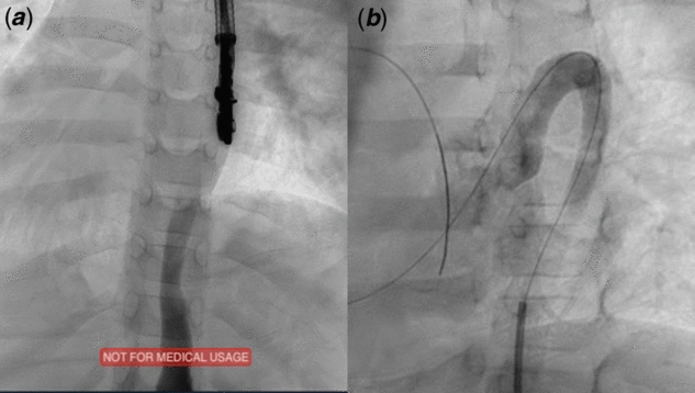
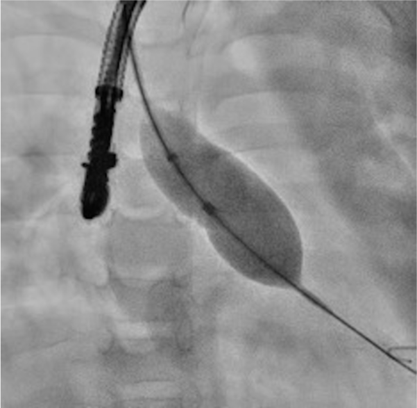
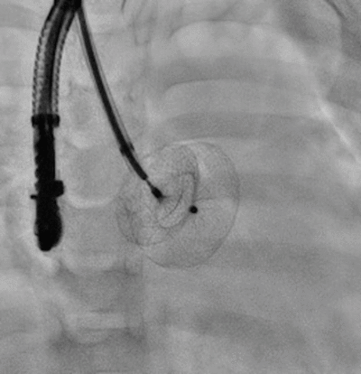
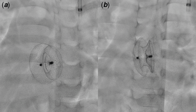
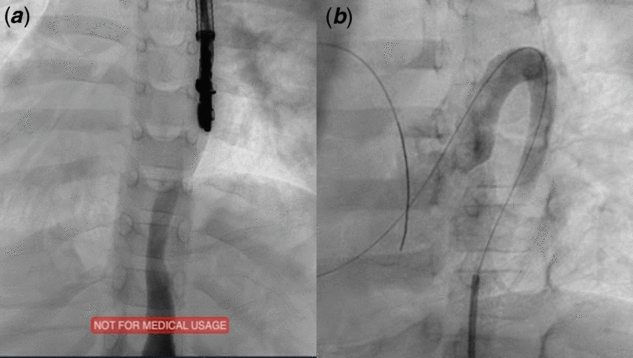
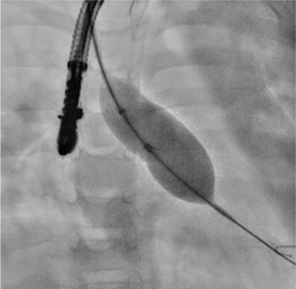
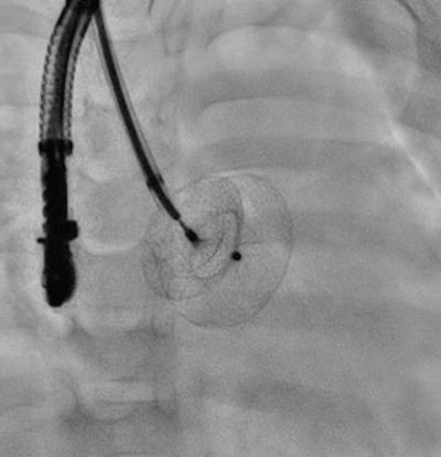
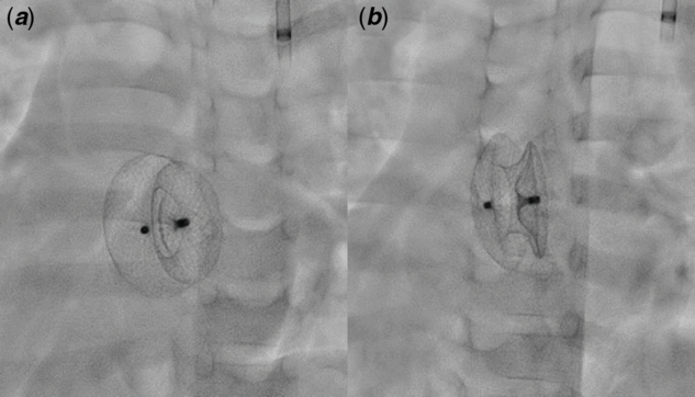
Male patient, 3-year-old, 12 kg, with previous diagnosis of situs inversus along with dextrocardia and ostium secundum atrial septal defect with progressive increase in size, from 5 mm initially to 15 mm, with a significant shunt and hemodynamic repercussion, was referred for elective percutaneous closure. After the cannulation of the right femoral vein, we observed agenesis of the infra-hepatic segment of the inferior caval vein, with a communication through the left superior caval vein by the azygos vein (Fig 1). Using ultrasound imaging and angiography with contrast injections in the right cubital and proximal femoral veins, the presence of double inferior caval vein and/or right superior caval vein was excluded. Therefore, we decided to use the left internal jugular vein approach for the intervention, following the conventional steps, and inverted the fluoroscopic image utilising the dedicated software of the X-ray system (Allura®; Philips Healthcare, the Netherlands) (Figs 2 and 3) to facilitate the manipulation and navigation of materials and devices. Ultrasound-guided (transversal view) internal left jugular vein puncture was performed. After the puncture, a 5-French sheath was introduced, followed by a Judkins-right catheter navigated through a hydrophilic guidewire to the pulmonary artery branches, pulled back to the right ventricle and right atrium and then sequentially placed in the left atrium and left ventricle through the atrial septal defect. The guidewire was then replaced by a support Stiff J-tip guidewire and maintained in left ventricle. The ASD diameter was 15 mm in basal transesophageal echo with adequate rims, except for the anterior that was 2 mm. When a 24-mm Amplatzer compliant balloon (Figure 2) was inflated until the interruption of left to right shunt, it was measured to be 18 mm. The balloon was then replaced by a long 9-French introducer sheath with the tip positioned above the mitral valve. An Amplatzer septal occluder (ASO19; St. Jude Medical, Inc., St. Paul, Minnesota, United States of America) – 1 ml larger than the balloon diameter to interrupt blood flow through the atrial septal defect – was mounted on the kit. The first disc of the device was released inside the left atrium, adjacent to the interatrial septum, immediately followed by the deployment of the right disk (Fig 3). After assuring good positioning, without interference with surrounding cardiac structures, it was completely released (Fig 4). Total procedure time was 38 minutes, with 18 minutes of fluoroscopy. The patient was observed for 24 hours, being discharged in good clinical conditions, and the favourable evolution was similar to those in which the procedure was performed by femoral approach. No complications were observed during follow-up. Sixty-month echocardiographic follow-up showed no residual shunt and normal right ventricle size and function.
( a ) Dextrocardia, agenesis of inferior caval vein and azygos vein connected to the left internal jugular vein. ( b ) Azygos vein connected to the left superior caval vein.
Compliant balloon for measurement of the stretched diameter of the atrial septal defect, with inverted fluoroscopic image.
Undeployed Amplatzer septal occluder 19, with inverted fluoroscopic image.
Deployed Amplatzer septal occluder 19 in ( a ) frontal and ( b ) oblique views, with catheter placed in the left internal jugular vein.
Discussion
Percutaneous closure of atrial septal defect utilising inferior caval vein access – especially the right side – is the standard approach in most scenarios and is proven to be safe and easy to perform. In the presence of an obstruction or agenesis of the inferior caval vein, a cautious evaluation of alternative access sites is necessary, especially in newborn and children. Unusual situations as situs inversus, when the position of cardiac structures is altered, the procedure becomes even more challenging, with a higher risk of complications. In this scenario, the hepatic veins usually have a great diameter, allowing the introduction of large sheaths for percutaneous procedures even in infants and newborns. Reference Shim, Lloyd, Cho, Moorehead and Beekman1 Transhepatic puncture has been used as an alternative venous access for the percutaneous atrial septal defect closure in other particular situations. Reference Gharib, Niazi, Hetta and Makkeyah2–Reference Oliveira, Pauperio, Oliveira, da Silva, Alves and Adjuto5 However, as an increased risk of complications – especially bleeding – was observed, this approach should be used only in the absence of other safer options, especially in patients with unusual liver anatomy, as in this case report.
The right internal jugular vein has also been occasionally used as an alternative venous access site, with good results reported in literature. Reference Hussain, Strumpf, Ghandforoush, Jamal and Diethrich3,Reference Baspinar, Al-Hadidy and Kervancioglu6–Reference Xie, Fang and Yang10 In our patient, in whom the superior caval vein (SCV) was on the left side due to the situs inversus and, in the absence of the inferior caval vein, the manipulation of the devices necessary to implant the prosthesis from the azygos vein or from the right internal jugular through the innominate vein down to the left-sided SVC and the right atrium would be considerably difficult, with great possibility of failure. For this reason, the left internal jugular vein was chosen. The puncture was guided by ultrasound in transversal view, and the inversion of the fluoroscopic image facilitated the navigation and positioning manoeuvres during the procedure. Furthermore, maintaining the guidewire in the left ventricle provided better support and facilitated the intervention, which was performed successfully, without major difficulties, using the usual fluoroscopy time. Xie et al has previously described an alternative atrial septal defect closure technique by this approach with a flexible guiding cuff. Reference Xie, Fang and Yang10 Interestingly, Butera et al reported atrial septal defect cases with bilateral femoral vein occlusion requiring right internal jugular access, in which a venous–arterial circuit with exchange and standard guidewires placed through the defect allowed for adequate balloon sizing and safe over-the-wire device deployment. Reference Butera, Lovin and Basile7
Another issue that should be considered during the evaluation of alternative access sites for this type of procedure is the possibility of prosthesis embolisation and the difficulties for its retrieval must be anticipated. Dedicated devices for this purpose should be promptly available. However, with experience and awareness of the risks and possible complications, and predefined strategies to overcome them, these alternative venous sites can be successfully used for paediatric structural interventions. With no major issues during our procedure, the patient’s favourable evolution and early hospital discharge was similar to those undergoing the procedure with the conventional femoral access.
The association of situs inversus and the absence of inferior caval vein in a patient with atrial septal defect is an extremely rare situation, making the procedure even more challenging, especially in small children with large septal defects, as our patient. To the best of our knowledge, this is the first case in which the left internal jugular vein approach was applied for percutaneous closure of atrial septal defect.
Conclusion
Alternative access sites for percutaneous atrial septal defect closure, such as the left internal jugular vein, can be safely used, provided that the interventional team is experienced with the technique and aware of its limitations, risks and adequate approaches to procedural complications. The impossibility of using standard inferior caval vein access is not a reason to disregard percutaneous ostium secundum atrial septal defect closure as the best treatment option.
Acknowledgements
None.
Financial Support
This research received no specific grant from any funding agency, commercial or not-for-profit sectors. ALPR receives research grants from CNPq/Brazil (grants 465518/2014-1 and 310679/2016-8) and from the Fundação de Amparo à Pesquisa de Minas Gerais (FAPEMIG, Brazil; PPM-00428-17).
Conflicts of Interest
The authors have no conflicts of interest to disclose regarding this manuscript.
Ethical Standards
No specific ethical approval from Institutional Reviews Boards are necessary for this type of publication. The authors assure that all patient data provided in this case report are anonymized.