Case
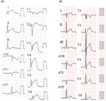
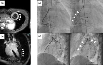
A 13-year-old boy with a medical history of IgA vasculitis was referred to our hospital because of recurrent chest pain and persistent low-grade fever. Early morning chest pain suddenly occurred at rest 36 hours after his second dose of BNT162b2 vaccination, and it continued for a few minutes. His intermittent chest pain persisted for 3 days. At referral 3 days after vaccination, body temperature was 38°C and vital signs were stable. Laboratory tests revealed elevated cardiac enzyme: creatinine kinase 1003 U/L, troponin I 15,805 pg/mL. C-reactive protein was 2.68 mg/dL, and brain natriuretic peptide was 22.3 pg/mL. The electrocardiogram during chest pain showed marked ST elevation in I, II, aVF, V3-V6 (Fig 1a), and then it improved with the remission of chest pain (Fig 1b). There was no cardiomegaly on the chest X-ray. The transthoracic echocardiography demonstrated slightly reduced motion of the posterior wall with generally preserved cardiac function (ejection fraction 61%). Cardiac MRI showed subepicardial linear late gadolinium enhancement (Fig 2a) and high-intensity signal in T2W black blood with fat suppression at the posterolateral wall and apical wall (Fig 2b). These findings determined the diagnosis of acute myocarditis. Polymerase chain reaction test for SARS-CoV-2 was negative, and the degree of anti-SARS-CoV-2 IgG was 1397.1 AU/mL. Coronary angiography showed no anomalous coronary artery. Coronary angiography with acetylcholine provocation test demonstrated diffuse vasospasm of both coronary arteries with 50–75% stenosis (Fig 2c and d) with temporary complete atrioventricular block and ST elevation in V2–V5. These findings and the characteristic chest pain suggested the presence of vasospastic angina. Intravenous immunoglobulin for myocarditis was administered, and oral calcium blocker for vasospastic angina was initiated. After these treatments, cardiac enzymes decreased gradually. The patient was discharged home 8 days after the COVID-19 vaccination without any neurological complications. He has no longer presented with chest pain.
(a) Electrocariogram during chest pain. ST elevation was shown in I, II, aVF, V3-V6. (b) Electrocardiogram at the remission of chest pain. ST elevation improved.

(a, b) Cardiac MRI showed positive late gadolinium enhancement. (white arrows and white arrow heads) (c) Right coronary angiogram (d) left coronary angiogram Achetilcholine provocation test demonstrated diffuse vasospasm of both coronary arteries. (white arrows and white arrow heads).

Discussion
COVID-19 vaccine-related myocarditis is diagnosed based on the symptoms and laboratory and image findings, accounting for 12.6 cases per million with second-dose mRNA vaccine. Reference Bozkurt, Kamat and Hotez1 Symptoms include chest pain, dyspnoea, palpitation, and syncope. Elevated troponin, abnormal electrocardiogram or rhythm monitoring, abnormal ventricular systolic function, and cardiac magnetic resonance findings are used as diagnostic tools. The median onset age and hospitalisation length were 15.8 years and 2 days, respectively. Reference Truong, Dionne and Muniz2 Although chest pain was the major symptom in 99.3%, most of which occurred 2 days after the second dose of vaccination, the mortality rate was extremely low. Reference Truong, Dionne and Muniz2 Several recent studies demonstrated the molecular mimicry between the spike protein and human peptide protein or reaction to adjuvant, nanoparticles, or other components of the vaccine as the mechanism of myocarditis, Reference Truong, Dionne and Muniz2,Reference Vojdani and Kharrazian3 although it is not fully understood.
On the other hand, vasospastic angina is characterised by night or early morning chest pain persisting for several to about 15 minutes. 4 It was endorsed by abnormal electrocardiogram findings suggesting myocardial ischaemia and acetylcholine provocation test at coronary angiogram. 4 The pathophysiology of vasospastic angina includes vascular endothelial dysfunction and vascular smooth muscle abnormality, resulting in unresponsiveness of endothelial nitric oxide synthase. 4
A previous study demonstrated the strong association between myocarditis, especially caused by human parvovirus B19, and coronary vasospasm. Reference Yilmaz, Mahrholdt and Athanasiadis5 It was suggested that coronary vasospasm played an important role in the appearance of atypical chest pain in clinically suspected and biopsy-proven myocarditis. They assumed that myocarditis might cause severe endothelial dysfunction with decreased bioavailability of nitric oxide leading to coronary vasospasm.
Our current case met the diagnostic criteria for COVID-19 vaccine-related myocarditis contemporary with vasospastic angina. It was assumed that vasospastic angina occurred following COVID-19 vaccine-related myocarditis. The chest pain never recurred after the initiation of calcium blocker. Vasospastic angina might contribute to the development of chest pain, the major clinical symptom in COVID-19 vaccine-related myocarditis.
Acknowledgements
None.
Financial support
This research received no specific grant from any funding agency, commercial, or not-for-profit sectors.
Conflicts of interest
None.