Superior caval vein syndrome can occur in children with CHD. It is most commonly seen in single-ventricle patients status-post bidirectional Glenn surgeryReference Tzifa, Marshall, McElhinney, Lock and Geggel 1 . It is also seen post cardiac transplantReference Tzifa, Marshall, McElhinney, Lock and Geggel 1 , Reference Aldoss, Arain and Vinocur 2 , with or after extracorporeal membrane oxygenation cannulationReference Tzifa, Marshall, McElhinney, Lock and Geggel 1 , Reference Kazanci, McElhinney and Thiagarajan 3 , and after surgery involving manipulation or transection of the superior caval veinReference Tzifa, Marshall, McElhinney, Lock and Geggel 1 , Reference Said, Burkhart and Dearani 4 – Reference Bottega, Silversides and Oechslin 6 . Superior caval vein syndrome would not be expected in a post-operative congenital cardiac surgery not directly involving the Superior caval vein but can be observed as a result of central line placement during these proceduresReference Tzifa, Marshall, McElhinney, Lock and Geggel 1 , Reference Johnson, Saltzman, Suh, Dahms and Leonard 7 – Reference Manlhiot, Menjak and Brandao 10 . We report a case of unexpected superior caval vein syndrome after Tetralogy of Fallot surgery.
Case report
A 7-month-old male patient with DiGeorge and Tetralogy of Fallot and Pulmonary Atresia with multiple aorto-pulmonary collaterals was referred to our institution for repair. Preoperative catheterisation demonstrated five multiple aorto-pulmonary collaterals. In the operating room, the anaesthesia team placed bilateral three French internal jugular lines given the patient’s history of difficult intravenous line placement. The intraoperative anaesthetic and surgical course was uneventful. The surgery included a complete repair with right ventricle–pulmonary artery conduit, ventricular septal defect closure, and bilateral pulmonary artery unifocalisation. Post-operative haemodynamics showed good right ventricular pressure (<½ systemic). Three 24-Fr chest tubes were placed before closure, and the patient returned to the Cardiac intensive care unit in stable but critical condition.
Upon arrival to the intensive care unit, the patient was noted to have head, neck, and upper extremity discolouration and swelling. Given the clinical picture, the left internal jugular line was removed post-op day 1, with the belief that possibly having lines in both internal jugular veins of this small infant could be the cause for the upper-body swelling. The right internal jugular line was retained for infusion of milrinone and sedatives. On post-op day 3, the patient continued to have head and neck swelling. An echocardiogram showed flow disturbance in the superior caval vein, but was unable to define the anatomic cause of the obstruction. Subsequently, a heparin drip was started, given the concern for possible clot formation as a result of having a central line in place.
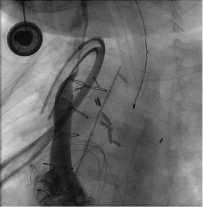
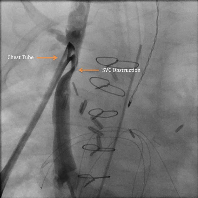
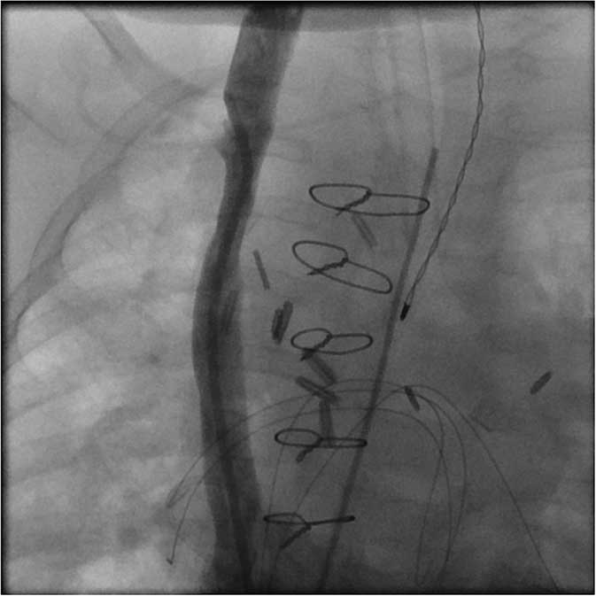
For further diagnosis, the patient was brought to the catheterisation laboratory for evaluation and possible intervention. The initial angiogram showed significant narrowing of the superior caval vein. Multiple views and angles demonstrated that the external compression by the chest tube was the likely source of anatomic obstruction (Fig 1). A paediatric cardiac surgical fellow pulled back the chest tube, but the superior caval vein obstruction remained unchanged (Fig 2). The chest tube was then removed completely with resolution of the superior caval vein obstruction as demonstrated by repeat angiography (Fig 3).
Cardiac catheterisation: pre-intervention supine view. Significant obstruction of contrast flow in the superior caval vein by the surgically placed chest tube is demonstrated.

Cardiac catheterisation: supine view. After repositioning of the surgical chest tube, continued obstruction of contrast flow in the superior caval vein (SVC) by the surgically placed chest tube is still present.

Discussion
After review of the literature, superior caval vein syndrome secondary to chest tube placement has not been previously reported, and this case highlights the importance of investigating unexpected post-operative findings.
Post-operative superior caval vein syndrome can occur in some subgroups of congenital heart patients. Superior caval vein syndrome can occur after having Glenn – superior cavopulmonary anastomosis – or Fontan surgery. In both these instances, passive flow from the superior caval vein fills the pulmonary arteries. Any cause of elevated pulmonary artery pressure resulting in elevated superior caval vein pressure can lead to superior caval vein syndrome. Patients status post atrial switch can also develop superior caval vein baffle obstruction. In these cases, surgeons tend to compromise on the size of the systemic venous baffle in favour of the pulmonary venous baffleReference Bender, Stewart, Merrill, Hammon and Graham 5 , Reference Bottega, Silversides and Oechslin 6 . Cardiac transplant patients are also at risk for developing superior caval vein syndrome secondary to stenosis at the site of the superior caval vein anastomosis. A publication reported that patients with previous cavopulmonary anastomosis are at a higher risk for superior caval vein obstruction post cardiac transplantReference Aldoss, Arain and Vinocur 2 . In the adult literature, there are case reports of haematoma post cardiac surgery causing superior caval vein syndrome.
Previous studies have reported thrombogenic catheter-associated superior caval vein syndrome in adults and children. Short-term haemodialysis catheters even have high rates of pericatheter sleeve and thrombus formationReference Johnson, Saltzman, Suh, Dahms and Leonard 7 , Reference Rinat, Ben-Shalom, Becker-Cohen, Feinstein and Frishberg 8 . In this particular case, this infant arrived from the operating room with superior caval vein syndrome. Placement of a single internal jugular line is standard of care for our patients undergoing cardiac surgery for two-ventricle repair. We do not frequently see superior caval vein syndrome owing to central line placement, even in our smallest post-operative patientsReference Miller, Vu and Chai 9 , Reference Manlhiot, Menjak and Brandao 10 . This patient, however, had bilateral internal jugular lines given the difficulty with intravenous access, making the clinical picture less clear. Persistent superior caval vein syndrome after removal of the left internal jugular lines showed that there was still an undiscovered cause for the patient’s clinical condition. This finding highlights the need to further investigate unexplained findings in post-operative congenital heart surgery patients (Fig 3).
Cardiac catheterisation: post-intervention supine view. Upon complete removal of the chest tube, unobstructed contrast flow could be observed, confirming patency of the superior caval vein. No gradient is measured across the superior caval vein.

Acknowledgements
The study was supported by the Heart Center at Lucile Packard Children’s Hospital.
Financial Support
None.
Conflicts of Interest
None.