Cardiac disease is the commonest type of congenital anomaly. Moderate to severe congenital cardiac malformations are present in from 4 to 8 per 1000 liveborn neonates,Reference Hoffman and Kaplan1 They constitute a major source of fetal and neonatal mortality, accounting for one-fifth of all stillbirths, and almost one-third of neonatal deaths due to congenital defects.2
Prenatal detection of such congenitally malformed hearts is important, as it may improve the planning for pregnancy, perinatal management, and even survival, particularly in duct-dependent lesions such as hypoplastic left heart syndrome and transposition.Reference Kumar, Newburger, Gauvreau, Kamenir and Hornberger3, Reference Tworetzky, McElhinney, Reddy, Brook, Hanley and Silverman4 In addition, early detection of fetal congenital cardiac disease may prompt a closer search for associated non-cardiac anomalies, permit planning of perinatal treatment, and facilitate more informed counseling. Despite improvements in prenatal diagnosis, detection remains imperfect, and better methods are needed to target screening of the fetuses as highest risk.
Increased thickness of the nuchal translucency on an ultrasound scan performed at 11 to 14 weeks of gestation has been shown to be associated particularly with chromosomal anomalies, but also with congenital cardiac disease.Reference Atzei, Gajewska, Huggon, Allan and Nicolaides5–Reference Wald, Morris, Walker and Simpson14 Its use for the detection of Down’s syndrome was first reported in 1985, with 43 percent sensitivity, and a very low false-positive rate.Reference Benacerraf, Barss and Laboda15–Reference Benacerraf, Gelman and Frigoletto18 The normal nuchal thickness in fetuses of 11 to 14 weeks gestation varies slightly with age,Reference Snijders, Noble, Sebire, Souka and Nicolaides11, Reference Pandya, Goldberg and Walton19 but the upper limit of normal is often set at 2.5 millimetres, and the 99th percentile at 3.5 millimetres.Reference Hafner, Schuller, Metzenbauer, Schuchter and Philipp6, Reference Hyett, Perdu, Sharland, Snijders and Nicolaides7, Reference Chaoui, Koerner and Bommer20–Reference Hyett, Moscoso, Papapanagiotou, Perdu and Nicolaides22 Chromosomal abnormalities are found in about one-third of late-first or early-second trimester fetuses with increased nuchal translucency, with trisomy 21 or 18 discovered in about three-quarters of affected cases.Reference Snijders, Noble, Sebire, Souka and Nicolaides11
Congenital cardiac malformations have been reported in from one-eighth to half of fetuses with increased nuchal translucency, albeit that some have also suggested that there is no link between increased nuchal translucency and specific congenital cardiac malformations.Reference Kumar, Newburger, Gauvreau, Kamenir and Hornberger3, Reference Atzei, Gajewska, Huggon, Allan and Nicolaides5, Reference Gembruch, Bald and Redel21, Reference McAuliffe, Hornberger, Winsor, Chitayat, Chong and Johnson23 Others, in contrast, report a disproportionate association with left heart disorders and atrioventricular septal defects.Reference Hyett, Perdu, Sharland, Snijders and Nicolaides7, Reference Orvos, Wayda, Kozinszky, Katona, Pal and Szabo24, Reference Westin, Saltvedt, Bergman, Almstroem, Grunewald and Valentin25 Thus, existing data are inconclusive as to whether increased nuchal translucency is associated with specific congenital cardiac malformations. Our aim in this study, therefore, was to estimate the prevalence of increased nuchal translucency in fetuses with a normal karyotype that were subsequently shown by fetal echocardiography to have congenitally malformed hearts, and to assess differences in prevalence among fetuses with different types of prenatally diagnosed congenital cardiac disease.
Methods
Patients
We reviewed all patients referred for detailed fetal cardiac echocardiography between January 1998 and December 2007 to two large perinatal centres in London, United Kingdom, namely King’s College Hospital and the Evelina Children’s Hospital. All patients had undergone screening including measurement of nuchal translucency prior to the fetal echocardiogram, the measurements being made at between 11 weeks and 13 weeks plus 6 days gestation by a specialist experienced in fetal medicine. Fetuses were included regardless of the reason for referral for fetal echocardiogram, which included family history of congenital heart disease, a suspected cardiac defect, increased nuchal translucency, known non-cardiac anomalies, or a combination of multiple indications. Specific indications for referral for fetal echocardiography were not tracked systematically. Patients were excluded from the analysis when they were shown to have an abnormal karyotype or other genetic evaluation, such as deletion of chromosome 22q11, an unknown karyotype, or no measurement of nuchal translucency available from the 11 through the 14th weeks of gestation. Karyotypic or genetic data may have come from amniocentesis, chorionic villous sampling, postnatal analysis, or postmortem examination.
The gestational age was calculated by the reported last menstrual period, correcting this estimate if there was a difference of 7 days or more from the gestational age calculated on the basis of crown-rump length. For analysis, we calculated the 95th to 99th centile for the given crown-rump length. Although the 95th percentile for nuchal translucency varies slightly with crown-rump length, we made the standard approximation that a nuchal translucency of 2.5 millimetres or more was higher than normal.Reference Hafner, Schuller, Metzenbauer, Schuchter and Philipp6, Reference Hyett, Perdu, Sharland, Snijders and Nicolaides7, Reference Pandya, Goldberg and Walton19
Fetal echocardiography and classification of congenital cardiac malformations
All fetal echocardiograms were performed by a fetal or paediatric cardiologist. Fetal echocardiography was carried out transabdominally, and included imaging of the four chamber view, outflow tracts, arterial duct, and aortic arch. Images were obtained using cross-sectional imaging and colour and pulsed Doppler.
Congenital heart disease was categorized according to the primary cardiac diagnosis, providing cohorts of fetuses with tetralogy of Fallot, atrioventricular septal defect, hypoplastic left heart syndrome, coarctation of the aorta, interrupted aortic arch, transposition, double outlet right ventricle, pulmonary stenosis, pulmonary atresia with intact ventricular septum, and ventricular septal defect Those groups containing fewer than 20 fetuses were then combined either with others having a similar diagnosis, for example pulmonary stenosis and pulmonary atresia with intact ventricular septum, or into non-specific groups such as other complex anomalies, which included functionally univentricular anomalies not otherwise classified, common arterial trunk, totally anomalous pulmonary venous connection, and similar lesions, or other non-complex anomalies, which included anomalies such as atrioventricular valvar regurgitation and interruption of the inferior caval vein. Prenatal cardiac diagnosis was confirmed on postnatal evaluation or, if applicable, postmortem examination. Patients with discordant pre- and postnatal cardiac diagnostic data were evaluated according to the corrected postnatal diagnosis.
Data analysis
We calculated the proportions of chromosomally normal fetuses with congenitally malformed hearts, both overall and with specific types of malformation, with increased nuchal translucency, estimating also the 95% confidence intervals. These estimates were compared with known proportions using the binomial test.
Results
The overall cohort of fetuses diagnosed with congenital cardiac disease by prenatal echocardiography numbered 2133. Of those, 707 were excluded due to abnormal karyotype or other genetic testing, and 690 were excluded because of an unknown karyotype. This meant that 736 with normal chromosomes were eligible for inclusion. A total of 631 fetuses were known to have congenitally malformed hearts in the absence of measurement of nuchal translucency, including 255 with known normal chromosomes.
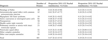
Among the 481 fetuses with documented congenital cardiac disease and normal chromosomes in whom nuchal translucency measurements were available, accounting for 23% of the overall cohort, the proportion with increased nuchal thickness, defined as equal or greater than 2.5 millimetres, was 0.47, 95% confidence intervals being 0.42 to 0.51. The number of fetuses was 224. Setting the threshold at equal or greater than 3.5 millimetres, corresponding approximately to the 99th percentile of the normal population, the proportion of fetuses with increased nuchal translucency, numbering 139, was 0.29, with 95% confidence intervals from 0.25 to 0.33. These proportions were significantly higher than the expected normal population proportions of 0.05 and 0.01, respectively (both p < 0.001). Among the entire cohort of 481 fetuses, the median nuchal translucency thickness was 2.2 millimetres, with an interquartile range of 1.5 to 3.8 millimetres. Among the 224 fetuses with nuchal translucency equal or greater than 2.5 millimetres, the median thickness was 4.0, with an interquartile range of 3.1 to 5.8 millimetres.
Proportions of fetuses with increased nuchal translucency according to cardiac diagnosis are summarized in Table 1. For all diagnostic categories, the proportions of nuchal translucency equal or greater than 2.5 millimetres and nuchal translucency equal or greater than 3.5 millimetres were higher than in the normal population. The only diagnosis for which the proportion of fetuses with nuchal translucency equal or greater than 2.5 millimetres was higher than the others was atrioventricular septal defect, at 0.62, with 95% confidence intervals from 0.47 to 0.77. This gave an odds ratio of 1.97, with 95% confidence intervals from 1.02 to 3.78, and a p value equal to 0.038. There were no diagnoses for which the proportion of nuchal translucency equal or greater than 3.5 millimetres was higher, or for which the proportion of either nuchal translucency threshold was lower.
Summary of nuchal translucency proportions according to cardiac diagnosis.

Discussion
We studied a subset of fetuses that underwent prenatal echocardiography at 2 large tertiary perinatal cardiology centers in London over a period of 9 years. Among 2133 fetuses that underwent echocardiography during this period, and were documented to have congenitally malformed hearts, 481 were known to have normal chromosomes, and also had a measurement of nuchal translucency performed from the 11 through the 14th week of gestation available for evaluation. Among this subset, the prevalence of increased nuchal translucency, defined at greater than 2.5 millimetres, was substantially higher than in the normal population, with an overall prevalence of 0.41, and prevalence ranging from 0.35 to 0.62 in fetuses with different types of congenitally malformed hearts. Nearly three-tenths of our studied fetuses with congenitally malformed hearts had nuchal translucency of 3.5 millimetres or greater.
These findings support that conclusion that increased nuchal translucency discovered on ultrasonic screening during the 11th through the 14th week of gestation is associated with congenital cardiac disease, independent of the presence of chromosomal or other genetic anomalies, and that increased nuchal translucency is not concentrated in fetuses with certain anomalies. This conclusion supports the recommendation that fetuses found to have increased nuchal translucency should be referred for fetal echocardiography regardless of the existence of chromosomal or non-cardiac abnormalities.
There is conflicting evidence about the association of increased nuchal translucency and specific congenital cardiac malformations. Some report no such association, while others report an association with left heart disease and septal defects.Reference Atzei, Gajewska, Huggon, Allan and Nicolaides5–Reference Makrydimas, Sotiriadis and Huggon8, Reference Michailidis and Economides10, Reference Orvos, Wayda, Kozinszky, Katona, Pal and Szabo24, Reference Westin, Saltvedt, Bergman, Almstroem, Grunewald and Valentin25 Although the prevalence of increased nuchal translucency was higher in our fetuses with atrioventricular septal defects, our findings cannot resolve this conflict, since fetuses with different congenital cardiac malformations tend to be diagnosed with differing frequency, and we cannot be certain that biased referral did not contribute to the similarities and differences in prevalence found across the diagnostic spectrum.
There are several potential limitations to our study. As one of the reasons for referral for fetal echocardiography was increased nuchal translucency found on obstetric ultrasonic screening, there is potential selection bias in our sample. The purpose of the study, however, was to report the prevalence of increased nuchal translucency in a population of patients without chromosomal anomalies referred for fetal echocardiography and found to have congenital cardiac disease. We acknowledge that this may not reflect the population of all fetuses with diagnosed and undiagnosed congenital cardiac malformations. Also, because the cohort screened was not an all-inclusive population, and we included only fetuses with documented congenital cardiac malformations, it is not possible to estimate the sensitivity or specificity of our measurements for the purpose of screening fetuses for congenital cardiac disease. The relatively high observed prevalence of increased nuchal translucency in fetuses with all forms of congenital cardiac disease, nonetheless, suggests that the association between increased nuchal translucency and congenitally malformed hearts spans the spectrum of congenital cardiac disease.
On the basis of our study, therefore, we recommend referral of all fetuses with increased nuchal translucency for fetal echocardiography, Others have reported that up to half of fetuses with increased nuchal translucency have congenitally malformed hearts.Reference Hyett, Perdu, Sharland, Snijders and Nicolaides7–Reference Marvridis, Cobian-Sanchez, Tekay, Moscoso, Campbell, Thilaganathan and Carvalho9, Reference Wald, Morris, Walker and Simpson14, Reference Westin, Saltvedt, Bergman, Almstroem, Grunewald and Valentin25 We endorse their statements, since we found that nearly half of our cohort of prenatally diagnosed fetuses with congenital heart disease were found on first or early-second trimester ultrasound to have increased nuchal translucency. Thus, although measurement of nuchal translucency may not have high sensitivity or specificity as a screen for congenital cardiac disease, it may be a more useful method of targeting high risk fetuses for prenatal echocardiography in the absence of universal fetal echocardiography. Historical factors currently used as indications for fetal echocardiography, such as a family history of congenital cardiac disease, generally produce a very low yield,Reference Hoffman and Kaplan1 and increased nuchal translucency may facilitate early diagnosis and decision making for those fetuses with congenitally malformed hearts. True determination of the association between increased nuchal translucency and specific types of congenital cardiac malformation in the fetus would require a population-based study with both universal obstetric ultrasound and specialist fetal echocardiography, with sufficient sample size to represent a broad spectrum of congenitally malformed hearts. In the absence of such an undertaking, our findings should help expand the collective understanding of the utility of screening for congenital cardiac disease on the basis of nuchal translucency.