Introduction
Tapeworms belonging to the genus Taenia are associated with serious medical and economic concerns in humans and animals. Taenia hydatigena is a cosmopolitan intestinal parasite of domestic and wild canids (dog, wolf, fox and coyote) that can infect a wide spectrum of ruminants, such as sheep, buffalo, yak, cattle and goats with its larval stage (Cysticercus tenuicollis) (Murrell et al., Reference Murrell, Dorny, Flisser, Geerts, Kyvsgaard, McManus, Nash and Pawlowski2005; Samuel and Zewde, Reference Samuel and Zewde2010). The prevalence of the adult and larval stages of T. hydatigena is ranged from 6–80% (in dog) and 12.8–37.3% (in sheep and goat) in Iran, respectively (Gholami et al., Reference Gholami, Mobedi, Ziaee and Sharif1999; Dalimi et al., Reference Dalimi, Sattari and Motamedi2006; Eslami et al., Reference Eslami, Ranjbar-Bahadori, Meshgi, Dehghan and Bokaie2010).
Cysticerci are localized in the omentum, mesenterium, brain, visceral surface of the liver (known as hepatitis cysticercosa), subcutaneous tissue, skeletal muscle, eyes, lungs and heart and they become pathogenic with the diameter of 6–8 cm (Kilinc et al., Reference Kilinc, Kesik and Simsek2019). It has been shown that the cysticerci of T. hydatigena can be fatal in female lambs due to severe hepatitis (Scala et al., Reference Scala, Urrai, Varcasia, Nicolussi, Mulas, Goddi, Pipia, Sanna, Genchi and Bandino2016). An epidemiological study on sheep samples in Sardinia, Italy indicated the total economic losses related to T. hydatigena cysticercosis amounted to almost € 330 000 (Scala et al., Reference Scala, Pipia, Dore, Sanna, Tamponi, Marrosu, Bandino, Carmona, Boufana and Varcasia2015).
Clinical manifestations of C. tenuicollis infection in intermediate hosts are dependent on the severity of cysticercosis. Accurate differentiation of the family Taeniidae strengthens our knowledge for better understanding of their taxonomic status, transmission dynamics, control programs and also evolutionary biology and epizootiology of zoonotic diseases (Shariatzadeh et al., Reference Shariatzadeh, Spotin, Gholami, Fallah, Hazratian, Mahami-Oskouei, Montazeri, Moslemzadeh and Shahbazi2015; Galeh et al., Reference Galeh, Spotin, Mahami-Oskouei, Carmena, Rahimi, Barac, Ghoyounchi, Berahmat and Ahmadpour2018). To identify the adult and larval stages of T. hydatigena, morphological characters regarding large and small hook lengths, have been previously used to differentiate cyclophyllidean cestodes (Verster, Reference Verster1969; Edwards and Herbert, Reference Edwards and Herbert1981; Loos-Frank, Reference Loos-Frank2000). However, the accurate diversity and identification of cyclophyllidean cestodes are almost based on molecular biology techniques (Gasser et al., Reference Gasser, Zhu and McManus1999). DNA sequencing methods inferred by evolutionary mitogenome markers can provide reliable information on evolutionary biology, intra-and inter-species diversity, gene migration (gene flow) and population structure of parasites (Gasser et al., Reference Gasser, Zhu and McManus1999; Avise, Reference Avise2000; Spotin et al., Reference Spotin, Gholami, Nasab, Fallah, Oskouei, Semnani, Shariatzadeh and Shahbazi2015, Reference Spotin, Mahami-Oskouei, Harandi, Baratchian, Bordbar, Ahmadpour and Ebrahimi2017). Several studies on the phylogeny of C. tenuicollis and its relatedness to other Taenia species have been conducted (Gasser et al., Reference Gasser, Zhu and McManus1999; Lavikainen et al., Reference Lavikainen, Haukisalmi, Lehtinen, Henttonen, Oksanen and Meri2008; Jia et al., Reference Jia, Yan, Guo, Zhu, Wang, Shi, Chen, Zhan, Zhang, Fu, Littlewood and Cai2010). However, limited studies have been done on the genetic variation of T. hydatigena (Boufana et al., Reference Boufana, Stidworthy, Bell, Chantrey, Masters, Unwin, Wood, Lawrence, Potter and McGarry2012, Reference Boufana, Scala, Lahmar, Pointing, Craig, Dessì, Zidda, Pipia and Varcasia2015; Rostami et al., Reference Rostami, Salavati, Beech, Babaei, Sharbatkhori, Baneshi, Hajialilo, Shad and Harandi2015; Luo et al., Reference Luo, Zhang, Li, Rehman, Mehmood, Lan, Huang and Li2017; Kilinc et al., Reference Kilinc, Kesik and Simsek2019). Understanding of the genetic diversity within this economically important type of parasites is essential for epidemiological investigations and also the implementation of control measures against parasitic infections. The present study was aimed to identify the larval stage of T. hydatigena based on morphological and phylo-molecular characterizations targeting mitochondrial DNA markers; cytochrome C oxidase subunit 1 (Cox1) and 12S ribosomal RNA (12S rRNA) genes in sheep and goat samples from northern Iran to integrate its genetic variation information.
Materials and methods
Study area and sample collection
This study was carried out at different geographical regions of the Mazandaran province (East, central and west), northern Iran. Mazandaran province is divided into three parts; plains, coastal and mountainous areas and located at the southern coast of the Caspian Sea area in the Central Alborz mountain (35°47′N, 50°34′E). The province is one of the most densely populated provinces in Iran and has diverse natural features including prairies, rainforests and other natural ecosystems.
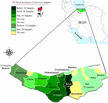
Eighty-eight samples of C. tenuicollis were collected from infected sheep (n = 44) and goats (n = 44) of industrial and traditional slaughterhouses during 2015–2016 (Fig. 1). All samples were divided into two groups according to a slaughtered animal (indigenous and imported). The collected cyst samples from animals were transferred to the parasitology lab of Mazandaran University of Medical Sciences, under cold chain condition. The morphological characteristics of C. tenuicollis scolex were measured by the Nikon light microscope (using eyepiece micrometre) and preserved in 70% ethyl alcohol at −70°C before DNA extraction (Radfar et al., Reference Radfar, Tajalli and Jalalzadeh2005; Ebrahimi Behrestaghi et al., Reference Ebrahimi Behrestaghi, Sarvi, Alizadeh, ZiaeiHezarjaribi and Gholami2018).
Map of Iran presenting study counties in bordering the Caspian Sea, northern Iran.

Hook morphometric study
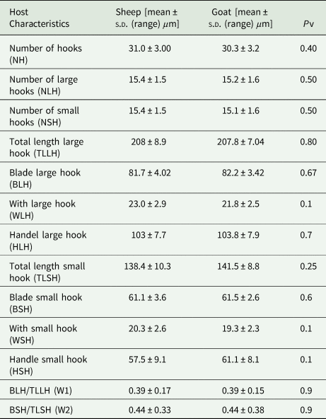
Larval rostellar hooks were mounted on slides in lactophenol mounting medium, and sufficient pressure was applied to protoscoleces under the cover slip to flattened them but not damage the hooks (Gomez-Puerta et al., Reference Gomez-Puerta, Pacheco, Gonzales-Viera, Lopez-Urbina, Gonzalez and Peru2015). The hook parameters were measured according to Hobbs et al. (Reference Hobbs, Lymbery and Thompson1990). Measurements of the total length of large and small hooks, as well as the blade length of large and small hooks, were performed using a calibrated eyepiece micrometre under oil immersion (Table 1). Morphometric data were collected from five large hooks and five small hooks per rostellum from sheep and goat isolates and their size characters (mean ± standard deviation) were determined by a calibrated microscope.
The average morphometric criteria in T. hydatigena larval stage isolated from sheep and goat in the present study

Genomic DNA extraction and polymerase chain reaction
The DNAs of all specimens were extracted using a DENAZIST genomic DNA isolation kit (Catalog number; S-1033-1) based on the manufacturer's instructions. For this purpose, all samples were grinded and lysed by a freeze–thaw method for six times. The DNA concentration of each extraction was measured using a Nanodrop (Thermo Scientific Inc., Wilmington, DE) and stored at −20 °C. The extracted DNA was amplified using single-round polymerase chain reaction (PCR) by targeting the Cox1 (JB3/JB4.5) and 12S rRNA (12SRF/12SRR) markers. Two primers, JB3 (forward): 5′-TTTTTTGGGCATCCTGAGGTTTAT-3′ and JB4.5 (reverse): 5′-TAAAGAAAGAACATAATGAAAATG-3′, were used to amplify a part of the Cox1 gene (Bowles et al., Reference Bowles, Blair and McManus1992, fragment size: 444 bp) under the following conditions: 5 min at 94 °C as an initial hot start step, followed by 35 cycles of 30 s at 94 °C, 45 s at 51 °C, 30 s at 72 °C and a final extension step of 10 min at 72 °C. Two primers, 12SRF (forward): 5′-AGGGGATAGGACACAGTGCCAGC-3′ and 12SRR (reverse): 5′-CGGTGTGTACATGAGCTAAAC-3′ were used to amplify the 12S rRNA gene (Rostami et al., Reference Rostami, Salavati, Beech, Babaei, Sharbatkhori, Baneshi, Hajialilo, Shad and Harandi2015, fragment size: 500 bp) under the following conditions: 5 min at 94 °C as an initial hot start step, followed by 35 cycles of 30 s at 94 °C, 45 s at 59 °C, 30 s at 72 °C and a final extension step of 10 min at 72 °C.
2.4. DNA sequencing, diversity indices, pairwise sequence distances and multiple alignments
Amplicons of C. tenuicollis isolates were sequenced (Bioneer. Company, Korea) by targeting Cox1 and 12S rRNA using the JB3/JB4.5 and 12SRF/12SRR primers. The ambiguity sites of sequences were edited according to the reference sequence (RefSeq) using the Sequencher Tm v.4.1.4 software. According to the analysis of molecular variance, the diversity indices (nucleotide diversity (π) and haplotype diversity (H d), and neutrality indices (Fu's F s statistic and Tajima's D)) were calculated using DnaSP software version 5.10 (Rozas et al., Reference Rozas, Sánchez-DelBarrio, Messeguer and Rozas2003). To compare the genetic structure of C. tenuicollis obtained from this study with southeastern populations of Iran, 12S rRNA sequences were directly retrieved from the GenBank database for FASTA format. Fixation indices (F-statistics: F st, a scale of gene migration and number of migrants per generation (N m)) were used to estimate a genetic differentiation of C. tenuicollis metapopulations (regional population) between northern and southeastern isolates of Iran. The pairwise distances (percent identity (%) and intra-species diversity) of C. tenuicollis, among geographical sequences of the 12S rRNA were built using the DNASTAR's MegAlign program. To identify the probable haplotypes, multiple alignments were performed based on the ClustalW method (BioEdit software, version 7.0.5).
Phylogenetic analysis and haplotype network
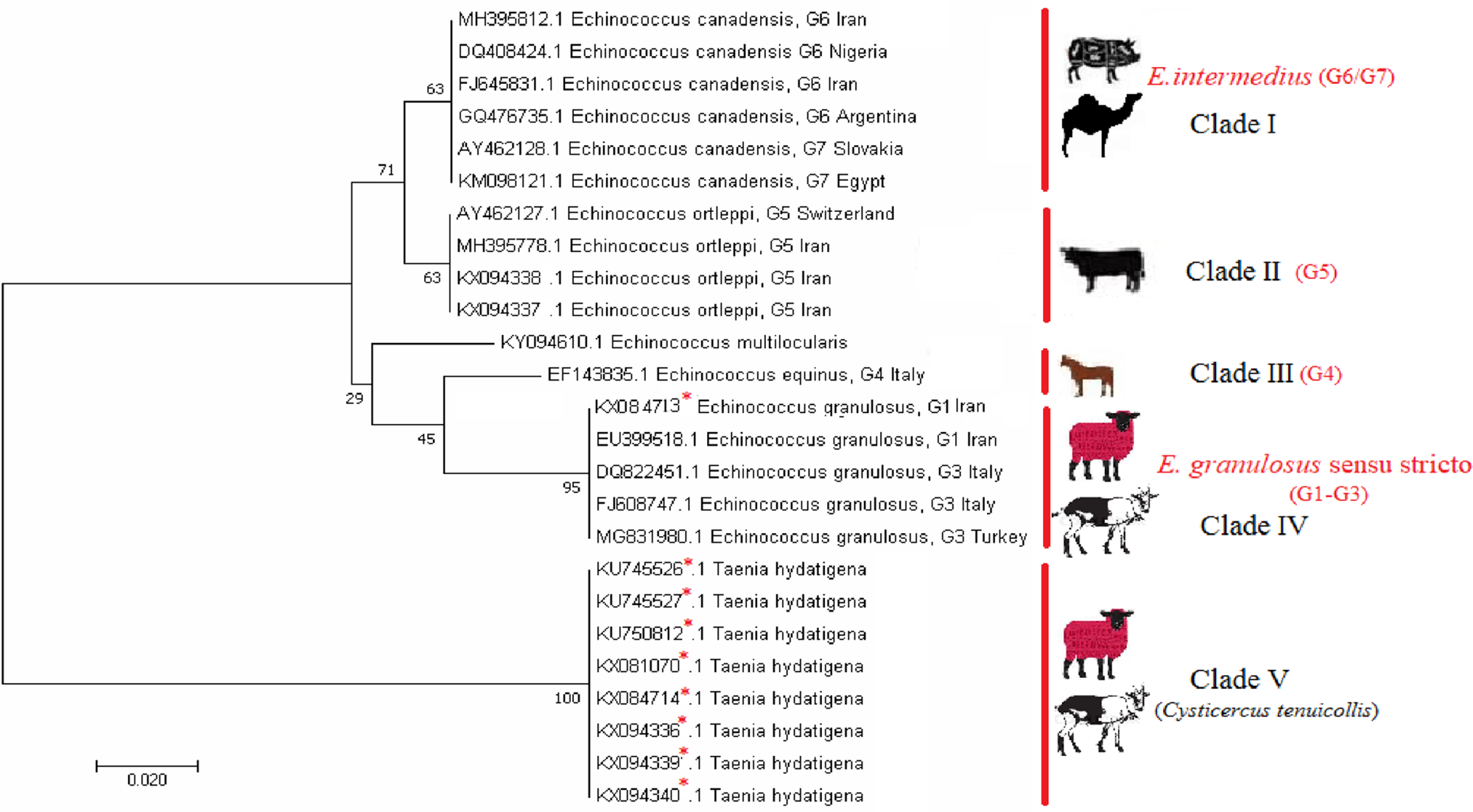
To authenticate genetic associations among identified genotypes of family Taeniidae (C. tenuicollis and Echinococcus spp.), a phylogenetic tree was generated by MEGA 5.05 software based on Maximum Likelihood algorithm and Kimura 2-parameter model. The distance scale was estimated 0.020. The T. hydatigena larval stage was considered as an out-group. Bootstrap values of higher than 60% were considered supportive of branching.
To demonstrate the genealogical relationships at intra-genetic diversity of C. tenuicollis isolates, a haplotype network was constructed by PopART software using the Median Joining algorithm (Bandelt et al., Reference Bandelt, Forster and Röhl1999). One-way ANOVA was used to determine the statistical significance of differences among the rostellar hook length in different haplotypes.
Results
Morphological findings
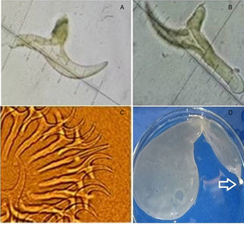
Eighty-eight cyst samples of C. tenuicollis were collected from sheep and goat (n = 44 for each) and subjected to morphometric characterization of larval rostellar hooks and PCR assay inferred by 12S rRNA and Cox1 genes. The biometric characteristics of C. tenuicollis based on larval rostellar hook size are shown in Table 1. The total numbers of large and small hooks of the C. tenuicollis for sheep and goat isolates were 31.0 ± 3.00 and 30.3 ± 3.2, respectively. The mean total length of the large and small hooks size was 208 ± 8.9 μm, 138.4 ± 10.3 µm in sheep samples and 207.8 ± 7.04 µm, 141.5 ± 8.8 µm in goat samples, respectively (Table 1). In all the samples, the arrangement of hooks was alternate in large and small hooks in two rows and the shape of the hooks was smooth in their outline (Fig. 2). No significant differences were found between the size and shape of large and small hooks in C. tenuicollis isolated from sheep and goat samples (P > 0.05), however a significant difference was found in the mean handle width of large hooks in the native and nonnative isolates (P < 0.05). In addition, an intra-species variation was found within the Iranian T. hydatigena isolates based on the total length of large and small hook size.
Small hook (A), large hook (B) rostellar hooks of T. hydatigena larval stage (C), a C. tenuicollis cyst. Arrow: appearance of the invaginated scolex (D).

PCR findings, multiple sequence alignments, diversity indices and pairwise sequence distance matrix
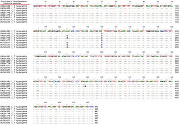
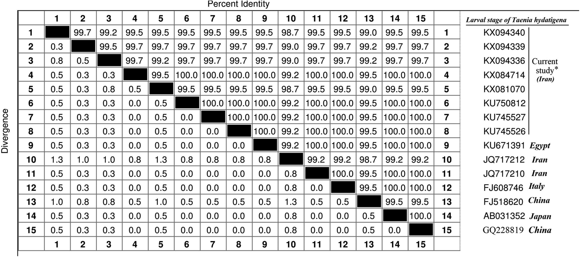
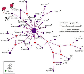
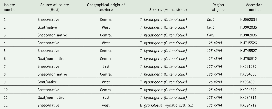
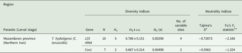
The fragments of 444 and 500 bp were successfully amplified by targeting Cox1 and 12S rRNA genes, respectively. Based on the sequence analysis of Cox1 and 12S rRNA genes, C. tenuicollis (accession numbers listed in Table 2) and Echinococcus granulosus (G1 genotype; accession number; KX084713) were explicitly identified (Table 2). The diversity and neutrality indices of C. tenuicollis isolates are shown in Table 3. Heterogeneity analysis of C. tenuicollis 12S rRNA sequences indicated a genetic diversity (H d: 0.786) including five haplotypes (Table 3), however the nucleotide differences in the 12S rRNA and Cox1 genes were 0.00290 and 0.00498, respectively. Tajima's D (−0.72673) and Fu's F s (−2.169) indices of C. tenuicollis isolates demonstrated negative values, indicating a considerable divergence from neutrality. The multiple nucleotide alignments of C. tenuicollis 12S rRNA sequences indicated the occurrence of synonymous substitutions (transition and/or transversion mutations) at the positions 125, 150, 304 and 338 (Fig. 3). However, no nonsynonymous substitutions were identified in the coding region of 12S rRNA sequences. Pairwise differences amongst the sequences of C. tenuicollis showed an intra-species diversity of 0.3–0.5% and an identity of 99.5–100% (Fig. 4). The genealogical relationship for the identified haplotypes of C. tenuicollis (northern Iran, marked by red asterisk*) and southeastern haplotypes (marked by black asterisk*) were constructed based on 12S rRNA sequences (Fig. 5).
The multiple nucleotide alignments of the T. hydatigena 12S rRNA sequences based on identified isolates. The transition or transversion mutations occurred at the positions 125, 150, 304 and 338.

The pairwise sequence distances between identified isolates of T. hydatigena 12S rRNA with previously reported sequences.

Median joining haplotype network of T. hydatigena 12S rRNA sequences obtained from Iran. Identified haplotypes in the present study (Northern isolates) marked by red asterisk (*), while, haplotypes of southeastern Iran marked by black asterisk (*). Violet circles are relative to the frequency of each haplotype. Red circles represent the hypothetical haplotypes. Each line between haplotypes indicates single mutational step.

Taenia hydatigena larval stage (C. tenuicollis) and E. granulosus G1 genotype from sheep and goat in Mazandaran province, northern Iran

Diversity and neutrality indices of T. hydatigena larval stage based on 12S rRNA and Cox1 genes. N: number of isolates; H n: number of haplotype; H d: haplotype (gene) diversity; N d: nucleotide diversity

*Not significant, P > 0.10.
**P < 0.02.
ND: Not determined
Haplotype network and phylogenetic tree
The haplotype network exhibited star-like characteristics in metapopulation of C. tenuicollis including Th1* as the most common haplogroup (Fig. 5). These findings indicated the sharing of the identical haplotypes of C. tenuicollis between the northern and southeastern parts of Iran. On the one hand, the results of F st (0.05421) and N m (4.36) demonstrated that C. tenuicollis haplotypes originating from the Iranian livestock experienced a relative genetic differentiation between two studied distinct populations (northern haplotypes Th2*–Th4* and southeastern haplotypes Th5*–Th9*) (Fig. 5). To authenticate the taxonomic status of sequenced isolates, a maximum likelihood phylogenetic tree was constructed inferred from 12S rRNA gene. The topology of identified taeniid cysts showed that the E. granulosus (G1 genotype, accession number; KX084713, clade IV) and T. hydatigena larval stage (clade V) were in their specific clades (Fig. 6).
Phylogenetic analysis of E. granulosus G1 genotype and T. hydatigena 12S rRNA sequences based on maximum likelihood algorithm with the kimura 2-parameter model. Taenia hydatigena was addressed as out-group branch. The identified taeniid species marked by red asterisk (*) in this study.

Discussion
Due to the increasing prevalence of infections caused by T. hydatigena and E. granulosus sensu lato in animals (livestock), their genetic data sets and also effective anti-parasitic drugs in endemic regions of the Middle East have widely considered (Mirbadie et al., Reference Mirbadie, Nasab, Mohaghegh, Norouzi, Mirzaii and Spotin2019). Limited studies are available on the intra-species diversity of T. hydatigena larval stage based on the morphological, biochemical and molecular methods in different regions of the world and Iran, as well (Abidi et al., Reference Abidi, Nizami, Khan, Ahmad and Irshadullah1989; Boufana et al., Reference Boufana, Stidworthy, Bell, Chantrey, Masters, Unwin, Wood, Lawrence, Potter and McGarry2012, Reference Boufana, Scala, Lahmar, Pointing, Craig, Dessì, Zidda, Pipia and Varcasia2015; Rostami et al., Reference Rostami, Salavati, Beech, Babaei, Sharbatkhori, Baneshi, Hajialilo, Shad and Harandi2015; Mirbadie et al., Reference Mirbadie, Nasab, Mohaghegh, Norouzi, Mirzaii and Spotin2019).
In this study, the moderate haplotype diversity, low nucleotide diversities and the negative neutrality indices were found for 12S rRNA sequences for C. tenuicollis from sheep and goat samples of Iran, which is consistent with the findings of studies on E. granulosus worldwide (Nakao et al., Reference Nakao, Li, Han, Ma, Xiao, Qiu, Wang, Yanagida, Mamuti, Wen, Moro, Giraudoux, Craig and Ito2010; Casulli et al., Reference Casulli, Interisano, Sreter, Chitimia, Kirkova, La Rosa and Pozio2012; Boufana et al., Reference Boufana, Lahmar, Rebai, Ben Safta, Jebabli, Ammar, Kachti, Aouadi and Craig2014, Reference Boufana, Scala, Lahmar, Pointing, Craig, Dessì, Zidda, Pipia and Varcasia2015).
The obtained mean of the large and small hook lengths was similar to the hook lengths reported by other researchers (Verster, Reference Verster1969; Loos-Frank, Reference Loos-Frank2000; Rostami et al., Reference Rostami, Salavati, Beech, Babaei, Sharbatkhori, Baneshi, Hajialilo, Shad and Harandi2015). On the other hand, 12S rRNA haplotypes showed significant differences in the hook length. It has been shown that the 12S rRNA gene, in particular, is variable in Taenia spp. (Jia et al., Reference Jia, Yan, Guo, Zhu, Wang, Shi, Chen, Zhan, Zhang, Fu, Littlewood and Cai2010) and has to be more polymorphic nature in T. hydatigena than Cox1 (Dai et al., Reference Dai, Liu, Song, Lin, Yuan, Li, Huang, Liu and Zhu2012; Rostami et al., Reference Rostami, Salavati, Beech, Babaei, Sharbatkhori, Baneshi, Hajialilo, Shad and Harandi2015).
Biometric findings indicated that there was no significant difference in the size and shape of the large and small hooks in C. tenuicollis collected from sheep and goat samples of the northern Iran (P > 0.05), however the morphological characteristics of C. tenuicollis from sheep and goat samples of the southeastern Iran (Kerman) and India were significantly different in cysticerci (P < 0.05) and it was concluded that these cysticerci are possibly related to two different strains and probably follow the same pattern of speciation as reported for E. granulosus (Radfar et al., Reference Radfar, Tajalli and Jalalzadeh2005; Singh et al., Reference Singh, Sharma, Gill and Sharma2015).
The present study indicated the relatively high-genetic diversity (H d: 0.667–0.786) and nucleotide divergence of 0.3–0.5% (Nd: 0.00290–0.00498) within the 12S rRNA/Cox1 C. tenuicollis sequences collected from sheep and goat isolates, however morphometric findings and topology (branch lengths) of the maximum likelihood phylogenetic tree did not confirm this variability within the isolates (Fig. 6). In terms of sheep intermediate hosts, a significant haplotype diversity of Cox1 C. tenuicollis sequences has previously been recorded from Iran (H d; 0.997) followed by Sardinia, Italy (H d; 0.806) and Palestine (H d; 0.647) (Boufana et al., Reference Boufana, Scala, Lahmar, Pointing, Craig, Dessì, Zidda, Pipia and Varcasia2015; Rostami et al., Reference Rostami, Salavati, Beech, Babaei, Sharbatkhori, Baneshi, Hajialilo, Shad and Harandi2015). Rostami et al. (Reference Rostami, Salavati, Beech, Babaei, Sharbatkhori, Baneshi, Hajialilo, Shad and Harandi2015) reported a 0.3–3.4% pairwise nucleotide variation between individual Cox1 haplotypes. In addition, the intra-specific variation of T. hydatigena isolates from Ukraine, Wales and Poland was 0.4–5.5% based on the NADH dehydrogenase subunit 1 (Nad 1) gene (Kedra et al., Reference Kedra, Tkach, Swiderski and Pawłowski2001). Such observed discrepancy in haplotype diversities can be related to the farming practices, parasite prevalence, host mobility, transmission rate and even semi-conservative nature of the 12S rRNA gene compared to Cox1 and Nad 1 markers.
Using the sequence data from 12S rRNA we found a moderate genetic difference (F st; 0.05421) in C. tenucollis from livestock isolates collected from the northern and southeastern regions of Iran. This would suggest the sharing of a limited number of haplotypes of C. tenuicollis from one population to another one and also through the natural migration of host mammals. Moreover, it probably seems that the lack of bottleneck effects in T. hydatigena populations and the long-term geographic segregation into the mentioned regions are reasonable heterogeneity hypotheses.
The negative Tajima's D and Fu's neutrality indices (−2.169 to −0.72673) to support T. hydatigena populations indicated the excess of low-frequency haplotypes compared with the expectations in neutral developments, such as purifying selection, equilibrium population size and population expansion following the bottleneck event (Spotin et al., Reference Spotin, Mahami-Oskouei, Harandi, Baratchian, Bordbar, Ahmadpour and Ebrahimi2017, Reference Spotin, Boufana, Ahmadpour, Casulli, Mahami-Oskouei, Rouhani, Javadi-Mamaghani, Shahrivar and Khoshakhlagh2018; Mirbadie et al., Reference Mirbadie, Nasab, Mohaghegh, Norouzi, Mirzaii and Spotin2019).
Since T. hydatigena is a cosmopolitan tapeworm from taeniid species found in animals (livestock and canids), this cestode has been distributed throughout the world through animal transportation over a long period of time (Rostami et al., Reference Rostami, Salavati, Beech, Babaei, Sharbatkhori, Baneshi, Hajialilo, Shad and Harandi2015). Therefore, pairwise comparison of nucleotide variation between individual 12S rRNA haplotypes of T. hydatigena showed a close identity (99.5–100%) with countries, such as China, Italy, Egypt and Japan (Fig. 4). However, more extensive samplings from other regions of Iran are needed to confirm this evolutionary assumption.
In conclusion, the parsimony network analysis showed a common haplogroup (Th1*) for C. tenuicollis isolates. We concluded that the genetic variants of T. hydatigena larval stage are undoubtedly distributing mostly between the northern and southeastern regions of Iran. Further studies with a larger number of T. hydatigena isolates collected from various intermediate and definitive hosts are needed to study this evolutionary assumption and also to determine the apparent genetic differences observed in the studied regions. The complete mitochondrial genomes, such as NADH dehydrogenase subunit 2, ND3, ND5 and ND6 may be helpful in scrutinizing the population genetics and molecular ecology of T. hydatigena isolates in the region.
Acknowledgments
We thank Dr Hamidi Kish and Mohammad Ahmadzadeh who have been involved in the process of doing this research.
Financial support
This work was as thesis of Ms. Laya Ebrahimi Behrestaghi supported financially by the Deputy of research and technology.
Conflict of interests
None.
Ethical standards
The research project was approved by the Toxoplasmosis Research Center of Mazandaran University of Medical Sciences at number 1080 in 2014.