Introduction
The microbial burden of numerous closed systems such as the International Space Station (ISS) (Castro et al. Reference Castro, Thrasher, Healy, Ott and Pierson2004), submarines (Boyden Reference Boyden1962; Morris Reference Morris1972; Upsher et al. Reference Upsher, Fletcher and Upsher1994; Thomas et al. Reference Thomas, Hooper, Camarca, Murray, Sack, Mole, Spiro, Horn and Garland2000), environmental chambers (Levine & Cobet Reference Levine and Cobet1970; Pierson et al. Reference Pierson, Ott and Groves2002), and office buildings (Burge et al. Reference Burge, Pierson, Groves, Strawn and Mishra2000; Ferguson et al. Reference Ferguson, Taylor and Mieszkuc1975; Stenberg et al. Reference Stenberg, Eriksson, Hansson, Höög, Sandström, Sundell and Wall1993) has previously been evaluated using traditional culture-dependent methods. In all of these culture-based studies humans were the predominant sources of bacteria, with other sources arriving with hardware materials (Pierson Reference Pierson2001). Data obtained from Apollo (Ferguson Reference Ferguson, Taylor and Mieszkuc1975), Skylab (Taylor et al. Reference Taylor, Graves, Brockett, Ferguson, Mieszkuc, Johnston and Dietlein1977), the space shuttles (Koenig & Pierson Reference Koenig and Pierson1997; Pierson Reference Pierson2001), and the Russian space station Mir (Kawamura et al. Reference Kawamura, Li, Liu, Huang, Li and Ezaki2001) demonstrated the capability to provide and maintain space environments compatible with human occupation. Conventional microbiological examinations of several space flights demonstrated that microorganisms were ubiquitous throughout the habitable modules of spacecraft (Pierson Reference Pierson2001). However, the ISS presents substantial challenges to limiting microbial contamination and preserving the health and safety of the crews and the integrity of the ISS. Since it is difficult to collect air samples periodically from the ISS in a controlled fashion, the Regenerative Enclosed Life Support Module Simulator (REMS) system was sampled and the microbial burden and diversity were evaluated utilizing both conventional and molecular microbiological techniques.
Regenerative enclosed life-support technologies represent the most economical and effective systems for the long-term operation of orbiting spacecraft and are the only option for interplanetary craft (Samsonov et al. Reference Samsonov, Bobe, Gavrilov, Novikov, Farafonov, Grigoriev, Zaitsev, Romanov, Grogoriev and Sinjak2000). Designed to revitalize and reclaim (i.e. regenerate) important constituents from the internal atmosphere and water supplies, these systems must function without input from external sources and are therefore the basis for sustainable and habitable space facilities. Although the current technologies comprising the environmental control and life support systems (ECLSS) aboard the ISS represent the most advanced systems used to date in any space flight mission, upgrades of the oxygen regeneration and water reclamation systems are to be implemented in 2008 (Carasquillo Reference Carasquillo2005).
The REMS was constructed to simulate the conditions aboard the ISS and to create an environment for testing technologies for future ISS implementation. A water processor assembly (WPA) designed to reclaim and treat waste water aboard the ISS was evaluated from September 2004 to March 2005 (operational phase) using humidity condensate and simulated waste water collected from REMS (Carter et al. Reference Carter, Tabb, Tatara and Mason2005). For optimal testing of the WPA, REMS was designed and constructed copying the ISS, including structural design, materials and overall conditions. Crew activity was simulated using volunteers who exercised, performed prescribed hygienic activities (e.g. face washing and brushing their teeth using carefully measured quantities of water) and prepared meals (Carter et al. Reference Carter, Tabb, Tatara and Mason2005). The REMS facility as a whole was maintained as a class 100K clean room (cumulative distribution of particles greater than 0.5 μm in one cubic foot of air) during its operational phase through stringent quality controls including controlled air circulation and filtration.
The results of the molecular microbial diversity study of the REMS atmosphere can be used to establish the criteria necessary for providing an environment promoting the health, safety and productivity of the ISS crew members. Lessons learned from previous studies (ISS, space shuttles, Skylab, Mir, etc.) were implemented into the ISS to ensure an environment capable of supporting human habitation for multiple years (Pierson Reference Pierson2001). For example, High Efficiency Particle Air (HEPA) filters were incorporated into the air-handling system of the ISS to reduce the levels of airborne bacteria, fungi, and particulates (Pierson Reference Pierson2001) and such measures capitulated levels of airborne bacteria consistently below the 10×104 colony forming unit (CFU) m−3 acceptability limit (Pierson Reference Pierson2001). Recently a 16S rDNA-based methodology was employed to identify strains isolated from the ISS (Castro et al. Reference Castro, Thrasher, Healy, Ott and Pierson2004). This allowed the identification of bacteria to the species level, and was a significant improvement over conventional biochemical analyses (Drancourt et al. Reference Drancourt, Bollet, Carlioz, Martelin, Gayral and Raoult2000; Tang et al. Reference Tang, Ellis, Hopkins, Smith, Dodge and Persing1998, Reference Suzuki, Taylor and DeLong2000). Likewise, state-of-the-art molecular microbiological techniques were recently employed to elucidate the microbial diversity of ISS drinking water (La Duc et al. Reference La, Nicholson, Kern and Venkateswaran2003; Newcombe et al. Reference Newcombe, Schuerger, Benardini, Dickinson, Tanner and Venkateswaran2005) and an integrated thermal coolant system (Benardini et al. Reference Benardini, Ballinger, Crawford, Roman, Sumner and Venkateswaran2005) intended for the ISS. Although some studies have been done on the molecular microbial communities within air samples collected from clean rooms (Venkateswaran et al. Reference Venkateswaran, La, Newcombe, Kempf, Koke, Smoot, Smoot and Stahl2004) and hospital surgical rooms (Edmiston et al. Reference Edmiston, Seabrook, Cambria, Brown, Lewis, Sommers, Krepel, Wilson, Sinski and Towne2005), molecular methods have not previously been used to examine the microbial community structure of the air inside the ISS-like closed systems.
The REMS provided an extreme, unique, encapsulated environment for humans and their associated microbes. Using molecular methods to examine the atmospheric microbial diversity of this facility under controlled (operational phase) and uncontrolled (post-operational phase) conditions allowed a first insight into microbial diversity irrespective of culture-based biases. Although we have not tested under microgravity, the results of this study can be projected onto the conditions aboard the ISS and other closed systems (e.g. submersibles, hospital environments, and the semi-conductor and pharmaceutical industries) leading ‘cleaner’ and potentially safer conditions.
Materials and methods
REMS facility
The REMS was built to simulate the conditions within the ISS and primarily to test the performance qualification of the WPA. This facility was operated and maintained at the Marshal Space Flight Center in Huntsville, Alabama. The REMS was constructed to be analogous to the ISS with respect to structural design, materials used and overall conditions, including the crew’s activity. During the qualification test of the WPA hardware, the REMS was a closed, controlled environment used for the generation and collection of humidity condensate from humans and human activities, such as respiration (breathing), perspiration (while exercising), hand washing, shaving, oral hygiene and food preparation. During the operational phase, the air within REMS was filtered using HEPA filters to remove airborne particulates. To prevent the contamination of the humidity condensate with chemicals not likely to be found in the ISS, the interior of the REMS is composed primarily of aluminium. As a result of these conditions, the REMS provided a unique, encapsulated environment for humans.
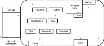
The main chamber (Fig. 1) of the REMS was a 12.8 m long cylinder with a diameter of 4.1 m. An elevated platform positioned 1.24 m from the bottom of the chamber served as the floor of the facility. Humidity in the facility was collected utilizing a condensing heat exchanger similar to that aboard the ISS. During the REMS operational phase relative humidity was controlled at 66.1±6.2% while the facility temperature was maintained at 23.5±0.88°C. In addition, the REMS was maintained as a class 100K clean room during this time (the maximum number of particles larger than 0.5 μm in one cubic foot of air; ISO (1999)). The period of sample collection spanned two months of the operational phase (February and March 2005) and one month of post-operational phase (April 2005).
Schematic drawing of the REMS facility at the Marshall Space Flight Center. The numbers indicate the sampling locations.

Sample collection
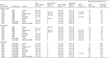
Samples were collected from the REMS during three different sampling trips in three consecutive months using a commercially available liquid impaction BioCapture air sampler. The BioCapture BT-550 (Mesosystems Technology Inc., Kennewick, WA) sampler employed in this study collects particles of size 0.5–10 μm from ambient air. The flow rate of this lightweight (4.5 kg) sampler is 150 l min−1. One sample cycle impinges a 750 l air parcel (roughly equivalent to the volume of air that human lungs exchange every 2 h) into 5 ml of sterile buffered saline over 5 min. During sampling, the BT-550 was centrally positioned to ensure the uniformity of the air parcels collected. The location and date of sampling are given in Table 1 and Fig. 1. Just prior to the sampling, the air sampler was sanitized using the manufacturer-defined protocols and cleaning cartridges. Samples were collected in duplicate using two separate units, side-by-side as per the manufacturer’s instructions. Collection fluids from the air sampler were stored at room temperature until sampling was complete (less than two hours). Samples were collected from the same locations during each sampling trip. After sample collection, duplicates were pooled together and subsequently distributed for various analyses. Samples were stored appropriately (−20°C for DNA extraction samples, 4°C for all others) and shipped overnight to the lab (JPL, Pasadena, CA, USA) for analysis, except for the ATP-based assay that was performed within 1 h of aliquot preparation. Since the original samples were portioned out for numerous assays, no cryoprotectant was used.
Locations and microbiological examination of various air samples collected at REMS

* Selected for clone library analysis.
Culture-dependent assay
The cultivable bacterial burden (cultivable aerobic heterotrophs) was estimated using standard plate count methods. Briefly, 100 μl of each sample was spread on R2A medium (Difco Laboratories, Detroit, MI) and incubated at 25°C. Plates were spread in triplicate and the direct counts averaged to estimate the total heterotrophic bacterial population. CFUs were enumerated after seven days. Estimates of spore-forming bacteria were obtained using the NASA standard assay (NASA 1980). The samples were heat shocked (80°C, 15 min), placed in a sterile petri dish, and mixed with Trypticase Soy Agar (TSA, Difco) using pour-plating techniques. Pour-plated samples were incubated at 32°C and CFUs enumerated at two- and seven-day intervals. When fewer than five colonies arose on a plate, all colonies were picked, whereas when more than 10 colonies were present, 10 colonies were randomly picked for further identification. Colony morphology, colour and shape were considered while selecting the isolates for further characterization. Isolates were picked, purified, cultured and stored at −80°C for further processing and analysis. The identification of purified strains was determined via 16S rDNA sequence analysis (see below) or by the use of specific primers for Bacillus pumilus (600F, TGA AGA TGT GCG AGA AGG CT, and 980R, AGG ATC TTC CCT CTT AAC GG); Satomi (Reference Satomi, La and Venkateswaran2006).
ATP-based bioluminescent assay
Commercially available kits utilizing firefly luciferase to catalyze the reaction of luciferin with ATP were used to quantify the amount of ATP present in each sample. Both total and intracellular ATP levels were measured to gauge the total bioburden and viable cells, respectively. Bioluminescence generated by the reaction was directly proportional to the amount of ATP in the sample. The dynamic range of this assay was from 5×10⁻¹² to 10−7 M ATP, with one relative luminescence unit (RLU) corresponding to 2×10⁻¹⁴ M ATP as determined by linear regression analysis of standard curves with known ATP concentrations (Venkateswaran et al. Reference Venkateswaran, Hattori, La and Kern2003). To determine the total ATP, 0.1 ml of each sample was assayed using a Checklite HS Plus kit (Kikkoman Corp., San Francisco, CA) as previously described by Venkateswaran et al. (Reference Venkateswaran, Hattori, La and Kern2003) and the amount of bioluminescence was measured with a luminometer (Lumitester K-200; Kikkoman Co., Japan). To determine the intracellular ATP, exogenous ATP was enzymatically removed from 0.5 ml of each sample using a reagent provided by the manufacturer after which the assay for ATP was carried out as described above. All samples were measured in quadruplicate and the results averaged for final estimates. Estimates of the bioburden were calculated by multiplying ATP-assay results by a dilution factor to represent RLU per cubic metre of air sampled.
DNA extraction and DNA-based bioburden analysis
In order to reduce the shearing of DNA during this process, and to minimize the loss of DNA product while transferring between several reaction tubes, samples were not concentrated. DNA was extracted from the samples following standard lysozyme/organic solvent extraction protocols (Johnson Reference Johnson, Gerhardt, Murray, Costilaw, Nester, Wood, Krieg and Phillips1981). Briefly, samples were treated with lysozyme (final concentration 10 mg ml−1) to degrade cell walls, followed by Proteinase K and RNAse treatment to remove unwanted bioploymers, and finally by phenol-chloroform extraction to clean-up cellular debris. DNA was precipitated with two volumes of ice-cold ethanol and DNA was re-suspended in a TE buffer (30 μL) before storing at −80°C. Ribosomal RNA gene copy numbers were estimated by quantitative polymerase chain reaction (Q-PCR). All experiments were done in triplicate using universal eubacterial primers targeting the 16S rRNA gene, 1369F (5′-CGGTGAATACGTTCYCGG-3′), 1492R (5′-GGWTACCTTGTTACGACTT-3′) and the fluorescent-labelled probe TM1389F (5′-FAM-CTTGTACACACCGCCCGTC-TAMRA-3′) (Suzuki et al. Reference Suzuki, Taylor and DeLong2000). Each 50 μl PCR reaction consisted of 25 μl of 2X Taqman Universal PCR Master Mix (Applied Biosystems Inc, Foster City, CA), 0.8 μM of each oligonucleotide primer, 0.5 μM of oligonucleotide probe, and 1 μl of template DNA. Reaction conditions were as follows: 95°C denaturation for 15 min, followed by 40 cycles of denaturation at 95°C for 15 s and a combined annealing and extension at 60°C for 1 min 30 s. The Q-PCR reaction was performed in an MJ Research PTC-100 thermal cycler (Bio-Rad Lab., Waltham, MA).
Bacterial diversity and identification
Bacterial 16S rRNA genes were PCR-amplified with the forward primer 27F (5′-GAGTTTGATCMTGGCTCAG-3′) and the reverse primers 1406uR (5′-ACGGGCGGTGTGTRCAA-3′); Lane (Reference Lane, Stackebrandt and Goodfellow1991). The PCR was performed under the following conditions: 95°C for 4 min; 33 cycles of 95°C for 50 s, 55°C for 50 s and 72°C for 1 min 30 s; and a final incubation at 72°C for 10 min. If necessary, amplification products were purified with a gel excision kit (Qiagen, Chatsworth, CA). Selected products were ligated into the pCR4-TOPO® cloning vector (Invitrogen, Carlsbad, CA) and transformed into chemically competent E. coli Top10 cells (Invitrogen, Calsbad, CA) according to manufacturer’s protocols. For each of the samples, at least 100 randomly picked clones were either sequenced directly (Agencourt, Beverly, MA) or subjected to restriction fragment length polymorphism (RFLP) analyses. The presence of inserts of the expected size was analysed by direct PCR screening of 100 transformants without plasmid extractions. Inserts from each clone were amplified as described above with T7 and M13R primers targeted to vector regions flanking the insert. Amplicons were digested with HhaI restriction endonuclease (Promega, Madison, WI) for 3 h at 37°C and analysed on a 2% low melting point agarose gel (Shelton Scientific, Prosta, IA). Clones were grouped according to similarity of RFLP banding patterns and representative purified plasmids (Qiaprep kit, Qiagen, Chatsworth, CA) of each group were bi-directionally sequenced.
Appropriate controls were employed for all analyses. The buffer used to collect air particulates was sterilized by irradiation by the manufacturer and such steps eradicated all microbes as confirmed by an absence of traces of culturable microbes and an absence of intracellular ATP. For the PCR amplification several sterile water-blank, reagent-blank (DNA extraction step and PCR reaction step) and appropriate DNA-free negative controls were employed to avoid any laboratory contamination. None of the controls showed positive signals; however, the unused sterile buffers revealed slight positive 16S rDNA fragment amplification when the more sensitive PCR primer set 27F-1406uR was used. Since most of our air samples showed a very low concentration of DNA, the usage of the primer set 27F-1406uR was necessary to obtain consistent positive amplifications. To overcome this problem, a total of three unused buffers (one per month) were subjected to all of the assays performed in this study. DNA was extracted as carried out for all of the sample libraries and described above. Subsequent cloning followed by sequencing of 100 clones each was carried out. The clone sequences obtained from blank samples were removed from the results and not included in phylogenetic analyses.
Phylogenetic analyses
All sequences were submitted to the CHECK_CHIMERA program of the Ribosomal Database Project (Maidak et al. Reference Maidak2000) to detect possible chimeric artifacts. The phylogenetic relationships of organisms were determined by the comparison of individual 16S sequences to sequences in the GenBank database (http://www.ncbi.nim.nih.gov/). For more extended phylogenetic analyses, an alignment of approximately 20 000 homologous full and partial sequences available in public databases was used with the ARB program (Ludwig et al. Reference Ludwig2004). The new 16S rRNA gene sequences were integrated in the 16S rRNA alignment using the corresponding automated tools of the ARB software package (Ludwig et al. Reference Ludwig2004) and the resulting alignment was checked manually and corrected if necessary. For tree reconstruction, methods were applied as implemented in the ARB software package.
Controls and lower detection limits of assays employed
Appropriate controls were used whenever necessary. Unopened Mesosystems cartridges served as negative buffer controls and were included in every sampling/analysis. To address problems associated with microfluidics, a 0.2 μm sterile Millipore disc filtre was placed at the mouth of the Mesosystem air sampler and the collection fluid assayed with all methodologies utilized in this study. Pure ATP (Sigma, MO) was decimally diluted and served as a standard curve. Purified DNA from B. pumilus ATCC 7061 was included in the PCR amplification protocols as a positive control. In addition, water blanks free of ATP and DNA were used as negative controls in all molecular assays. In order to elucidate whether any PCR inhibitory substances were present in the sample, a known amount of DNA was extracted from B. pumilus and spiked (1 pg per reaction mixture) as an internal standard. None of the DNA extracts in this study inhibited the PCR reaction. The lower (below) detection limits (BDL) of various assay were: 1 CFU for the cultivable plate count assay, 10 RLU for ATP analysis and 100 copies for Q-PCR analysis when molecular grade Sigma water was used as negative control. However, the unused blank cartridge exhibited 8–15 RLU when 100 μl of the buffer was measured for ATP and 1.0×10² rRNA gene copies μl−1 when DNA extracted from the 5 ml buffer and concentrated to 100 μl.
Statistical analyses were performed as per the manufacturers’ instructions, as well as with the MS Excel software package. For example, for the ATP analyses, when the coefficient of variation exceeded 10%, an additional measurement was performed on that sample and standard deviations were calculated from four individual replicates of each sample. Software included with the MJ Research PTC-100 thermal cycler was used to generate standard deviations from three replicates of each Q-PCR amplified sample. The averages of two measurements were calculated for each sample that underwent plate-count analysis.
Nucleotide sequence accession number
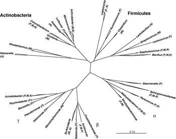
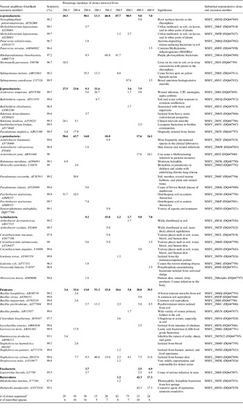
The 16S rRNA gene sequences of isolates and clones were deposited in the NCBI nucleoide sequence database. The accessions numbers are given in Fig. 2 (cultivable isolates) and Table 3 (clone sequences).
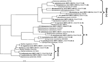
Phylogenetic analysis of cultivable microbial diversity isolated from various locations of the REMS. The phylogram was computed by maximum-parsimony analysis using the ARB program. The 16S rDNA sequence of the nearest-neighbour-type strain was included. The strain number and GeneBank accession numbers were given after the strain name. The letter in parenthesis denotes months during which the strain was isolated (February, F; March, M; April, A).

Results
Cultivable aerobic heterotrophs
The number of heterotrophs capable of growth on R2A varied between sample location and sampling trip (Table 1). In general, cultivable heterotrophic bacterial populations ranged from BDL to 103 CFU m−3 of air during the REMS operational phase (February and March) and were one log higher during the non-operational phase (April). It is worth mentioning that three out of 18 samples collected during the REMS operational phase did not exhibit any cultivable population and approximately 72% of the samples (13 out of 18 samples) showed less than 8.9×10² CFU m−3 of air. Similarly, only one air sample harboured greater than 103 CFU m−3 (the changing room during February). In contrast, samples collected in April had a range of 8.4×10² to 1.0×10⁴ CFU m−3. A lower bioburden was witnessed in locations 8 and 9 (Table 1) where the air circulation was not controlled in all three months compared with the high human activity that occurred in the confined area (locations 1–7). The incidence of cultivable spore-forming microorganisms was significantly lower (seven out of 27 samples), than the cultivable heterotrophs (Table 1). Samples from February and March revealed between BDL to 8.1×10² and to 5.5×10² CFU m−3 of air, respectively. None of the nine samples from April showed any bacterial growth in TSA following heat-shock, although higher numbers of cultivable microbes were documented.
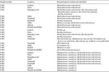
The diversity of cultivable bacteria and their phylogenetic affiliations are given in Fig. 2 and Table 2. Seven of 14 isolates picked from February air samples were affiliated to different species. Likewise, the air samples from March (all 15 isolates) and April (15 of 125 isolates) exhibited 12 and two bacterial species, respectively. Methylobacterium radiotolerans was repeatedly isolated from samples of each month (15 out of 27 samples) and Sphingomonas yanoikuyae was grown from every sample taken in April (Table 2). More than 70% of the bacterial species (12 out of 17 species; see Fig. 2) isolated from REMS were Gram-positives, including five Bacillus strains. Bacillus species isolated from the samples collected during February and March were identified as B. megaterium, B. luciferensis, Bacillus sp. (Fig. 2) by 16S rDNA sequencing and one B. pumilus isolate using species specific primer sets (Satomi et al. Reference Satomi, La and Venkateswaran2006). Among all 44 isolates identified, the majority of them (27 strains comprising three Gram-negative species) belonged to members of the α-proteobacteria. Two novel Gram-positive species were also isolated (less than 97% 16S rDNA sequence similarities to Microbacterium sp. and Microlunatus sp.) and more detailed taxonomical studies will be required to describe them.
Culturable bacterial diversity of the air samples collected from various locations of REMS

Microbial population as estimated by molecule-based assays
One of the culture-independent methods, microscopy, is a powerful tool and is invaluable for qualitatively assessing microbial populations. However, because of the drastically low number of microbes present in these types of environment (La Duc et al. Reference La, Nicholson, Kern and Venkateswaran2003, Reference La, Kern and Venkateswaran2004), and the limitations associated with the microscopy technique (autofluorescence, false-positives, non-specific binding of stain, insufficient penetration resulting in false negatives (Stackebrandt & Embley Reference Stackebrandt, Embley, Colwell and Grimes2000)) means that it is impractical. We did not perform in situ nucleic acid hybridizations since previous attempts have shown that such techniques necessitate greater than 107 cells per reaction (data not shown).
Estimates of the amount of ATP present in collected samples were consistent (with respect to the order of magnitude) concerning the total and intracellular ATP content (Table 1). The ATP content of the buffer blank did not exceed values of any of the 27 air samples collected during this study. The total microbial population as estimated by the total ATP averaged approximately 104 cells m−3 in all of the three months sampled. The levels of total viable microbes as estimated by the intracellular ATP were one to two orders of magnitude lower than estimated total microbes and were as follows: samples collected in February ranged between 2.63×10² and 1.23×10³ RLU m−3 (average of 4.95×10²); March samples had a range of 5.10×10² and 2.82×10³ RLU m−3 (average of 1.81×10³); and estimates from April were between 2.73×10³ and 2.22×10⁴ RLU m−3 (average of 7.53×10³). A comparison of the estimated total and intracellular ATP levels suggests that about 10% of the ATP present in the REMS atmosphere can be attributed to viable organisms (Table 1). Estimates were in the range 0.8–6.0% (average of 3.1%), 1.1–7.8% (average of 4.4%) and 3.1–60.3% (average of 17.1) for February, March and April, respectively. Interestingly, the total viable population average was lower than 5% when the REMS facility air circulation was purified when compared with the April estimates (∼17%) during which the facility was not maintained. The estimate of total microbial population in terms of 16S rRNA copy numbers varied considerably between sampling periods. Quantification estimates similar to the blank in eight of nine of the April samples was noteworthy. Likewise, only one-third of the samples during February consisted of measurable 16S rDNA copies (∼1 to 3×10⁶ copies m−3). In contrast, seven out of nine samples collected during March showed at least 1.5×10⁷ copies m−3.
Cultivable versus viable
To estimate the levels of cultivable cells with respect to viable cells in the air of the REMS, the number of CFU per cubic metre for a given sample location was compared with the suggested number of viable cells based on intracellular ATP levels (Table 1). The percentages obtained for the February and March (Table 1) sampling trips indicated that between 0–87% and 0–24% of viable cells were cultivable, respectively. Values for April suggested that at least 12% of viable cells were able to grow on the R2A plates provided. Likewise, four out of nine samples collected during April exhibited more cultivable organisms than predicted total organisms based on intracellular ATP measurements.
Molecular microbial diversity
Representative samples were chosen from each month for analysis of the 16S rRNA genes present. Care was taken to include four different categories (in terms of microbial community) of samples. Samples that showed less cultivable than viable bacteria (based on intracellular ATP; 2-M4, 3-M2, 3-M5, and 4-M9) fall under category 1. Category 2 samples consist of samples that documented a more or less equal number of cultivable and viable microbes (2-M3 and 4-M1). Category 3 samples showed more cultivable bacteria in R2A plates compared with intracellular ATP (4-M3). Finally, category 4 samples recorded no cultivable microbes but exhibited at least one to two logs higher viable microbes (2-M6 and 3-M1).
In total more than 900 clones were picked (at least 100 transformants per air sample), and 365 clones were further sequenced based on RFLP patterns. Since a phylogenetic study of the 16S rRNA gene sequences is not dependent on the culturing technique used, a much larger microbial diversity was revealed in the REMS samples (see Table 3 and Fig. 3). Six of the nine clone libraries comprised greater than 67% proteobacterial groups (Table 3) whereas the Gram-positive groups such as Firmicutes, Actinobacteria, Fusobacteria and Bacterioidetes constituted the remaining ∼30%. Among proteobacterial groups, α-proteobacterial clones were abundant (greater than 50%) in four and β-proteobacterial clones in two clone libraries. In total, 51 different sequences were retrieved, which also included γ- and β-proteobacteria. These groups were not represented in the isolates obtained utilizing the cultivation approach. Some of the microbes detected by the molecular assay were obtained in parallel as isolates during the culturing (e.g. Sphingomonas yanoikuyae, Bacillus megaterium). The highest diversity of molecularly detected microbes occurred in the February samples (27 different sequences), with 19 and 17 different sequences detected in April and March, respectively. When category-wise samples were analysed no unique pattern was noticed. While numerous examples of the clone sequences retrieved were originally isolated from soil- or plant-associated samples (e.g. Methylobacterium), other clone sequences belonging to human-borne microbes were also detected, some of which are known as opportunistic pathogens (e.g. Acinetobacter, Leptotrichia, Staphylococcus and Streptococcus).
Phylogenetic analysis of bacterial 16S rDNA sequences of various air samples collected from several locations of the REMS. The cladogram was computed by maximum-parsimony analysis using the ARB program. The letter in parenthesis denotes months during which clone sequences were retrieved (February, F; March, M; April, A).

Molecular microbial diversity of various locations and samplings of REMS

1 96 clones were picked per sample and based on RFLP pattern, representative clones were sent for sequencing.
Within the α-proteobacteria, a total of nine 16S rRNA gene sequences were identified. Clone libraries 3-M2 and 3-M5 contained high percentages of sequences that were putatively identified as Rhodopseudomonas rhenobacensis (60% and 82%, respectively). Likewise, 4-M1 was dominated by the sequences of S. yanoikuyae (88%; Table 3). In general, Sphingomonadaceae seemed very prominent in REMS, with sequences detected in samples from all three months, and one representative of this family successfully cultivated. In addition, seven β-proteobacteria (Acidovorax, Burkholderia, Ralstonia, etc.) and 10 γ-proteobacteria sequences were retrieved. Three different 16S rRNA gene sequences from the Acinetobacter genus, which is known to include opportunistic pathogens, were also identified. Members of the Actinobacter genus were detected frequently throughout the three months sampled, but appear only in very low percentages among the clones sequenced. Thirteen firmicute sequences were found, including five Bacillus sequences (B. borophilicus, B. cereus, B. megaterium, B. psychrodurans and B. pumilus). The DNA of Staphylococcus xylosus was retrieved from every site and month except from 2M-3.
Discussion
The ISS was built to allow orbital living and research, and will be constantly manned during operation. In conjunction with human habitation come human-associated microbes, many of which are capable of colonizing locales of this unique environment and have been previously identified as problematic organisms (Novikova Reference Novikova2004; Novikova et al. Reference Novikova, De, Poddubko, Deshevaya, Polikarpov, Rakova, Coninx and Mergeay2006). In addition to growing on the surfaces and within water systems, some colonizing microbes may also affect human health, for example Legionella (Ott Reference Ott1994). Research from the late 1960s indicated that ‘the primary source of microbial contamination within clean rooms is humans, and the level of contamination depends on personnel activity and density’ (Favero et al. Reference Favero, McDade, Robertsen, Hoffman and Edwards1968). The monitoring of overall microbial diversity and the presence of opportunistic pathogens is therefore an important focus to provide optimal and safe working conditions for astronauts, particularly as activity and crew sizes increase. As direct sampling of the ISS is difficult, it is advantageous to identify potential contaminants prior to the implementation of new ‘house-keeping’ technologies. The REMS was designed to be analogous to the ISS with respect to spatial dimensions, internal operations, and conditions. The facility therefore provided a unique opportunity to study the microbial diversity of a habitat utilizing an ECLSS, namely the protoflight WPA, and to evaluate the effectiveness of cleaning procedures and environmental maintenance utilized aboard the ISS. The effects of microgravity on the microbial community structure were not accounted for during this investigation.
The study of natural microbial communities using solely culture-based methods has been shown to be limited by the selective nature of the enrichment media used (Ward et al. Reference Ward, Weller and Bateson1990a; Amann et al. Reference Amann, Ludwig and Schleifer1995). As a result, only a minor portion of the microbes present in an environment can be readily cultured (Torsvik & Ovreas Reference Torsvik and Ovreas2002). To encompass the broadest microbial diversity present in REMS air samples, it was therefore necessary to combine a cultivation approach with cultivation-independent methods. Molecular 16S rDNA-based techniques have been successfully used to describe the microbial diversity in numerous environments (Britschgi & Giovannoni Reference Britschgi and Giovannoni1991; Liesack & Stackebrandt Reference Liesack and Stackebrandt1992; Ward et al. Reference Ward, Weller and Bateson1990a, Reference Ward, Weller and Batesonb; Wise et al. Reference Wise, McArthur and Shimkets1997) and these methodologies were employed to examine the phylogenetic diversity within the REMS. The results of these phylogenetic analyses were coupled with culture-based methodology and an ATP-based quantification assay to provide the most complete insight to date regarding the microbial diversity and bioburden of an enclosed space habitat system.
The DNA-based molecular community assay revealed a significantly broader biodiversity (51 species, including β- and γ-proteobacteria), than the culture-dependent approach (only Gram-positive and α-proteobacteria were isolated). The prevalence of Gram-positive bacterial sequences was high in three out of nine clone libraries, while Gram-negative sequences comprised a substantial percentage (greater than 67%) of the remaining six. Analyses of DNA-based assays suggested the presence of some opportunistic pathogens, including Acinetobacter, Acidovorax, Staphylococcus, Streptococcus, and Leptotrichia. While the presence of viable Staphylococcus sp. was demonstrated by culture-based assays, the viability of other putative pathogens could not be verified. However, it has been shown previously that opportunistic pathogens entered into a viable but non-cultivable (VBNC) state when ISS-bound water samples were treated with NASA recommended biocides (Stuecker et al. Reference Stuecker, Newcombe, Murdock, Sumner and Venkateswaran2005). By partaking in this dormant-like VBNC condition, cells may be able to escape detection via culture-dependent methods and should not be ignored (Oliver Reference Oliver2005; Oliver & Bockian Reference Oliver and Bockian1995; Oliver et al. Reference Oliver, Dagher and Linden2005).
While 16S rRNA genes of Staphylococcus species were found in eight of nine samples, and the highest clone percentage was detected in the March samples. Previous studies have also suggested the predominance of Staphylococcus species (13 of the 36 colony types isolated) in the ISS air habitat (Novikova Reference Novikova2004; Novikova et al. Reference Novikova, De, Poddubko, Deshevaya, Polikarpov, Rakova, Coninx and Mergeay2006), space shuttle (Pierson Reference Pierson2001), Mir Space Station (Kawamura et al. Reference Kawamura, Li, Liu, Huang, Li and Ezaki2001) and closed environments on Earth (Pierson et al. Reference Pierson, Ott and Groves2002). Given that all of the staphylococci isolated during flight were S. aureus and S. epidermidis, the contamination was thought be the result of a clean system ‘infected’ by human occupation. In addition, molecular microbial studies from controlled clean rooms, surgical operating rooms of hospitals and indoor environments reported the predominance of staphylococci (Edmiston et al. Reference Edmiston, Seabrook, Cambria, Brown, Lewis, Sommers, Krepel, Wilson, Sinski and Towne2005; Novikova et al. Reference Novikova, De, Poddubko, Deshevaya, Polikarpov, Rakova, Coninx and Mergeay2006). The presence of opportunistic pathogens other than Staphylococcus in the ISS has also been shown (Novikova Reference Novikova2004) and, coupled with previous reports suggesting the suppression of crew immune systems during flight (Mehta et al. Reference Mehta, Stowe, Feiveson, Tyring and Pierson2000, Reference Mehta, Cohrs, Forghani, Zerbe, Gilden and Pierson2004), this finding remains concerning.
A substantial portion of the microbes was judged to be viable but not cultivable (VBNC) when REMS samples were measured for viable microbial populations using quantitative ATP analyses. A comparison of the two operational phase months indicated lower ATP amounts (both total and intracellular) during the February sampling. It is well known that ATP content is directly proportional to the size of the microbe (Karl Reference Karl1980; Venkateswaran et al. Reference Venkateswaran, Hattori, La and Kern2003). However, the microbial diversity of the REMS air samples suggests the eukaryotic portions of these communities to be negligible (failed 18S-biased PCR, culture plates devoid of fungal colonies), and suggests that these samples contained primarily smaller prokaryotes (1–5 RLU per 1 CFU). It can also be presumed that in an environment such as REMS, microbial cells might exist in a less active metabolic state, and therefore microbial cells might contain less ATP when compared with actively growing cells (Karl Reference Karl1980). Hence, oligotrophic microbes such as M. radiotolerans and S. yanoikuyae might not be active in the nutrient-limited conditions. The higher observed percentage of cultivable microbes versus ATP during the April sampling period may be the result of the lower ratio of ATP per CFU of the bacteria present in these samples. The two strains predominately isolated in the April samples, M. radiotolerans and S. yanoikuyae, possessed a low ratio of ATP to CFU (1 RLU per 10 CFU) compared with copiotrophic E. coli or Pseudomonas sp. (1 RLU per 1 CFU); Venkateswaran et al. (Reference Venkateswaran, Hattori, La and Kern2003). Overall, the low cultivable microbial diversity of the April samples is in striking contrast to the CFU and ATP data, which indicated significantly higher biomasses. The absence of human activity during the month of April might be the reason for the lower biomass, but the presence of indigenous oligotrophic microbes that can be cultured could not be ruled out in this closed habitat.
Culture-based assays of ISS air samples (Novikova et al. Reference Novikova, De, Poddubko, Deshevaya, Polikarpov, Rakova, Coninx and Mergeay2006) documented up to 7.1×10² CFU m−3. The average CFU per cubic metre in the REMS air was less than 1.5×10² CFU m−3 during the operational phase (February and March 2005). Among 18 samples measured for the cultivable microbes during this study, only 27% (five samples) exhibited a cultivable microbial density of greater than 102 CFU m−3 during this phase. Nevertheless, the amount of CFU was significantly higher during post-operation (up to 104 m−3). It appears that the stringent protocols implemented by NASA within the REMS were effectively decreasing the quantity of airborne microbes, and are therefore well suited to reduce the total microbial contamination of enclosed habitats.
S. yanoikuyae and M. radiotolerans were the two main isolates obtained during this study. Members of these genera were also described as dominant cultivable contaminants in potable water of the ISS (Novikova Reference Novikova2004; Novikova et al. Reference Novikova, De, Poddubko, Deshevaya, Polikarpov, Rakova, Coninx and Mergeay2006) while other studies reported the occurrence of the aforementioned organisms within the ISS drinking water system (Castro et al. Reference Castro, Thrasher, Healy, Ott and Pierson2004). Furthermore, S. yanoikuyae, the most prominent cultivable organism from the April samples, was also detected in some clone libraries in a high percentage (∼88%, sample 4M-1) and Sphingomonas species are known components of biofilms in drinking water distribution systems (Koskinen et al. Reference Koskinen, Ali-Vehmas, Kampfer, Laurikkala, Tsitko, Kostyal, Atroshi and Salkinoja-Salonen2000). In addition, as member of this genera have been isolated from clinical specimens, these microbes represent putative opportunistic pathogens (Gilchrist et al. Reference Gilchrist, Kraft, Hammond, Connelly and Myers1986). Although not considered pathogenic, numerous Methylobacterium species, typified as pink-pigmented bacteria, have been found in oligotrophic freshwater environments such as the surface water of reservoirs and drinking tank water and have been shown to exhibit high levels of chlorine resistance (Hiraishi et al. Reference Hiraishi, Furuhata, Matsumoto, Koike, Fukuyama and Tabuchi1995). Elevated levels of resistance to typical chlorine-based cleaning agents may have played a roll in this organism’s ability to persist in the REMS environment and warrants further investigation.
Culture-dependent analyses indicated a significantly lower bacterial diversity in the samples collected in April. Interestingly, a significantly higher total heterotrophic count was observed in addition to higher intracellular ATP estimates (viable microbes). This suggests that only a few bacterial species (namely Sphingomonas and Methylobacterium) could adapt in an enclosed facility when the clean room was not inhabited by humans, irrespective of its maintenance. To date little work has been carried out on microbial communities subsisting in regions of nutrient scarcity although it is well accepted that microbes have interesting strategies to persist under severe environmental conditions; for example, spore-formers are able to form spores in desiccated controlled environments or at elevated temperatures (La Duc et al. Reference La, Nicholson, Kern and Venkateswaran2003, Reference La, Kern and Venkateswaran2004; Venkateswaran et al. Reference Venkateswaran, Satomi, Chung, Kern, Koukol, Basic and White2001). In this study, spore-forming microbes were isolated from the REMS air during the operation phase (February and March), whereas no spore formers were isolated from the post-operation phase. It is conceivable that any spore formers present germinated under the moist uncontrolled conditions and the vegetative cells were killed by the heat shock protocols employed in this assay. Similarly, the air samples collected from a clean-room facility during the assembly of the Mars Exploration Rovers revealed no spore-formers whereas spores were repeatedly isolated from the metallic spacecraft surfaces (Venkateswaran et al. Reference Venkateswaran, La, Newcombe, Kempf, Koke, Smoot, Smoot and Stahl2004). Microbes that can withstand low nutrient levels (oligotrophs) can survive during controlled conditions and subsequently become established when the facility is not maintained. This is a plausible explanation for the low cultivable diversity obtained from the April samples. Similar observations were made from samples of Mars Odyssey spacecraft surfaces that were significantly cleaner than the spacecraft assembly facility floor surfaces where the spacecraft was constructed (La Duc et al. Reference La, Nicholson, Kern and Venkateswaran2003).
Conclusion
The microbial burden during REMS operation phase was demonstrably lower than during the post-operation phase. The regimented maintenance procedures and controlled atmospheric conditions within the REMS (45% humidity, 20°C temperature, HEPA filtration and periodic cleaning) created a challenging environment for microbes and appeared to be an effective means of reducing the microbial burden within the REMS. Utilizing similar procedures and controls in other enclosed systems (e.g. the ISS, hospital and pharmaceutical environments) may be equally effective with respect to controlling microbial contamination. The approach taken in this study of coupling molecular community analyses with culture-based methodology and molecule-based quantification assay(s) was necessary to provide the most complete insight with regards to the microbial diversity and bioburden of an enclosed system.
Acknowledgements
The authors thank D. Newcombe, S. Osman for technical assistance, and D. Jan for continued managerial support. The research described in this publication was carried out at the Jet Propulsion Laboratory, California Institute of Technology, under a contract with the National Aeronautics and Space Administration.