Introduction
Tympanoplasty is the treatment of choice for perforation of the tympanic membrane. The technique has evolved over time. The concept of tympanoplasty is credited to Berthold, who is thought to have performed the first true tympanoplasty in 1878. Zollner and Wullstein began their work on tympanoplasty in 1952. The work of these two surgeons integrated all previous developments, and formed the basis for modern otological practice.Reference Rizer1
Techniques for repairing tympanic membrane perforation have undergone many changes, with the use of different graft materials and methods. Based on the early work of Zollner and Wullstein, most otological surgeons in the 1950s used the overlay technique. In 1959, Shea and Tubb employed vein as an undersurface graft, while Austin and Shea (1961) elaborated the technique and reported additional cases.Reference Glasscock2 Although the underlay and overlay techniques developed in parallel, the overlay method was the procedure of choice until the 1970s. However, with time overlay grafting began to be associated with problems, such as lateralisation of the graft, blunting of the anterior sulcus, epithelial pearls and delayed healing.Reference Packer, Mackendrick and Solar3
Soon, there was a general shift towards the underlay technique of grafting. This technique avoided the complications of the overlay method and was quick and easy to perform. Subsequently developed methods included the sandwich technique, Crown-Cork tympanoplasty, swinging door technique, fascial pegging, laser ‘spot welding’, microclip techniques, and over-under tympanoplasty based on the earlier two basic techniques.Reference Kartush, Michaelides, Becvarovski and LaRouere4
Although the underlay technique is presently the most widely accepted method, surgeons continue to improvise on the basic techniques, in order to achieve greater success with graft uptake, hearing improvement and prevention of complications.
One of the problems observed with tympanic membrane repair is that the anterior part of the graft is not well stabilised, especially in cases of large anterior and subtotal perforations, due to the acute angle between the tympanic membrane and the anterior canal wall. In these cases, a lack of medial support causes the graft to become unstable, resulting in its medialisation.
Anterior tagging is a technical modification which has been developed in an attempt to deal with this problem. In this technique, in addition to placing the graft under the remnant of the tympanic membrane using the underlay technique, a part of the graft is pulled out through a small window created in the skin of the anterior wall of the external auditory canal.
There are very few studies comparing the efficacy of underlay graft placement with or without anterior tagging of the graft material. The present study was undertaken to compare these two techniques with respect to graft acceptance, hearing improvement and complications.
Materials and methods
This was a prospective study carried out over a period of two years from July 2006 to June 2008. The study was conducted on patients with the mucosal type of chronic otitis media, who had perforations in the pars tensa.
We included in the study patients aged over 12 years who had dry, central perforations and who underwent type one tympanoplasty with simple mastoidectomy. Ears were dry for at least six weeks before surgery.
We excluded from the study any cases with polypoidal middle-ear mucosa, ossicular discontinuity or fixity, intracranial complications, nasal allergy, cleft palate, or systemic co-morbidity.
All patients selected for the study underwent detailed history-taking and clinical examination. The nose, paranasal sinuses, oral cavity and pharynx were examined to exclude any foci of infection. Otoscopy, otomicroscopic examination and tuning fork tests were performed. Pure tone audiometry was conducted in all cases. Before surgery, all patients underwent X-ray of the mastoids, blood investigations (e.g. haemoglobin level, and bleeding and clotting time), total and differential white blood cell counts, and urine analysis.
All patients were admitted to hospital one day prior to surgery. Type one tympanoplasty was undertaken using the postaural approach and underlay technique, under local anaesthesia, in all cases. Cases were divided into two groups using the lottery method of simple randomisation. In group one, anterior tagging of graft material was performed, while in group two the standard underlay method was used, without anterior tagging.
Temporalis fascia was the only graft material used in all cases. A T-shaped periosteal incision was made over the mastoid bone. After freshening the margins of the perforation, a standard posterior tympanomeatal flap was raised by making two radial incisions, one at 6 o'clock and the other at 12 o'clock, plus a lateral circumferential incision connecting them. The middle ear was examined; at this stage, cases with unhealthy middle-ear mucosa and/or ossicular discontinuity or fixity were excluded from the study. The graft was placed using the underlay method. These standard steps were followed for all patients in both groups.
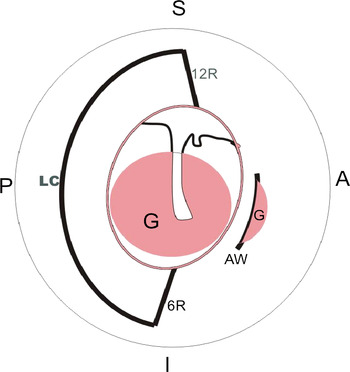
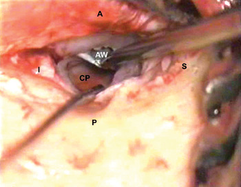
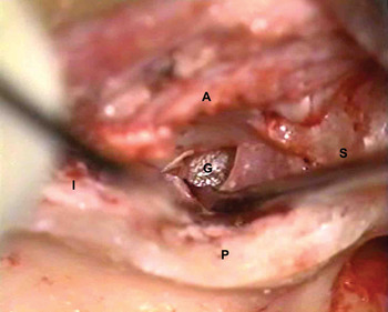
In addition to the above steps, patients in group one underwent anterior tagging (Figure 1). In these patients, after elevation of the tympanomeatal flap, a circular knife was used to create an anterior window by making a circumferential incision of 3–4 mm, approximately 4 mm lateral to the anterior annulus (Figure 2). The skin and the annulus below the incision were then elevated to create a tunnel that connected the anterior window with the middle-ear cavity. While placing the graft, using the underlay technique, the anterior edge of the graft was pulled through the tunnel and the window (Figures 1 and 3). The parts of the anterior annulus and canal skin which had been elevated were then replaced and supported with absorbable gelatine sponge® in the external auditory canal.
Diagrammatic representation of anterior tagging (right ear). 12R = radial incision at 12 o'clock; 6R = radial incision at 6 o'clock; LC = lateral circumferential incision; AW = anterior window; G = temporalis fascia graft; A = anterior; P = posterior; S = superior; I = inferior

Operating microscope photograph of creation of an anterior window (right ear). A = anterior; P = posterior; S = superior; I = inferior; CP = central perforation; AW = anterior window

Operating microscope photograph of anterior tagging (right ear). G = graft seen through anterior window; A = anterior; P = posterior; I = inferior; S = superior

Following graft placement, the middle-ear cavity and ear canal were packed with absorbable gelatine sponge®. After closure of the wound, a mastoid pressure bandage was applied.
Patients received post-operative antibiotics for one week. Sutures were removed after one week.
All cases were regularly followed up once weekly for one month, then fortnightly for three months, then at six and 12 months post-operatively. Pure tone audiometry was performed three, six and 12 months after surgery. The follow-up period ranged from 12 to 22 months, with a mean of 16 months.
We evaluated both groups' results for graft uptake, hearing outcome and complications. Pre-operative and post-operative pure tone audiograms were compared. Hearing gain and mean air–bone gap were calculated for the speech frequencies 0.5, 1, 2 and 4 kHz.
Statistical analysis
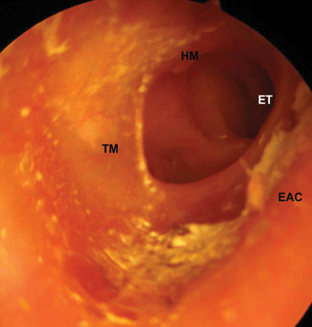
The results were tabulated and presented as percentages, means and standard deviations. For statistical analysis, anterior (Figure 4) and subtotal perforations were considered together, since the anterior remnant of the tympanic membrane was either absent or minimal (i.e. less than 4 mm) in these cases. Similarly, central and posterior perforations were considered together. The chi-square test was used to assess the statistical significance of graft uptake, and the Student t-test to compare the hearing outcomes of the two groups. Hearing results followed a normal distribution in both groups. The level of significance was set at 5 per cent.
Endoscopic photograph of anterior perforation (right ear). EAC = external auditory canal; TM = tympanic membrane; HM = handle of malleus; ET = eustachian tube opening

Results and observations
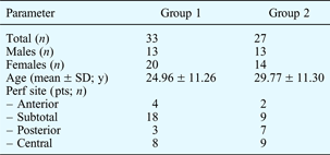
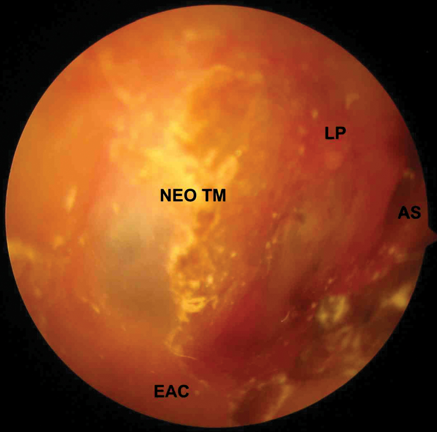
A total of 60 patients were included in the study, after obtaining ethics committee clearance and informed consent. General characteristics of these patients are shown in Table I. There were 33 patients in group one (type one tympanoplasty with anterior tagging) and 27 patients in group two (type one tympanoplasty without anterior tagging). Success with graft uptake was defined as an intact ear drum confirmed by otomicroscopy, 12 months after surgery (Figure 5).
Endoscopic photograph of tympanic membrane 15 months after tympanoplasty with anterior tagging (right ear). Neo TM = neotympanum; LP = lateral process of malleus; EAC = external auditory canal; AS = anterior sulcus
General patient characteristics

SD = standard deviation; y = years; perf = perforation; pts = patients
In group one, 32 of the 33 ears (96.96 per cent) showed successful graft uptake, while one ear with a subtotal perforation (3.04 per cent) had residual perforation. In group two, 22 ears (81.5 per cent) showed successful graft uptake, while five ears (18.5 per cent) had residual perforations (two with anterior and three with subtotal perforations).
In group one, 22 of the 33 cases had anterior or subtotal perforations. Of these 22 cases, 21 had successful graft uptake following surgery, giving a success rate of 95.45 per cent in this subgroup. The remaining 11 cases had central or posterior perforations; all of them had successful outcomes.
In group two, 16 of the 27 cases had central or posterior perforations; all of these 16 cases showed successful graft uptake following surgery. The remaining 11 cases had anterior or subtotal perforations. Of these 11 cases, six (54.54 per cent) had successful outcomes, while the other five showed residual perforations.
In patients with anterior and subtotal perforations (i.e. remnant tympanic membrane either absent or minimal), tympanoplasty with anterior tagging (i.e. group one) resulted in a higher incidence of successful graft uptake (94.45 per cent), compared with tympanoplasty without tagging (i.e. group two; 54.54 per cent). This difference was statistically significant (p < 0.05).
Hearing gain following the two surgical procedures was studied in patients with successful outcomes. The six patients (one from group one and five from group two) who had residual perforations were excluded. In the 32 successful cases in group one, the mean pre-operative air–bone gap was 24.84 dB, with a mean air conduction threshold of 40 dB and mean bone conduction threshold of 15.15 dB. The mean post-operative air–bone gap in this group was 9.4 dB, with a mean air conduction threshold of 24.55 dB and mean bone conduction threshold of 15.15 dB. Therefore, there was a mean gain of 15.45 dB in air conduction threshold, and no change in bone conduction threshold, comparing pre- and post-operative values.
In the 22 successful cases in group two, the mean pre-operative air–bone gap was 24.65 dB, with a mean air conduction threshold of 37.13 dB and mean bone conduction threshold of 12.48 dB. The post-operative mean air–bone gap was 9.5 dB, with a mean air conduction threshold of 21.98 dB and mean bone conduction threshold of 12.48 dB. Thus, there was a mean gain of 15.15 dB in air conduction threshold, and no change in bone conduction threshold, comparing pre- and post-operative values.
In our study, patients with residual perforation were not included in the hearing outcome analysis. Assessment of hearing in patients with successful graft uptake revealed a statistically significant improvement, comparing mean pre- and post-operative air conduction thresholds (p < 0.05), irrespective of the procedure used. The difference in hearing gain between the two groups was not statistically significant (p > 0.05).
The distribution of mean air–bone gap was calculated as the difference between mean post-operative air conduction and post-operative bone conduction thresholds. In group one, of the 32 successful cases, we found a mean air–bone gap of 0–10 dB in 26 cases (81.25 per cent), of 11–20 dB in five cases (15.625 per cent), and of 21 dB or more in one case (3.125 per cent). The percentage of group one patients with a mean air–bone gap within 20 dB was 96.83 per cent. In group two, of the 22 successful cases, we found a mean air–bone gap of 0 to 10 dB in 16 cases (72.7 per cent), of 11–20 dB in five cases (22.7 per cent) and of 21 dB or more in one case (4.6 per cent). The percentage of group two cases with a mean air–bone gap within 20 dB was 95.4 per cent.
In group one, there was one residual perforation (3.04 per cent). Of the 32 successful cases, two cases had tympanosclerosis (6.25 per cent) and one case had retraction (3.125 per cent). In group two, there were five residual perforations (18.5 per cent). Of the 22 successful cases, two cases (9 per cent) had tympanosclerosis and two cases (9 per cent) had retraction.
Discussion
The underlay technique is commonly used. However, its disadvantages include reduction of middle-ear space (due to graft medialisation) and increased failure rates in cases of large perforations (due to the limited bed size for the graft).Reference Gibb and Chang5 Underlay grafts have a tendency to fall away from the anterior annulus into the deepest portion of the middle ear at the eustachian tube orifice, despite the middle ear being adequately packed with absorbable gelatine sponge®.Reference Primrose and Kerr6 On the other hand, elevation of a large part of the fibrous annulus, in order to sufficiently fix the graft, causes significant anterior blunting.Reference Primrose and Kerr6
As a result, techniques to improve anchorage of the graft have been developed. In 1975, Gerlach described a quilting technique in which buttons of graft were pulled through tiny holes in the remnant tympanic membrane and fixed with a drop of histoacryl glue, in order to prevent graft medialisation.Reference Tos7 Based on this technique, Primrose and KerrReference Primrose and Kerr6 developed the anterior hitch method in 1986. Subsequently, GoycooleaReference Goycoolea, Goycoolea, Paparella and Nissen8 pioneered the use of a small anterior flap to support the graft. Better results have been obtained using these techniques for underlay grafts.Reference Sharp, Terzis and Robinson9–Reference James, Ludman and Wright12
Anterior tagging is another such technical modification of tympanoplasty, which is designed to fix the underlay graft under the drum remnant.Reference Tos7 In the present study, no difference was observed in the graft uptake of patients with posteriorly and centrally placed perforations, comparing the two surgical techniques: 100 per cent graft uptake was seen in all these patients. In patients with anterior or subtotal perforations, graft uptake was significantly better in group one (i.e. with anterior tagging) compared with group two.
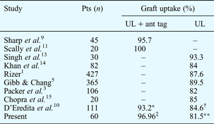
Table II compares published results for standard underlay technique versus underlay with anterior tagging.
Graft uptake: underlay with anterior tagging vs underlay
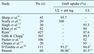
*n = 59; †n = 52; ‡n = 33; **n = 27. Pts = patients; UL = standard underlay technique; ant tag = anterior tagging
Many authors have reported lower success rates for standard underlay tympanoplasty in patients with anteriorly placed and larger perforations.Reference Chopra, Munjal and Mathur15–Reference Onal, Uguz, Kazidas, Gursoy and Gokee19 While the general reported incidence of post-tympanoplasty reperforation is 3–10 per cent,Reference Yuasa, Takasaki, Alpaer and Bluestone20 this is influenced by the surgeon's technique and the type of procedure performed; immediate failure is usually due to technical failure.Reference Yuasa, Takasaki, Alpaer and Bluestone20
The greater incidence of reperforation in patients with anterior and subtotal perforations may be due to reduced vascularity of the anterior aspect of the tympanic membrane compared with the posterior, with a greater risk of necrosis and reabsorption of the graft material. There is also delayed healing due to the lack of residual tympanic membrane and a resulting lack of nourishment of the graft tissue.Reference Schraff, Dash and Strasnick21, Reference Papas and Simpson22 Another factor contributing to failure of the underlay technique is inadequate anchorage of the graft due to improper insertion into the external auditory canal wall, with resulting graft medialisation.Reference Jung and Park16, Reference Goldenberg and Edelstein23 The presence of an anterior bony overhang also makes it difficult to visualise and repair anterior perforations.Reference Jung and Park16, Reference Schraff, Dash and Strasnick21
Some authors have suggested the use of a combination of overlay and underlay grafting methods, in order to achieve better results in cases of anterior and subtotal perforation.Reference Jung and Park16, Reference Schraff, Dash and Strasnick21, Reference Papas and Simpson22 An anteriorly based flap has also been described for the repair of anterior perforations.Reference Goldenberg and Edelstein23 However, these methods carry a risk of anterior blunting.Reference Goldenberg and Edelstein23 In contrast, some authors have found that the size and site of perforation do not affect the graft outcome.Reference Rizer1, Reference Saito, Tanaka and Tokuriki24, Reference Wasson, Papademetrion and Pau25
• Underlay type I tympanoplasty failure in large perforations may be due to small graft bed size and inadequate support
• Anterior tagging anchors part of the anterior graft within an anterior canal wall window
• In this study, graft success was 97 per cent for tympanoplasty with anterior tagging (33 patients) and 82 per cent for tympanoplasty alone (27 patients)
• Patients with anterior and subtotal perforations had higher graft success rates after underlay plus anterior tagging (95 per cent) vs underlay alone (55 per cent)
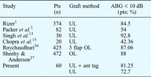
In our study, the two surgical groups showed no statistically significant difference in their mean hearing gain, making the two procedures comparable with respect to hearing improvement. This also indicates that there was no lateralisation or significant anterior blunting in group one, in which anterior tagging was performed. Table III shows reported hearing results for standard and modified underlay and overlay methods.
Hearing outcome for various graft methods

Pts = patients; ABG = air–bone gap; UL = underlay; OL = overlay; ant tag = anterior tagging
In our study, complications were minimal and were encountered in similar numbers in both groups. There were no cases of anterior blunting, tympanic membrane lateralisation or sensorineural hearing loss in either group.
Conclusion
Anterior tagging is a reliable technique to prevent medialisation of the graft material, performed during type one tympanoplasty using the underlay technique. It appears to be of greater benefit in patients with anterior or subtotal pars tensa perforations in which the anterior remnant tympanic membrane is very small or absent. It is not technically difficult, and is not associated with more complications than the standard technique.