Introduction
The South American tomato pinworm, Tuta absoluta (Meyrick) (Lepidoptera: Gelechiidae), is native to highlands of Peru (Vargas, Reference Vargas1970) and has been regarded as one of the most devastating pests of tomato (Solanum lycopersicum L.) (Desneux et al., Reference Desneux, Luna, Guillemaud and Urbaneja2011, Reference Desneux, Wajnberg, Wyckhuys, Burgio, Arpaia, Narváez-Vasquez, González-Cabrera, Catalán Ruescas, Tabone, Frandon, Pizzol, Poncet, Cabello and Urbaneja2010; Guedes and Picanço, Reference Guedes and Picanço2012). T. absoluta has spread to various Latin American countries, including Argentina, Bolivia, Brazil, Chile, Colombia, Ecuador, Panama, Paraguay, Peru, Uruguay, and Venezuela, from the 1960s to the 1980s (USDA–APHIS, 2014). Researchers such as Biondi et al. (Reference Biondi, Guedes, Wan and Desneux2018) and Shashank et al. (Reference Shashank, Twinkle, Chandrashekar, Meshram, Suroshe and Bajracharya2018) confirmed its presence in the Middle East and Africa, signalling a formidable threat to agriculture. T. absoluta was first recorded in the YiLi Prefecture of China’s Xinjiang Uygur Autonomous Region in August 2017 (Wang et al., Reference Wang, Gao and Guan2021a), followed shortly by sightings in March 2018 in Midu County, Dali Prefecture, Yunnan (Yin et al., Reference Yin, Zheng, Li, Ma, Song, Chen, Chen, Liu and Chen2021). This tiny but destructive pest is wreaking havoc on tomato crops worldwide, has voracious larvae that can devour leaves, buds, stems, and fruits (Desneux et al., Reference Desneux, Wajnberg, Wyckhuys, Burgio, Arpaia, Narváez-Vasquez, González-Cabrera, Catalán Ruescas, Tabone, Frandon, Pizzol, Poncet, Cabello and Urbaneja2010). Without effective management, this invasive pest has caused up to 80–100% crop loss (Kinyanjui et al., Reference Kinyanjui, Khamis, Ombura, Kenya, Ekesi and Mohamed2021). Tuta absoluta is a highly adaptable pest with a greedy appetite for Solanaceous crops, which poses a major challenge for farmers and researchers (Desneux et al., Reference Desneux, Wajnberg, Wyckhuys, Burgio, Arpaia, Narváez-Vasquez, González-Cabrera, Catalán Ruescas, Tabone, Frandon, Pizzol, Poncet, Cabello and Urbaneja2010). Biopesticides, such as spinetoram, offer a promising management option for organic farming and help to minimise potential harm to beneficial insects and other non-target organisms (Biondi et al., Reference Biondi, Zappalà, Stark and Desneux2013; Sağlam et al., Reference Sağlam, Çelik, Işıkber, Bozkurt, Sakka and Athanassiou2022).
Spinetoram, a natural insecticide derived from the fermentation of the actinomycete Saccharopolyspora spinosa, consists of spinosyn (XDE-175-L) and spinosyn (XDE-175-J) in a 1:3 ratio, respectively (Yang, Reference Yang2016). Spinetoram works by targeting the nervous system of insects, triggering continuous muscle contractions that lead to paralysis and eventual death. It achieves this by interfering with γ-aminobutyric acid and nAChR receptors, key neurotransmitters, causing excessive nerve activity and overwhelming the insect’s system (Wang et al., Reference Wang, Guo, Liu, Wang and Wang2022). The reduced sensitivity of pest target sites to insecticides leads to the development of resistance to spinosad (Campos et al., Reference Campos, Rodrigues, Silva, Silva, Silva, Guedes and Siqueira2014), but it remains unclear whether similar resistance mechanisms apply to spinetoram. And the mechanisms of insect resistance to spinetoram are multifaceted, and similar patterns have been documented across various insect species in existing literature, such as the corn weevil (Wang et al., Reference Wang, Xiao, Gao and Liu2014), grain beetle (Wang et al., Reference Wang, Guo, Liu, Wang and Wang2022), and diamondback moth (Tamilselvan et al., Reference Tamilselvan, Kennedy and Suganthi2021). The overexpression of three enzymes – glutathione S-transferases (GSTs), cytochrome P450 monooxygenases (P450s), and carboxylesterases – can degrade or detoxify insecticides before they reach their target sites (Li et al., Reference Li, Schuler and Berenbaum2007).
GSTs are a diverse family of enzymes found in both prokaryotes and eukaryotes (Koirala et al., Reference Koirala, Moural and Zhu2022). The main function of GSTs is to catalyse the conjugation of reduced glutathione (GSH) to harmful xenobiotic compounds, transforming them into water-soluble, less toxic products (Hayes et al., Reference Hayes, Flanagan and Jowsey2005), and to detoxify various types of pesticides (Pavlidi et al., Reference Pavlidi, Vontas and Van Leeuwen2018). Specific GSTs like MseGSTe2 and MseGSTs6 in Mythimna separata can metabolise the insecticide chlorantraniliprole (Li et al., Reference Li, Zhang, Liu, Liu, Zhang, Wang, Sun, Toufeeq and Rao2022). Similarly, CpGSTe3 and CpGSTs2 in Cydia pomonella may metabolise pyrethroid insecticides (Hu et al., Reference Hu, Wang, Ju, Chen, Tan, Mota-Sanchez and Yang2020a, Reference Hu, Wei, Li, Harwood, Li and Yang2020b). HaGST-8 from Helicoverpa armigera can break down a wide range of insecticides, including dichlorvos, chlorpyrifos, and cypermethrin, thus conferring high resistance to these chemicals in this pest (Labade et al., Reference Labade, Jadhav, Ahire, Zinjarde and Tamhane2018). Genome sequencing and molecular biology studies have provided valuable insights into the GST gene families of several model insect species, including Drosophila melanogaster, Aedes aegypti, Bombyx mori, and Tribolium castaneum, and from which the researchers have gained a deeper understanding of GST-mediated detoxification pathways (Rane et al., Reference Rane, Ghodke, Hoffmann, Edwards, Walsh and Oakeshott2019). For insect species lacking a sequenced genome, RNA sequencing (RNA-seq) has been successfully applied to various insect species, including M. separata (Li et al., Reference Li, Zhang, Liu, Liu, Zhang, Wang, Sun, Toufeeq and Rao2022), Glyphodes pyloalis (Liu et al., Reference Liu, Xing, Liang, Ding, Li, Shao, Wu, Wang and Sheng2022), Pieris rapae (Liu et al., Reference Liu, Zhang, Wang, Zhang and Li2017b), and Hyphantria cunea (Sun et al., Reference Sun, Yin, Du, Liu and Cao2020). To combat the growing resistance to traditional insecticides, researchers are exploring innovative approaches like RNA interference (RNAi). By targeting specific genes essential for insect growth and development, RNAi-based biopesticides offer a promising solution to control pests like T. absoluta while minimising environmental impact (Zhang et al., Reference Zhang, Khan, Heckel and Bock2017).
RNAi has become a powerful tool for studying the role of GSTs in insecticide resistance. Researchers have observed that several insects showed increased sensitivity to various insecticides when specific GST genes were silenced, including Nilaparvata lugens (Zhou et al., Reference Zhou, Liang, Xu, Gurr, Bao, Zhou, Zhang, Cheng and Zhu2013), Bactrocera dorsalis (Lu et al., Reference Lu, Wang, Huang, Dou, Chen, Wei and Wang2016), Diaphorina citri (Yu and Killiny, Reference Yu and Killiny2018), and Sitobion avenae (Zhang et al., Reference Zhang, Su, Xie, Zhen, Hu, Jiang, Huang, Liu, Gao, Chen and Gao2020), exposed to chlorpyrifos, malathion, thiamethoxam, and imidacloprid, respectively. These findings highlight the potential of RNAi-based technologies as a novel approach for controlling insect pests and mitigating the development of insecticide resistance.
Based on our previous transcriptome data analysis, we compared the GST genes from two different strains of T. absoluta collected from YiLi and Alaer regions in Xinjiang province, China. We identified five significant GST genes and analysed their evolutionary relationship with GST genes from other insect species. To understand the role of these genes in spinetoram resistance, we investigated their expression levels in T. absoluta larvae exposed to spinetoram and found that one gene, TaGSTs1, was significantly upregulated in response to spinetoram. To further explore the function of TaGSTs1, we cloned the gene and analysed its sequence. We then used RNAi to silence the gene in T. absoluta larvae and observed its impact on spinetoram detoxification and development. Our findings provide valuable insights into the role of TaGSTs1 in detoxification and development of T. absoluta. This research provides a foundation for further investigations into the molecular mechanisms underlying T. absoluta insecticide resistance and the development of novel control strategies.
Materials and methods
Maintenance of T. absoluta populations and tomato plants
Strains of T. absoluta were collected from commercial tomato fields in YiLi and Alaer, Xinjiang, China, in 2018. After being maintained for 6 years without exposure to pesticides, the strains were identified as a relatively susceptible population for laboratory use. The T. absoluta individuals were reared on 4-week-old Maofen tomatoes (802) at 25°C, with a 14 (light): 10 (dark) hour photoperiod and 65% relative humidity. Insects were laboratory-maintained in plastic cages (50 × 50 × 50 cm) with insect mesh. The host plants (Maofen tomatoes) were individually grown in 9-cm diameter pots under the same conditions as the tomato leaf miner.
Insecticides exposure
The insecticide used was spinetoram (C42H69NO10, CAS: 935545-74-7), which was purchased from Corteva Agriscience China Holding Co., Ltd. (Shanghai, China), and the concentration of the active ingredient was 98%. The lethal concentration (LC50) of spinetoram at 48 h was 0.044 mg/L. Clean leaves (1.5 cm × 1.5 cm) of Maofen tomatoes were immersed in the LC50 concentration of spinetoram solution for 10 s. The air-dried and treated tomato leaves were laid flat in plastic cups. Collected 2nd instar larvae of T. absoluta were carefully placed on the upper surfaces of the leaves and allowed to feed on the treated tomato leaves for 48 h, and the mortality rate was recorded. The exposure was biologically repeated three times. Individuals treated with the solvent Triton X-100 (0.05%) constituted the control group.
RNA extraction, cDNA synthesis, and TaGSTs1 cloning
Total RNA of T. absoluta was isolated and extracted using the TransZol Up Plus RNA Kit (TransGen Biotech, Beijing, China) according to the manufacturer’s instructions and stored at −80°C. The first-strand cDNA was synthesised using a TransScript All-in-one First-Strand cDNA Synthesis SuperMix for qRT-PCR (One-Step gDNA Removal) Kit (TransGen Biotech, Beijing, China). Based on transcriptome data for T. absoluta, Primer 5.0 was used to design primers for full-length amplification (Table S1). The amplified fragments were purified using a DNA Gel Extraction Kit (Genstone Biotech, Beijing, China). Single-band amplification products were cloned into the pEASY-T3 vector (TransGen) and sequenced.
Sequence analysis of TaGSTs1 in T. absoluta
Based on the transcriptome database, primers for amplifying the full-length open reading frame of GSTs were designed using Primer 5.0 software (Table S1). The full-length coding sequences were translated using DNAMAN (version 5.0; Lynnon BioSoft, QC, Canada). A phylogenetic tree was constructed using the neighbour-joining method in the MEGA software (version 11.0) to evaluate the molecular evolutionary relationships among various insects. Bootstrap majority consensus values for 1000 replicates were shown at each branch point, and the constructed tree was modified using the online tool iTOL (www.itol.embl.de).
Spatiotemporal-specific expression profile of TaGSTs1
The relative expression levels of TaGSTs1 were analysed, and the expression profiles were determined at different developmental stages, including 240 eggs, 36 1st instar larvae, 24 2nd instar larvae, 18 3rd instar larvae, 12 4th instar larvae, 15 pupae, and 10 adults. Additionally, different tissues such as 45 heads, 18 integuments, 36 midguts, and fat bodies were also collected from 3rd instar larvae. RNA was extracted from these samples and converted into cDNA. The reactions were performed using an ABI 7500 Real-Time PCR System (Applied Biosystems, Foster City, CA, USA). All amplifications were confirmed, and the specificity of RT-qPCR was estimated using melting curve analysis. PCR assays were prepared to a final volume of 20.0 μL with 1.0 μL of the cDNA template, 10.0 μL of Hieff qPCR SYBR Green Master Mix, 0.4 μL of the forward primer (10 μM), 0.4 μL of the reverse primer (10 μM), and 8.2 μL ddH2O. There were three replicates for each treatment or control, and each replicate was assessed in triplicate (technical replicates). Three genes were tested as references for qRT-PCR, including large subunit 5 ribosomal protein (RpL5), large subunit 23A ribosomal protein (RpL23A) and rRNA, which were used as internal references in recent studies (Askew et al., Reference Askew, Edwards and Gatehouse2024; Li et al., Reference Li, Huang and Li2024). The amplification efficiency was validated by constructing a standard curve using seven serial dilutions of cDNA. The relative mRNA expression was calculated using the 2-∆∆CT method (Livak and Schmittgen, Reference Livak and Schmittgen2001), where ∆∆CT is determined by the following formula:
 \begin{align*}^{\Delta \Delta }CT{\text{ }}& = {\text{ }}{\left( {C{t_{targe}}t - C{t_{reference}}} \right)_{treatment}}\nonumber\\
& \quad - {\text{ }}{\left( {C{t_{target}} - {\text{ }}C{t_{reference}}} \right)_{control}}.\end{align*}
Synthesis of dsTaGSTs1 and RNAi efficiency
To generate dsTaGSTs1, a linear DNA containing a segment of TaGSTs1 was constructed by PCR amplification using primers incorporating T7 RNA polymerase promoter sequences. as a template for dsRNA synthesis. A partial sequence of green fluorescent protein (GFP) was amplified to produce dsRNA for GFP (dsGFP) as a control. The target region was amplified by PCR using primers containing the T7 RNA polymerase promoter, and the PCR products were gel purified using the MinElute Gel Extraction Kit (QIAGEN, Germany). Next, dsRNA was synthesised using T7 RiboMAXTM Express RNAi system (P1700; Promega, Madison, WI, USA). After the transcription reaction, the DNA template was removed by digestion with DNase, and the synthesised dsRNA was purified by 3 M sodium acetate and isopropanol precipitation. Finally, the purity and yield of dsRNA were checked using 1.0% agarose gel and a microplate spectrophotometer (Spectramax 190), respectively. The synthesised dsTaGSTs1 and dsGFP solutions were diluted to final concentrations of 2.5 μg/200 μL, 5.0 μg/200 μL, and 10.0 μg/200 μL in 200 μL. Then, isolated, similarly sized-Maofen (802) tomato true leaves (approximately 5 cm × 7 cm) were collected after being cultured for 5 weeks at the seedling stage. Each petiole was wrapped with bandages and inserted into dsRNA solution for soaking until all the solution was absorbed (approximately 3-4 h). The whole set-up was maintained in the laboratory under a controlled environment of 25 ± 1°C, 60 ± 5% RH, and a 16 h: 8 h (Light: Dark) photoperiod. Immediately after uptake, 2nd instar larvae (n = 30) that had been starved for 4 h were gently placed onto a treated tomato leaf for continuous feeding for 48 h. Negative controls were set up in parallel, including treatment with dsGFP and with ddH2O. Each experiment had three biological replicates. After the initial dsRNA uptake (approximately 3–4 h), leaves were kept hydrated by replenishing the solution with DNase/RNase-Free Water daily. Individuals in the treatment and control groups were sampled at 12, 24, 36, and 48 h. The effect of silencing TaGSTs1 on the expression level in larvae was evaluated using qRT-PCR.
Effect of silencing TaGSTs1 on sensitivity of T. absoluta to spinetoram
Based on our previous research, the LC50 concentration of spinetoram for T. absoluta was 0.044 mg/L. Second instar larvae were first treated with 10 μg/200 μL dsTaGSTs1 for 48 h. They were then exposed to the LC50 concentration of spinetoram (0.044 mg/L) for a further 48 h, and the larval mortality was recorded. Each treatment had three biological replicates (n = 30 instar larvae per replicate).
Effect of RNAi on growth and development of T. absoluta
To investigate the effect of silencing TaGSTs1 on the growth and development of T. absoluta larvae, the larvae were exposed to each treatment (dsTaGSTs1, dsGFP, and ddH2O) through plant-mediated RNAi for 48 h. The leaf-mining time of the 2nd instar larvae was observed, larval mortality and any changes in larval stage were also recorded every 24 h. Each treatment included three replicates, with 30 larvae per replicate.
Data analysis
The relative expression of genes was calculated using the 2−ΔΔCt method (Livak and Schmittgen, Reference Livak and Schmittgen2001). Statistical significance was analysed using one-way analysis of variance (ANOVA) and Student’s t-test in SPSS 22.0 software (Chicago, USA). Differences between treatment and control groups were statistically analysed by Student’s t-test, and P values were considered significantly different (*P < 0.05; **P < 0.01). Different lowercase letters indicate significant differences (P < 0.05), and the same lowercase letters indicate no significant differences.
Results
GSTs in T. absoluta based on transcriptome analysis
Based on our transcriptome data analysis, we identified five significant differentially expressed genes in YiLi and Alaer. Following this, we performed a phylogenetic analysis to categorise these GST genes within T. absoluta. Our findings illustrated that these five TaGSTs grouped into three distinct clades: sigma, delta, and epsilon (fig. 1). As a result, we have classified these GSTs as TaGSTs1, TaGSTs2, TaGSTe1, TaGSTe3, and TaGSTd1, aligned with the established classification system. The predicted protein lengths for these TaGSTs ranged from 151 to 274 amino acid residues, with the majority exhibiting considerable amino acid similarity (60–84%) to their counterparts in other arthropod species.
Phylogenetic tree of the homologous amino acid sequences of GST of Tuta absoluta and other insects. The phylogenetic tree was generated via the test neighbour-joining tree method based on the bootstrap mode and was used to determine the relationships between the GSTs of different insects, including Heortia vitessoides (AVC05621.1), Ostrinia furnacalis (QIC35748.1), Pararge aegeria (BAA07890.1), Conogethes punctiferalis (UQQ66585.1), Cnaphalocrocis medinalis (AIZ46905.1), Amyelois transitella (XP_03195571.2), Galleria mellonella (XP_052755102.1), Maniola jurtina (XP_045775001.1), Bicyclus anynana (XP_023950273.1), Glyphodes pyloalis (QGZ00482.1), Pieris brassicae (CAH4029887.1), Glyphodes pyloalis (QEP99755.1), Pieris napi (XP_047521480.1), Hyposmocoma kahamanoa (XP_026318450.1), Melitaea cinxia (XP_045451455.1), Nymphalis io (XP_050347850.1), Pieris rapaea (XP_022116041.2), Leptidea sinapis (XP_050677250.1), Zerene cesonia (XP_038222288.1), Achroia grisella (XP_059048744.1), Vanessa cardui (XP_046969313.1), Spodoptera littoralis (AYM01153.1), Bombyx mori (NP_001036994.1), Spodoptera exigua (AHB18378.1), Danaus plexippus (OWR52256.1), Micromelalopha troglodyta (ANS56590.1), Grapholitha molesta (WDQ36044.1), Ectropis grisescens (WGO51733.1), Helicoverpa armigera (AIB07714.1), Holotrichia oblita (KAI4470440.1), Spodoptera frugiperda (QGA73325.1), Papilio machaon (XP_014360504.2), Achroia grisella (XP_059049156.1), and Eumeta japonica (GBP06710.1).

Effect of spinetoram on the expression level of TaGST genes in T. absoluta
To determine whether the expression levels of TaGST genes: TaGSTs1, TaGSTs2, TaGSTe1, TaGSTe3, and TaGSTd1 were induced by spinetoram, we measured the relative expression level of these genes after 24 h treatment with spinetoram at its LC50 concentration (fig. 2). Compared to the control group (ddH2O), the relative expression level of TaGSTs1 and TaGSTd1 in the spinetoram-treated group showed increases of 3.92-fold (P < 0.05) and 2.3-fold (P < 0.05) at 24 h (fig. 2) respectively. Conversely, TaGSTs2, TaGSTe1, and TaGSTe3 exhibited a response of less than twofold to spinetoram (fig. 2). These findings suggest a potentially significant role for these genes in spinetoram tolerance.
Relative expression level of TaGSTs1, TaGSTs2, TaGSTe1, TaGSTe3, and TaGSTd1 treated with spinetoram. Bars represent mean ± SD (n = 3). The different letters (a, b, c) indicate a significant difference among ddH2O group and spinetoram groups according to the ANOVA results (one-way ANOVA with Tukey’s test; P < 0.05), and the same letters indicate no difference among different groups.

Sequence analysis of TaGSTS1 in T. absoluta
The full-length cDNA sequence of TaGSTs1 was 465 base pairs and encoded a polypeptide of 154 amino acids. The structure of TaGSTs1 from T. absoluta is characterised by an N-terminal thioredoxin-fold domain and a C-terminal alpha-helical domain, with its active site located in a cleft between the two domains. Furthermore, conserved domain analysis highlighted the presence of GSH binding sites (G-sites) in the N-terminal regions and substrate binding sites (H-sites) in the C-terminal regions (fig. 3), underscoring their potential roles in detoxification mechanisms within T. absoluta.
The full-length cDNA sequence of TaGSTs1 and its deduced amino acid sequence. The full-length cDNA of T. absoluta TaGSTs1 was 465 bp and encoded a polypeptide of 154 amino acids. The green colour indicates the dimer-interface, red colour indicates the substrate binding pocket (H-site), and the blue colour indicates the N-terminal domain interface. Analysis of conserved domains revealed glutathione binding sites (G-sites) in the N-terminal regions and substrate binding sites (H-sites) in the C-terminal region of tagsts1 in T. absoluta.

Spatiotemporal expression profile of TaGSTs1 in T. absoluta
The expression pattern of TaGSTs1 was determined by qRT-PCR. The expression levels of TaGSTs1 in different tissues and developmental stages were investigated (fig. 4). The expression level of TaGSTs1 increased with T. absoluta development, becoming significantly upregulated from the first instar onwards, with the peak expression observed in the second instar, while exhibiting lower levels in the adult stage (fig. 4A). Regarding tissue-specific expression, the midgut displayed the highest expression, significantly exceeding that of the integument, fat body, and head (P < 0.05, fig. 4B).
Expression profiles of T. absoluta TaGSTs1 gene. (A) Represents of the expression during different developmental stages of larvae, including eggs – E, 1st instar, 2nd instar, 3rd instar, 4th instar, pupae – P, and adults – A. (B) Representation of different larval tissues, including the head – H, integument – I, midgut – M, and fat body – F. TaGSTs1 mRNA levels in different larval tissues and during different developmental stages are normalised relative to those in fat body, pupae and adults. Data were analysed by one-way ANOVA with Tukey’s test (p < 0.05). Different lowercase letters (a, b, c) represent significant changes in gene expression and same letters indicate no difference in different groups.

The expression level of TaGSTs1 after RNAi
To validate the efficiency of RNAi, the expression level of TaGSTs1 was assessed using qRT-PCR. We used different concentrations (2.5, 5.0, and 10.0 μg/200 μL) of dsTaGSTs1 to assess silencing efficiency. The results showed that the relative expression level of TaGSTs1 treated with 2.5 and 5.0 μg/200 μL was not significantly lower than the control groups at 12, 24, 36, and 48 h, respectively (fig. 5A and 5B). In the group treated with 10.0 μg/200 μL dsTaGSTs1, the relative expression level was significantly lower than the ddH2O and dsGFP control groups at 24, 36, and 48 h (P < 0.05). The 10.0 μg/200 μL dsTaGSTs1group had the highest silencing efficiency (62.29%) at 48 h compared to the other time points. These results suggested that TaGSTs1 in T. absoluta was effectively silenced by dsTaGSTs1 treatment at 48 h.
Feeding of dsTaGSTs1 (2.5 μg/200 μL). (A) Feeding of dsTaGSTs1 (5.0 μg/200 μL). (B) Feeding of dsTaGSTs1 (10.0 μg/200 μL). (C) Significant reduction of the expression level at 48 h in T. absoluta larvae as compared to the control individuals feeding with the dsRNA of green fluorescent protein (dsGFP) and nuclease-free water (ddH2O). Data are presented as means (n = 3) ± SE. The different lowercase letters (a, b, c) indicate a significant difference between three groups according to the ANOVA results (one-way ANOVA with Tukey’s test; P < 0.05), and the same lowercase letters indicate no difference among different groups.

Silencing of TaGSTs1 increases the sensitivity of T. absoluta to spinetoram
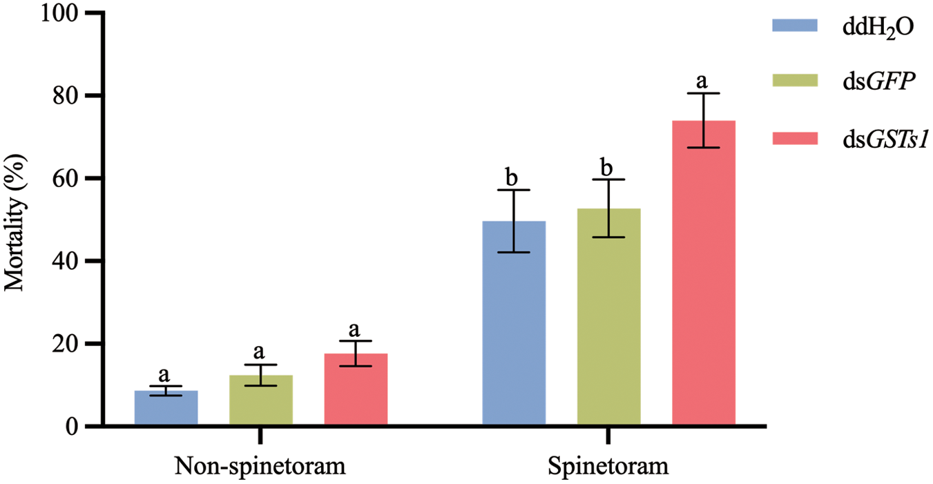
We observed that the dsTaGSTs1 group had no significant impact on mortality compared with the ddH₂O and dsGFP treated controls at 48 h after treatment (fig. 6, P > 0.05). However, upon subsequent spinetoram treatment, the mortality of the remaining T. absoluta treated with ddH2O, dsGFP, and dsTaGSTs1 was 48.6%, 51.1%, and 75.6% at 48 h post-spinetoram treatment, respectively (fig. 6), indicating that TaGSTs1 was involved in the detoxification of spinetoram in T. absoluta.
Effects of dsGSTs1 on Tuta absoluta larvae. (A) Silencing of TaGSTs1 via RNAi and its impact on cumulative insect mortality were investigated by evaluating the mortality rate of T. absoluta larvae subjected to different treatments (ddH₂O, dsGFP, and dsTaGSTs1) before and after exposure to an LC₅₀ dose of spinetoram. The data, presented as means (n = 3) ± standard error (SE), indicated notable differences in mortality among the treatment groups. Statistical analysis using one-way ANOVA with Tukey’s test (P < 0.05) revealed significant differences, with distinct letters (a, b, c), while groups sharing the same letter indicated no significant difference.

Effect of silencing TaGSTs1 on the growth and development of T. absoluta
We also investigated the effects of dsTaGSTs1 on the growth and development starting from the 2nd instar stage. The time to initiation of leaf piercing time for the 2nd instar larvae treated with dsTaGSTs1 was significantly longer than that of larvae treated with ddH2O and dsGFP, by 33.9% and 24.7%, respectively (fig. 7A), indicating an impact on the larval feeding behaviour. Additionally, dsTaGSTs1 group had a significantly longer developmental time from the 2nd to 4th instar larval stage compared to the ddH₂O and dsGFP-treated controls, respectively (fig. 7B). The mean developmental time for dsTaGSTs1, dsGFP, and ddH₂O treatment was 5.12 ± 0.27, 4.15 ± 0.14, and 3.96 ± 0.40 days, respectively (fig. 7B). Cumulative mortality results showed that by the 4th instar stage, mortality of the 2nd instar larvae was 38% and 33% after 7 days of feeding on ddH2O and dsGFP, respectively. In contrast, 58% of the larvae had died after feeding on dsTaGSTs1 (fig. 7B). These results indicated that silencing of TaGSTs1 by RNAi caused growth retardation and death in T. absoluta larvae. The phenotypic observations of T. absoluta larvae were carried out after RNAi and distinct changes were evident. Larvae treated with ddH₂O consistently exhibited no observed abnormalities, confirming a baseline of normal development. We focused the statistical comparison of abnormality rates on the dsGFP as control, larvae treated with dsGFP (fig. 7D(a) showed normal growth. In contrast, dsTaGSTs1 treated larvae displayed black spots on their epidermis (fig. 7D(b) some became slim due to not eating sufficient food (fig. 7(c, d)), some larvae showed ruptured epidermis due to swelling and decay (fig. 7E(d)), with some resulting in deformities (fig. 7D(f)) or, wrinkled tails (fig. 7D(g)). Moreover, the abnormality rate in the dsTaGSTs1 treated group was 18% compared with dsGFP (fig. 7E). These results suggested that lower expression of dsTaGSTs1 due to RNAi could cause growth retardation, deformities, and larval death.
The impact of silencing TaGSTs1 on the larval growth and development of T. absoluta. (A) Illustration of the leaf piercing or mining time of larvae after feeding with dsTaGSTs1. (B) Representation of the feeding duration of instar stages. (C) Presentation of the cumulative mortality of T. absoluta larvae after 7 days of feeding with dsTaGSTs1. Data are presented as means (n = 3) ± SE and ** above bars indicate significant differences between the treatment and the corresponding control (p < 0.01). (D) Phenotype of Tuta absoluta fed with, a dsGFP, and b–g phenotype of T. absoluta after feeding with dsTaGSTs1. (E) Representation of the abnormal rate of T. Absoluta after feeding of dsRNA (dsGSTs1) and dsGFP as control. **Above bars indicate significant differences between the treatment and the corresponding control (P < 0.01).

Discussion
Molecular research on the insecticide resistance of T. absoluta provided valuable insights into identifying potential target genes involved in pest resistance. These studies were also crucial for developing effective field prevention and control strategies. GSTs are a major superfamily of enzymes that catalyse the oxidation of a diverse range of endogenous and exogenous substrates in insects (Lu et al., Reference Lu, Song and Zeng2021). However, only a limited number of GST genes had been identified in T. absoluta. In this study, we selected five upregulated GST (TaGSTs1, TaGSTs2, TaGSTe1, TaGSTe3, and TaGSTd1) genes based on transcriptome data analysis. Among them, TaGSTs1 was the most significantly upregulated in response to spinetoram in T. absoluta. To investigate the functional role of the TaGSTs1 gene, we carried out RNAi experiments to silence its expression in T. absoluta. This silencing resulted in a significant increase in susceptibility to spinetoram, providing experimental evidence that this specific GST functionally contributes to spinetoram resistance. (Wang et al., Reference Wang, Rui, Wang, Nahiyoon, Huang, Zhu, Ji, Yang, Yuan and Cui2021b) reported that field-evolved resistance of H. armigera to spinetoram is associated with GSTs1 overexpression, which is consistent with its role in detoxification. Beyond TaGSTs1, the function of other four GST genes across various insects highlights the functional diversity. For instance, GSTe1 in Spodoptera littoralis is specifically involved in metabolising plant allelochemicals, contributing to host plant adaptation (Zou et al., Reference Zou, Xu, Zou, Liu, Chen and Feng2016). This demonstrates a role of GSTe1 in processing natural toxins from the insect’s diet. Furthermore, certain Delta-class GSTs, such as GmolGSTd1 and SzeaGSTd1, demonstrate highly specialised functions in the degradation of volatile compounds in insect antennae, suggesting a role in processing olfactory signals (Li et al., Reference Li, Chen, Xu and Wu2018; Xia et al., Reference Xia, Zheng, Huang, Lu and Tang2022). Furthermore, CpGSTs2 and CpGSTe3 in Cydia pomonella might contribute through alternative mechanisms such as sequestration of the insecticide lambda-cyhalothrin (Hu et al., Reference Hu, Liu, Wang, Mota-Sanchez, He and Shi2022). However, the specific functional roles of these other four TaGST genes in spinetoram resistance were not validated in this study and warrant further investigation.
In T. absoluta, TaGSTs1 consists of two main domains: an N-terminal thioredoxin-fold domain and a C-terminal alpha-helical domain. Its active site, where the detoxification reactions occur is located within a cleft between these two domains. Structurally, TaGSTs1 is characterised by an N-terminal domain that binds to GSH and a C-terminal domain that binds to hydrophobic substrates, which facilitates the detoxification process by bringing the two reactants together within the enzyme’s active site. The structural similarity of TaGSTs1 to GSTs from other insects, such as Anopheles gambiae AgGSTe2 (Wang et al., Reference Wang, Qiu, Ranson, Lumjuan, Hemingway and Setzer2008), Musca domestica MdGSTd1 (Sue and Yajima, Reference Sue and Yajima2018), and Mythimna separata MseGSTs6 and MseGSTe2 (Li et al., Reference Li, Zhang, Liu, Liu, Zhang, Wang, Sun, Toufeeq and Rao2022), suggests a potential role for TaGSTs1 in insecticide resistance in T. absoluta.
Insect GSTs are often induced in response to various stressors, including exposure to pesticides, and this rapid upregulation of gene expression leads to increased detoxification capacity (Amezian et al., Reference Amezian, Nauen and Le Goff2021). In our study, we found that TaGSTs1 was induced by spinetoram, with a 3.92-fold increase in expression after 24 h of exposure to an LC50 dose of spinetoram treatment. This suggests that TaGSTs1 plays a role in the response to spinetoram in T. absoluta. Similar research carried out on Aphis gossypii found that GST genes AgGSTs1, AgGSTs2, and AgGSTs3 were upregulated in response to an LC50 dose of imidacloprid treatment, suggests that the sigma family of GSTs plays a role in response to insecticide stress in this pest (Gawande et al., Reference Gawande, Subashini, Murugan and Subbarayalu2014). These findings indicated that TaGSTs1 might be involved in the response to spinetoram stress in T. absoluta. Additionally, dsTaGSTs1 significantly reduced the tolerance of T. absoluta larvae to spinetoram. However, further research is needed to confirm its direct role in this metabolic pathway.
The GST genes also play a crucial role in larval growth and development. For example, in Locusta migratoria, silencing GSTe1 and GSTe3 genes extended the larval development time by 0.5 days compared to control groups (Zhang et al., Reference Zhang, Ma, Yin, Park, Zhu, Zhang, Qin and Li2022). In Aedes aegypti mosquitoes, reducing the expression of the epsilon-3 transcript has been linked to a deformed wing phenotype, highlighting the importance of GSTs in normal wing development (Lumjuan et al., Reference Lumjuan, Rajatileka, Changsom, Wicheer, Leelapat, Prapanthadara, Somboon, Lycett and Ranson2011). This suggests that these epsilon GSTs are involved in regulating normal growth and development. In our results, TaGSTs1 also impacted the larval growth and development. dsTaGSTs1 led to prolonged leaf-mining time, delayed development from the 2nd to 4th instar, and increased cumulative mortality. Our results demonstrated that TaGSTs1 was involved in the growth and development of T. absoluta.
In conclusion, our research demonstrated that spinetoram induced the expression level of TaGSTs1, a key GST gene in T. absoluta. Using RNAi technology, we found that TaGSTs1 was involved in the detoxification of spinetoram and the growth and development of T. absoluta larvae. These findings enhance our understanding of the molecular mechanisms underlying spinetoram resistance in T. absoluta and provide valuable insights for developing effective pest management strategies.
Supplementary material
The supplementary material for this article can be found at https://doi.org/10.1017/S0007485325100321.
Author contributions
Amjad Ali carried out the studies, participated in data collection, acquisition, analysis, or interpretation of data, performed the statistical analysis, participated in experimental design, drafted the manuscript, and participated in paper review and editing. Jiaqi Wu and Jingang Xie participated in experimental design, acquisition, analysis, or interpretation of data, performed the statistical analysis, drafted the manuscript, and participated in paper review and editing. Yu Song participated in collecting data. Ning Liu participated in analysis, or interpretation of data, performed the statistical analysis, paper review and editing. Xiaoning Liu participated in funding acquisition, project administration, supervision, and experimental design, performed the statistical analysis, drafted the manuscript, and participated in paper review and editing.
Financial support
This work was supported by the ‘Tian Shan Talent’ Training Program ‘Agriculture, Rural Areas, and Farmers’ Backbone Talent Project (2024SNGGGCC011) and the Graduate Student Research and Innovation Projects in Xinjiang Autonomous Region (XJ2025G026).
Competing interests
None.