Introduction
Yunnan Province, situated in the south-west part of China, has always been a paradise for lichenologists. New species are described from this area regularly (Harada & Wang Reference Harada and Wang2008; Wei et al. Reference Wei, McCune, Wang and Wei2010), and still many remain to be discovered. During a field trip to Yunnan Province, a Pilophorus species was collected and identified in the field as P. robustus Th. Fr. Later critical examination in the laboratory, however, revealed it to be a different species, distinct from P. robustus and all other described species of this genus. After consulting the main accounts of this genus (Jahns Reference Jahns1970, Reference Jahns1981; Timdal Reference Timdal1988; Wang et al. Reference Wang, Joshi, Hur, Oh and Wang2010), we concluded that this specimen belonged to a previously unpublished species which we describe as P. fruticosus Li S. Wang & Xin Y. Wang. We provide here a detailed taxonomic description along with chemical and ecological data for this new taxon.
Materials and Methods
Specimens for this study were collected during surveys in the Yunnan Province, China, in July 2006. These specimens have been deposited both in the herbarium of Kunming Institute of Botany, China (KUN) and the Korean Lichen Research Institute, Sunchon National University, South Korea (KoLRI).
The specimens were examined using standard microscopic techniques and hand-cut sections under NIKON C-PS 1068908 dissecting microscope. All measurements were made on material mounted in water and stained with lactophenol cotton blue (LCB). Anatomical descriptions are based on observations of these preparations under a NIKON Eclipse E 200 microscope. For the size of the thallus and apothecia, and the thickness of the epihymenium, hymenium and subhymenium, we recorded five measurements for each specimen. Ten measurements per specimen were recorded for ascospore dimensions. The dimensions of ascospores are presented as smallest single value recorded – largest single value recorded. We conducted spot test reactions on hand-cut sections of thalli and apothecia under an OLYMPUS BX 50 microscope. Secondary metabolites were identified by TLC as described by White & James (Reference White and James1985), Elix et al. (Reference Elix, Johnston and Parker1987) and Orange et al. (Reference Orange, James and White2001). Terminology for tissues generally follows that of Jahns (Reference Jahns1981).
The Species
Pilophorus fruticosus Li S. Wang & Xin Y. Wang sp. nov
Mycobank No.: MB 519575
Similis P. robusto, sed differt in thallo dichotome ramoso, columella absenti et apotheciis solitariis.
Typus: China, Yunnan Province, Dali Co., Cangshan Mt., 25° 40′ 47·5″ N, 100° 06′ 02·8″ E, alt. 3570 m, on rock, 28 July 2006, L. S. Wang 06-26204 (kun—holotypus; KoLRI—isotypus).
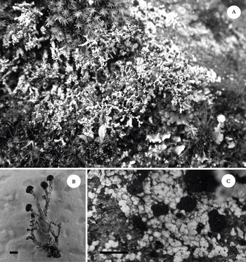
(Fig. 1)
Pilophorus fruticosus. A, habit; B, pseudopodetia; C, primary thallus and cephalodia. Scales: B & C = 1 mm.

Primary thallus persistent, tightly attached to the substratum, consisting of small granules (0·1–0·2 mm diam.) that usually become aggregated to form squamules (c. 1 mm diam.), grey to whitish grey when dry. Pseudopodetia dichotomously branched (from base to apex), 0·5–1 cm high and with branches less than 1 mm wide. Surface of the pseudopodetia covered with small granules, which are concolorous with the primary thallus. Pseudopodetia sometimes largely decorticate, exposing brown to dark brown medulla. Internal part of pseudopodetia solid, composed of strongly gelatinized hyphae, forming a dark pigmented central region. Photobiont green. Cephalodia frequent, sessile, mostly growing on the primary thallus but sometimes also present on the lower part of the pseudopodetia, 1–2 mm diam., verrucose to cerebriform, brown to dark brown, containing Nostoc.
Apothecia solitary and growing on the apices of the mature pseudopodetia, spherical, black, c. 1 mm diam. Epihymenium bluish green, c. 5 µm high. Hymenium non–amyloid, non-pigmented, 80–100 µm high. Subhymenium non-pigmented, 25–30 µm high. Excipulum absent. Vegetative and generative tissue separated by black to dark brown pigmented boundary. Columella absent. Boundary texture present. Asci clavate, with a well-developed amyloid tholus containing a darker-straining amyloid tube, 8-spored. Ascospores spindle-shaped, 18·0–22·5 × 5·0–7·5 µm.
Pycnidia numerous, cylindrical, in clumps (2–3), growing at the apex of the pseudopodetium, up to 0·3 mm long. Pycnoconidia sickle shaped, 4–5 × 1 µm. Photobiont green.
Chemistry. Spot test reactions: thallus K+ yellow, C−, P−. Secondary metabolites: atranorin and zeorin.
Etymology. The specific epithet fruticosus refers to the morphology of the pseudopodetia.
Ecology and distribution. The new species has been observed growing on siliceous rocks along with species of Cladonia and Stereocaulon in the alpine region, at altitudes of approximately 3500 m. The surrounding forest is dominated by Rhododendron and Abies georgei. We have observed this species only at two localitites in Yunnan Province.
Discussion. Pilophorus fruticosus can be easily identified by the dichotomously branched pseudopodetia, the partly decorticated surface of the pseudopodetia, the solitary apothecia at the apices of pseudopodetia, the presence of a boundary texture and the lack of a columella in the apothecia. It is not likely to be confused with P. robustus, which differs in having umbellately branched, massive and stout pseudopodetia; a well-developed columella, and in the lack of a boundary texture. Also the primary thallus in P. robustus is pulverulent or granular and usually evanescent. The occasional dichotomous branching of P. acicularis (Ach.) Th. Fr. and P. awasthianus Räsänen can make them easily mistaken for this new taxon. However, P. acicularis differs from P. fruticosus by having simple pseudopodetia that are occasionally forked at the tips, taller pseudopodetia (more than 1 cm), higher hymenium (up to 240 µm) and larger spores (21–29·5 µm long), whereas P. awasthianus differs by having a loosely, scattered primary thallus, aggregated apothecia, the lack of a boundary texture and in its restricted distribution in India.
Additional specimen examined. China: Yunnan Province: Dali Co., Cangshan Mt., 25° 40′ 49·8″ N, 100° 06′ 04·0″ E, alt. 3543 m, on rock, 2006, Hur Ch060323 (KoLRI).
The authors are grateful to the National Natural Science Foundation of China (No. 30870158) for providing financial assistance for carrying out this research. We thank Ms. Heidi Bissell for helping with the English text.