Introduction
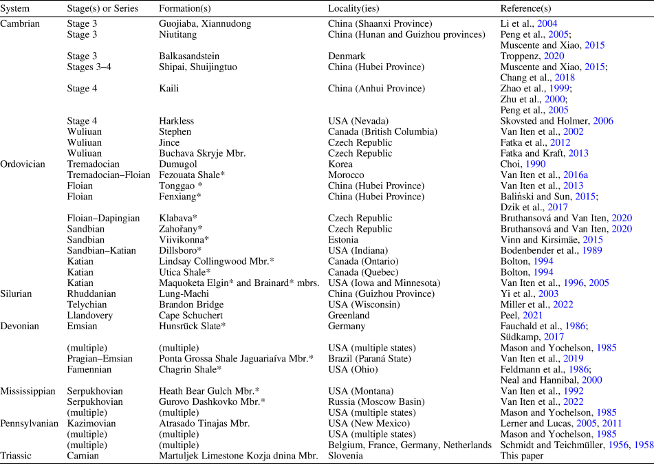
Sphenothallus Hall, Reference Hall1847, one of the most widely distributed and longest ranging genera in the fossil record, has been documented from all systems of the Paleozoic Erathem except the Permian (Table 1), although it has been stated (e.g., Choi, Reference Choi1990; Bolton, Reference Bolton1994; Fatka et al., Reference Fatka, Kraft and Szabad2012) that the genus also occurs in that system. At present the first appearance of this epibenthic, polypoid medusozoan cnidarian lies in Cambrian Stage 3, while the previously known youngest occurrences are in the Pennsylvanian System. Sphenothallus has been found in numerous formations on all continents except Australia and Antarctica. It occurs in a variety of marine facies ranging from shallow nearshore to deep offshore and has even been found in strata of coastal lacustrine origin, probably as an allochthonous element (Lerner and Lucas, Reference Lerner, Lucas, Sullivan, Lucas and Spielmann2011). Many of the rock units known to contain Sphenothallus also contain conulariids (Table 1), an extinct group of marine scyphozoans that may have been closely related to Sphenothallus (Van Iten et al., Reference Van Iten, Cox and Mapes1992, Reference Van Iten, Cox and Mapes1996). Van Iten et al. (Reference Van Iten, Cox and Mapes1992) interpreted Sphenothallus as a medusozoan cnidarian of uncertain class-level affinities, but later Dzik et al. (Reference Dzik, Baliński and Sun2017) documented internal peridermal structures that may be homologous to similar features in the periderm of coronate scyphozoans (see for example illustrations in Van Iten, Reference Van Iten1992, and Van Iten et al., Reference Van Iten, Fitzke and Cox1996).
Selected documented occurrences of Sphenothallus Hall, Reference Hall1847, in the Phanerozoic rock record. Asterisk next to a formation or member name indicates that the unit also yields conulariids.

The present article describes multiple, very well-preserved specimens of Sphenothallus carniolica (Kolar-Jurkovšek and Jurkovšek, Reference Kolar-Jurkovšek and Jurkovšek1997) from limestone strata of early Late Triassic (late Carnian) age in the Julian Alps of northwest Slovenia. Some of these specimens were originally described under the name Valvasoria carniolica Kolar-Jurkovšek and Jurkovšek, Reference Kolar-Jurkovšek and Jurkovšek1997, which was interpreted as a tubiculous worm of possible nematode or sipunculid affinities (see also Hitij et al., Reference Hitij, Gašparič, Zalohar, Jurkovšek and Kolar-Jurkovšek2019). Thus, this is the first report of Sphenothallus from the Mesozoic Erathem. Furthermore, Sphenothallus is now established as a long-ranging medusozoan cnidarian, which, together with conulariids, survived the End Permian Mass Extinction Event (MacLeod, Reference MacLeod2013).
Geological setting
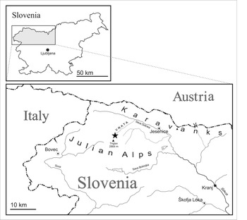
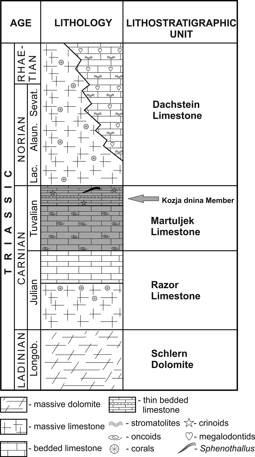
The studied Sphenothallus specimens were collected from thin, gray, laminated lime mudstones in the Kozja dnina Member of the Martuljek Limestone (early Late Triassic, late Carnian) in the northeast Julian Alps (Vrata Valley) of northwest Slovenia (Fig. 1). The late Carnian (Tuvalian) age of the section that yielded the specimens is based on conodont assemblages (Quadralella polygnathiformis Zone; Kolar-Jurkovšek, Reference Kolar-Jurkovšek1991). The section at Kozja dnina is ~80 m thick and represents a deep water paleoenvironment (Bitner et al., Reference Bitner, Jurkovšek and Kolar-Jurkovšek2010). The strata were deposited in an interplatform basin, where anoxic conditions and rapid sedimentation enabled exceptional preservation of both invertebrate and vertebrate fossils (Celarc and Kolar-Jurkovšek, Reference Celarc and Kolar-Jurkovšek2008). In addition to Sphenothallus, the Kozja dnina limestones also contain bivalves, brachiopods, echinoids, crinoids, asteroids, ammonites, belemnites, scleractinian corals, shrimp, lobsters, thylacocephalans, and fishes (Hitij et al., Reference Hitij, Gašparič, Zalohar, Jurkovšek and Kolar-Jurkovšek2019; Gašparič et al., Reference Gašparič, Audo, Hitij, Jurkovšek and Kolar-Jurkovšek2020).
Simplified geographical map of northwest Slovenia. The location of Kozja dnina in the Vrata Valley is marked by a star (figure adapted from Bitner et al., Reference Bitner, Jurkovšek and Kolar-Jurkovšek2010).

Lithostratigraphy of the Tuvalian section with platy limestones yielding Sphenothallus carniolica (Kolar-Jurkovšek and Jurkovšek, Reference Kolar-Jurkovšek and Jurkovšek1997) (arrow) at Kozja dnina (figure adapted from Celarc and Kolar-Jurkovšek, Reference Celarc and Kolar-Jurkovšek2008). Norian stage subdivisions are abbreviated as: Lac. = Lacian; Alaun. = Alaunian; Sevat. = Sevatian.

Material and methods
The present study is based on direct examination of 31 specimens of Sphenothallus carniolica (Kolar-Jurkovšek and Jurkovšek, Reference Kolar-Jurkovšek and Jurkovšek1997) from the Kozja dnina Member of the Martuljek Limestone. All specimens occur on exposed bedding planes, their preserved periderm appearing light to dark brown on the light gray weathered limestone, in some cases with a blueish hue resulting from vivianitization. Specimens were examined and photographed using reflected light and scanning electron microscopy (secondary electron mode). Material photographed under reflected light was whitened with ammonium chloride sublimate. Photographs were taken with a millimetric scale bar using a Nikon Z 6II digital camera equipped with a NIKKOR Z MC 105mm f/2.8 VR S lens. Photographs were edited in Photoshop CS6, and figures containing light photographs were assembled in CorelDRAW X8. Scanning electron microscopy was conducted using a JEOL JSM-649OLV. Finally, the elemental composition of the periderm of one of the specimens (T-1287) was determined using an Oxford INCA Energy 350 EDS under the following operating conditions: chamber vacuum 20 Pa, accelerating voltage 20 kV, spot size 48 μm, working distance 10 mm, and analysis time 60 seconds.
Repositories and institutional abbreviations
BJ = Paleontological collection of Jurkovšek, Dol pri Lubljani, Slovenia. T = Paleontological collection of Tomaž Hitij and Jure Žalohar, Godič, Slovenia. All collections are registered at the Slovenian Museum of Natural History in Ljubljana.
Systematic paleontology
Phylum Cnidaria Verrill, Reference Verrill1865
Subphylum Medusozoa Peterson, Reference Peterson, Larwood and Rosen1979
Class, Order, Family uncertain
Genus Sphenothallus Hall, Reference Hall1847
Type species
Sphenothallus angustifolius Hall, Reference Hall1847, originally described from the Upper Ordovician of eastern New York State, USA.
Sphenothallus carniolica (Kolar-Jurkovšek and Jurkovšek, Reference Kolar-Jurkovšek and Jurkovšek1997)
Figures 3–5
- Reference Kolar-Jurkovšek and Jurkovšek1997
Valvasoria carniolica Kolar-Jurkovšek and Jurkovšek, p. 1, pl. 1, figs. 1–4.
- Reference Hitij, Gašparič, Zalohar, Jurkovšek and Kolar-Jurkovšek2019
Valvasoria carniolica; Hitij et al., p. 21, figs. 16, 17.
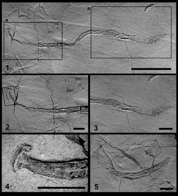
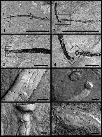
Sphenothallus carniolica (Kolar-Jurkovšek and Jurkovšek, Reference Kolar-Jurkovšek and Jurkovšek1997) from the Late Triassic (late Carnian) Kozja dnina Member of the Martuljek Limestone of northwest Slovenia (Vrata Valley). (1–4) BJ1286, the holotype and most complete specimen: (1) side view of the entire specimen, which preserves both the apical holdfast (left end of the specimen) and the apertural region of the main tube (right end of the specimen); (2) detail of the area in box (A) in (1), showing the apical region with berm-like marginal thickenings; (3) detail of the area in box (B) in (1), showing the heavily crumpled, relatively thin apertural region; (4) detail of the area in box (C) in (2), highlighting the sub-conical apical holdfast; (5) BJ1419, paratype, a nearly complete specimen with partially preserved apical holdfast. Scale bars = 30 mm (1) and 10 mm (2–5).

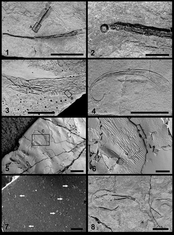
Sphenothallus carniolica (Kolar-Jurkovšek and Jurkovšek, Reference Kolar-Jurkovšek and Jurkovšek1997) from the Late Triassic (late Carnian) Kozja dnina Member of the Martuljek Limestone of northwest Slovenia (Vrata Valley). (1) T-1277, mostly complete specimen preserving the apical holdfast; (2) T-1270, two nearly contiguous specimens, both preserving the upper portion of the apical holdfast (basal membrane missing); (3) detail of the area in box (A) in (1), highlighting the apical region; (4) detail of the area in box (C) in (2), showing the apical regions of the two nearly contiguous specimens; (5, 7) scanning electron photomicrographs of T-1277: (5) detail of the area in box (B) in (1), showing the non-annulated external surface of the main tube; (7) detail of the area in box (D) in (3), showing the apicalmost portion of the periderm (7); (6, 8) scanning electron photomicrographs of T-1270: (6) detail of the area in box (E) in (4), showing the apicalmost portions of the specimens; (8) detail of the area in box (F) in (6), showing the wavy laminated microstructure of one the two holdfasts in transverse cross section (8). Scale bars = 10 mm (1, 2), 1 mm (3, 4), 500 μm (5, 6), 100 μm (7), and 50 μm (8).

Sphenothallus carniolica (Kolar-Jurkovšek and Jurkovšek, Reference Kolar-Jurkovšek and Jurkovšek1997) from the Late Triassic (late Carnian) Kozja dnina Member of the Martuljek Limestone of northwest Slovenia (Vrata Valley). (1) T-1273, two associated specimens, the shorter one of which preserves the apical holdfast; (2) detail of the box (A) in (1), highlighting the apical holdfast and the marginal thickenings, which are slightly elevated above the thinner periderm between them; (3) T-1257, portion of the main tube of a nearly complete specimen exhibiting pervasive irregular wrinkling of the thin periderm between the longitudinal thickenings; (4) T-1256, nearly complete specimen exhibiting pronounced curvature of the main tube in the apical region (left) and pervasive wrinkling of the thin periderm between the robust longitudinal thickenings (right); (5) detail (scanning electron micrograph) of the box (B) in (3), showing the absence of regular annulation of the main tube as well as areas (central part of the field of view) of possible “plywood [micro]structure” (Vinn and Mironenko, Reference Vinn and Mironenko2021); (6) detail (scanning electron micrograph) of the box (C) in (5), showing one of the areas of possible “plywood [micro]structure” (Vinn and Mironenko, Reference Vinn and Mironenko2021); (7) scanning electron micrograph of BJ1419, showing multiple, sub-circular micropores (arrows) on the surface of the periderm; (8) T-1271, assemblage of at least four specimens consisting entirely or predominantly of one or both longitudinal thickenings. Scale bars = 10 mm (1, 3, 4, 6, 8), 1 mm (2), 500 μm (5), and 20 μm (7).

Holotype
BJ1286 (originally designated as the holotype of Valvasoria carniolica Kolar-Jurkovšek and Jurkovšek, Reference Kolar-Jurkovšek and Jurkovšek1997; currently reposited in the collection of Bogdan Jurkovšek).
Paratypes
BJ1287, BJ1419 (originally designated as paratypes of Valvasoria carniolica; currently reposited in the collection of Bogdan Jurkovšek).
Diagnosis
Peridermal tube very gently tapered (angle of expansion mostly <2°), non-branching, lacking regular transverse annulations.
Occurrence
Thin, gray, laminated lime mudstones in the Kozja dnina Member of the Martuljek Limestone (early Late Triassic, late Carnian, Tuvalian; Paragondolella polygnathiformis conodont Zone) in the Vrata Valley in the northeast Julian Alps, northwest Slovenia. The geographic coordinates of the fossil locality are Lat. 46°24′08.1″N, Long. 13°50′46.2″E.
Description
Partial to nearly complete, compressed specimens lying parallel to bedding and ranging up to 129.5 mm in length and 5.5 mm in width. Specimens consist of one or more portions of the slender, originally sub-elliptical (transversely) main tube or of nearly the entire periderm, including the sub-conical attachment disc, although without the basal membrane. Attachment disc measures up to ~3.1 mm in diameter. Apertural margin not preserved; terminal schott (apical wall) absent. Main tube very gently tapered (mostly <2°) in the plane of the well-developed pair of oppositely situated, longitudinal thickenings, variably curved parallel to bedding, in some cases with the degree of curvature increasing toward the apical end. Relatively thin periderm between the longitudinal thickenings missing or, where present, exhibiting coarse, irregular, transverse to oblique wrinkles. Regular transverse annulations absent. Longitudinal thickenings terminate or thin near the apertural end of the main tube. Skeletal material phosphatic, very finely lamellar, light to dark brown or dark bluish gray, with exfoliated lamellae in some specimens exhibiting possible “plywood [micro]structure” (Vinn and Mironenko, Reference Vinn and Mironenko2021). Some specimens exhibit minute, shallow, sub-circular to sub-elliptical pores or pits ranging from ~2–4 μm in diameter.
Additional material
BJ2505 (currently reposited in the collection of Bogdan Jurkovšek); T-1256, T-1257, T-1270–T-1287 (currently reposited in the collection of Tomaž Hitij and Jure Žalohar).
Remarks
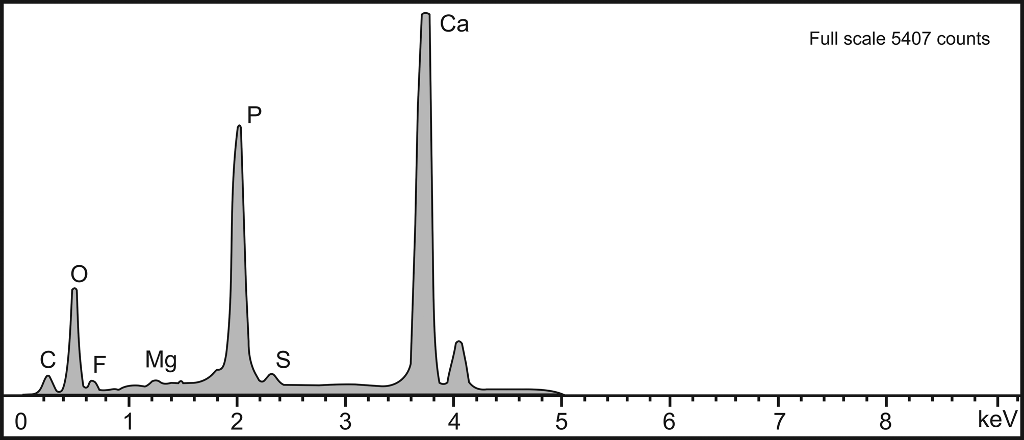
The principal diagnostic feature of Sphenothallus, namely the pair of longitudinal thickenings situated at the end points of the major diameter of the subelliptical main tube, is well developed in the Slovenian material (Figs. 3.1–3.3, 3.5, 4.1–4.3, 5.1–5.4, 5.8). As in Sphenothallus from Paleozoic formations, compression of the Slovenian specimens perpendicular to bedding has caused the longitudinal thickenings to form berm-like elevations rising above the thinner, deformed skeletal wall between them, which in some cases (Fig. 5.8) is now absent. Also well displayed in the Slovenian material is the characteristic, parallel lamellar microstructure (Figs. 4.8, 5.5, 5.6), which is commonly exfoliated. Lamellae in one specimen appear to exhibit “plywood [micro]structure” (Fig. 5.5, 5.6), discovered by Vinn and Mironenko (Reference Vinn and Mironenko2021, fig. 2C, D) in material from the Upper Mississippian of central Russia. A somewhat similar microstructure is present in the medusozoan Torellella Holm, Reference Holm1893, from the upper Cambrian of Estonia (Vinn, Reference Vinn2006, Reference Vinn2022). The presence of calcium phosphate (apatite), which constitutes the bulk of most Paleozoic specimens, is corroborated both by the brown to blue-gray color of the specimens and by the results of EDS analysis (Fig. 6), which yielded strong spectral peaks for Ca and P.
EDS spectrum of specimen T-1287, corroborating the presence of calcium phosphate in the periderm.

Turning to other characters, the very low rate of taper of the main tube of the Slovenian specimens is similar to that of S. angustifolius from the Upper Ordovician of Ontario and Quebec, Canada (see for example Bolton, Reference Bolton1994, pls. 1.1–1.3) and S. sica Clarke, Reference Clarke1913, from the Lower Devonian Ponta Grossa Shale of Brazil (see for example Van Iten et al., Reference Van Iten, Leme, Simões and Cournoyer2019, fig. 4). The size of the attachment disc (Figs. 3.1, 3.2, 3.4, 3.5, 4.1–4.4, 4.6, 4.7, 5.1, 5.2), both absolute and in proportion to the size of the main tube, matches closely that of the attachment disc of Sphenothallus sp. from the Bear Gulch Member of the Upper Mississippian Heath Formation of Montana, western USA (see Van Iten et al., Reference Van Iten, Cox and Mapes1992, fig. 1). Interestingly, specimens preserving both the attachment disc and the long slender tube above it may be attached to hard biological substrates (see for example Van Iten et al., Reference Van Iten, Leme, Simões and Cournoyer2019, fig. 6), or, like the Slovenian specimens and the Bear Gulch material referred to in the preceding sentence, they may be unattached, suggesting that their original substrate may have been plant or other organic matter susceptible to rapid decay. Finally, smooth curvature of the main tube, with the degree of curvature commonly decreasing in the direction of the apertural end (Figs. 3.2, 4.1, 4.2, 4.6, 5.3, 5.4, 5.6), is a feature exhibited by many Paleozoic specimens (see for example Van Iten et al., Reference Van Iten, Fitzke and Cox1996, pl. 2.1), as are thinning of the longitudinal thickenings in the vicinity of the aperture (Figs. 3.1, 3.3, 5.4) and the absence of well-defined annulation or other ornament (Fig. 4.5).
A noteworthy additional feature, previously documented in Sphenothallus cf. S. angustifolius from the Upper Mississippian of central Russia (Vinn and Mironenko, Reference Vinn and Mironenko2021, fig. 2f), is the presence in the main tube of some of the Slovenian specimens of microscopic, sub-circular to sub-elliptical pores or pits (Fig. 5.7), originally detected in specimen BJ1287 by Kolar-Jurkovšek and Jurkovšek (Reference Kolar-Jurkovšek and Jurkovšek1997, figs. 2, 3). These shallow perforations are similar in size, shape, areal density, and pattern of distribution to the microscopic circular pores, or micropores, of conulariids (see Van Iten et al., Reference Van Iten, Vhylasová, Zhu and Zhuo2005, Reference Van Iten, Lichtenwalter, Leme and Simões2006a, Reference Van Iten, Leme and Simõesb, Reference Van Iten, Mironenko and Vinn2022, and references cited therein). Conulariid micropores have been compared with microscopic borings and bioclaustrations, and they have been interpreted as primary anatomical features. If the latter hypothesis is true, then the presence of these features in conulariids and Sphenothallus may provide additional support for the hypothesis (e.g., Van Iten et al., Reference Van Iten, Fitzke and Cox1996) that the skeletons of these two taxa are mutually homologous.
Concluding remarks
Sphenothallus Hall, Reference Hall1847, an extinct genus of medusozoan cnidarians, is now known from the lower Upper Triassic of northwest Slovenia (Julian Alps). Moreover, the well-preserved specimens of S. carniolica from Slovenia are the only documented representatives of the genus from the Mesozoic Erathem. The present discovery extends the known age range of Sphenothallus by ca. 80 million years, from the Kazimovian to the late Carnian, and thus across the critical Permian-Triassic boundary. Additionally, since Sphenothallus has not yet been documented from any part of the Permian System, it may be a Lazarus taxon (Jablonski, Reference Jablonski1986). Nevertheless, we predict that the existence of Sphenothallus of Permian age, previously only asserted, will be confirmed and that additional discoveries of Triassic and maybe even younger Sphenothallus will be made, based in part on the ability of Sphenothallus to survive the most severe crisis in the history of life during the Phanerozoic Eon. Finally, given the presence of conulariids, which are commonly associated with Sphenothallus, in strata of terminal Ediacaran age (Van Iten et al., Reference Van Iten, Leme, Pacheco, Simões, Fairchild, Rodrigues, Galante, Boggiani, Marques, Goffredo and Dubinsky2016b; Leme et al., Reference Leme, Van Iten and Simões2022), we would not be surprised if the first appearance of Sphenothallus were extended downward into that system.
Acknowledgments
The authors are grateful to M. Miller (Geological Survey of Slovenia) for SEM photography and EDS analysis of Sphenothallus specimens and to the administration of the Triglav National Park for permission to perform paleontological research within its boundary. We also thank reviewers J.S. Peel (Department of Earth Sciences, Uppsala University, Sweden) and O. Vinn (Department of Geology, University of Tartu, Estonia) and associate editor R.J. Elias (Department of Earth Sciences, University of Manitoba, Winnipeg, Canada) for their constructive comments on and corrections of the original manuscript.
Declaration of competing interests
The authors declare none.