Introduction
The Brycon insignis (Steindachner, 1877), known as tiete tetra in English and piabanha in Portuguese, is a native and endemic species of the Paraíba do Sul River basin located at São Paulo, Minas Gerais and Rio de Janeiro States, Southeast Brazil (Hilsdorf & Petrere Jr, Reference Hilsdorf and Petrere2002). The genus Brycon belongs to the order Characiformes, family Characidae, subfamily Bryconinae and comprises more than 70 fish species with wide geographic distribution (Froese & Pauly, Reference Froese, Pauly and Editors2010). Many Brazilian fısh species, B. insignis migrate up-stream to spawn. This migratory behaviour is known as piracema and lasts from October to February. In the 1950s, B. insignis was the fourth most captured species in commercial fishing in this basin, therefore displaying an important role in the region's economy (Machado & Abreu, Reference Machado and Abreu1952). However, due to over-fıshing, pollution and changes in the Paraíba do Sul River course during the construction of the Hydroelectric Paraibuna-Paraitinga, B. insignis reproductive cycle was disrupted and its status is currently set as endangered (Andrade-Talmelli et al., Reference Andrade-Talmelli, Kavamoto, Romagosa and Fenerich-Verani2001).
Fertilization is a process of cellular fusion that encompasses the contact between the spermatozoon and the oocyte up to the union of the nuclei of both cells (Schatten, Reference Schatten, Knobil and Neill1999; Moore, Reference Moore2001). Fertilization promotes the activation of female gamete with the resumption of meiosis, formerly stuck at metaphase II and triggers a chain of events inside the oocytes (Brasil et al., Reference Brasil, Nakaghi, Santos, Grassiotto and Foresti2002). As fertilization in fish is generally monospermic (Kobayashi & Yamamoto, Reference Kobayashi and Yamamoto1981), after the first spermatozoon penetration, a series of events that prevents polyspermy takes place. These events include mechanical barriers such as: (a) closure of the micropyle (Kobayashi & Yamamoto, Reference Kobayashi and Yamamoto1981; Hart & Donovan, Reference Hart and Donovan1983, Kudo et al., Reference Kudo, Linhart and Billard1994); (b) formation of the fertilization cone (Kudo, Reference Kudo1980; Iwamatsu et al., Reference Iwamatsu, Onitake, Yoshimoto and Hiramoto1991; Linhart & Kudo, Reference Linhart and Kudo1997; Brasil et al., Reference Brasil, Nakaghi, Santos, Grassiotto and Foresti2002; Murata, Reference Murata2003); and (c) activation of the cortical reaction to eliminate any supernumerary spermatozoa (Iwamatsu & Ohta, Reference Iwamatsu and Ohta1981; Iwamatsu et al., Reference Iwamatsu, Ishijima and Nakashima1993). After cellular fusion, one diploid cell undergoes a process named embryonic development or embryogenesis. Knowledge of the embryonic development in fish species is useful to the management of fishery resources and to surveys related to fish culture as it provides additional information regarding species life cycles, for ontogeny studies, experimental modeling, evaluation of environmental quality and effects of toxic substances on aquatic fauna (Flores et al., Reference Flores, Araiza and Valle2002), as well as for experiments on ex situ species preservation.
Considering the great number of species, post-fertilization events and embryonic development studies on neotropical fısh species have received little attention. Reports on fertilization events and embryonic development in species of the order Characiformes are available for matrinxã Brycon cephalus (Lopes et al., Reference Lopes, Senhorini and Soares1995; Romagosa et al., Reference Romagosa, Narahara and Fenerich-Verani2001; Alexandre et al., Reference Alexandre, Ninhaus-Silveira, Veríssimo-Silveira, Buzollo, Senhorini and Chaguri2010), pirapitinga Brycon nattereri (Maria, Reference Maria2008), piracanjuba Brycon orbignyanus (Ganeco, Reference Ganeco2003; Reynalte-Tataje et al., Reference Reynalte-Tataje, Zaniboni-Filho and Esquivel2004; Ganeco et al., Reference Ganeco, Franceschini-Vicentini and Nakaghi2009), jatuarana Brycon spp. (Neumann et al., Reference Neumann, Mendes and Nakaghi2007), aimara Hoplerythrinus unitaeniatus, traírão Hoplias lacerdae and trahira Hoplias malabaricus (Gomes et al., Reference Gomes, Scarpelli, Arantes, Sato, Bazzoli and Rizzo2007), dorado Salminus brasiliensis (Nakaghi et al., Reference Nakaghi, Marques, Faustino, Ganeco and Senhorini2006) and streaked prochilod Prochilodus lineatus (Brasil et al., Reference Brasil, Nakaghi, Santos, Grassiotto and Foresti2002; Ninhaus-Silveira et al., Reference Ninhaus-Silveira, Foresti and Azevedo2006). Preliminary observations of some embryonic (cleavage, gastrula, neurula and embryo) and larval development stages of the B. insignis under stereomicroscopy were reported by Andrade-Talmelli et al. (Reference Andrade-Talmelli, Kavamoto, Romagosa and Fenerich-Verani2001). Oocytes, post-fertilization events, and the zygote and blastula stages of B. insignis however, have not yet been published. The use of scanning electron microscopy (SEM) to monitor morphological changes of oocytes and eggs allows a detailed observation of the external structures, provides a better understanding of the fertilization and embryonic development of a fish species, and completes the information gathered under histologic and stereomicroscopic analysis.
Thus, the aim of the present study was to evaluate the oocytes, post-fertilization events and embryonic development in Brycon insignis, under both SEM and stereomicroscopy.
Materials and methods
Fish, gamete collection and initial gamete evaluation
All fish were handled in compliance with published guidelines for animal experimentation (Van Zutphen et al., Reference Van Zutphen, Baumans and Beynen1993). Tiete tetra Brycon insignis broodfish was selected from earthen ponds at the Hydrobiology and Aquaculture Station of Hydroelectric Company of São Paulo (CESP) in Paraibuna (23°23′10″S; 45°39′44″W), São Paulo state, Brazil, during the spawning season (January and February).
Two males of 4–5 years of age (296 ± 28 g) with detectable running sperm under soft abdominal pressure were given a single intramuscular dose of carp pituitary extract (Argent Chemical Laboratory) at 3 mg/kg body weight and maintained at ~24 °C. Eight hours later, the urogenital papilla was carefully dried, and sperm was hand-stripped directly into test tubes. Sperm collection was carried out at room temperature (23–25 °C) and, soon after collection, the tubes containing sperm were placed in a Styrofoam box containing crushed ice (5 ± 2 °C). Contamination of sperm with water, urine or feces was carefully avoided. An aliquot of 5 μl of each sample was placed on a glass slide and observed under light microscope (Model L1000, Bioval, Jiangbei, China) at ×400 magnification. As fish sperm in seminal plasma should be immotile, any sperm motility was considered to have undergone premature induction resulting from urine or water contamination and the sample was discarded. In samples thusly selected, sperm motility was triggered in 25 μl of 1% NaHCO3 (Labsynth) as activating agent (Viveiros et al., Reference Viveiros, Amaral, Orfão, Isau, Caneppele and Leal2011) and subjectively estimated under light microscope. Sperm concentration (hemacytometer, Neubauer chamber) was also determined.
To harvest oocytes, eight females of ~12 years of age (814 ± 297 g) with detectable running oocytes under soft abdominal pressure, received two doses of carp pituitary extract (0.5 and 4.5 mg/kg body weight) at 12 h interval. Together with the second dose, females received a dose of human chorionic gonadotropin (Pregnyl™ Schering-Plough) at 1500 IU hCG/kg body weight and were hand-stripped 8 h later, at ~24 °C. The spawning weight, spawning index (spawning weight × 100/body weight), number of oocytes/g ova and the number of oocytes/female were calculated. Three aliquots of oocytes from each female were collected. The first aliquot (n = ~30 oocytes × 8 females) was fixed in Gilson's solution (50 ml 60% ethanol, 440 ml distilled water, 7 ml nitric acid, 10 g mercuric chloride, 9 ml glacial acetic) and the diameter of each oocyte was measured under a light microscope using a micrometric objective. The second aliquot (n = ~30 oocytes × 8 females) was fixed in Serra's solution (60 ml 90% ethanol, 30 ml 37% formaldehyde, 10 ml glacial acetic) and the germinal vesicle position (central, peripheric and breakdown) was examined under a stereomicroscope (TIM 2 T, Opton). Finally, the third aliquot (n = ~20 oocytes × 8 females) was fixed in modified Karnovsky (2.5% glutaraldehyde, 2.5% paraformaldehyde in 50 mM sodium cacodylate buffer, pH 7.2, 1 mM CaCl2) and transported to the Laboratory of Electron Microscopy and Ultrastructural Analysis at Federal University of Lavras (UFLA), in Lavras, Minas Gerais State, Brazil. Then oocytes were post-fixed in 1% osmium tetroxide for 4 h at room temperature, washed in 0.1 M cacodylate buffer (pH 7.4), dehydrated through grade acetone solutions (25, 50, 75, 90 and 100%), dried with CO2 in a PELCO CPD 030 critical point, coated with gold under vacuum conditions with SEM Coating Unit SCD 050 and examined with a scanning electron microscope (LEO EVO 40 XVP ESC Carl Zeiss, Santo Amaro, SP, Brazil) equipped with a digital camera.
Fertilization process
Freshly collected sperm was used to fertilize oocytes in a factorial of two males × two females, performing a total of four progenies. An approximate ratio of 5.0 × 10⁵ ± 1 × 10⁵ spermatozoa: oocyte was used, based on our previous study with another species of the genus Brycon (Maria et al., Reference Maria, Viveiros, Freitas and Oliveira2006). To achieve that ratio, 5 g of oocytes (~2835 oocytes) was fertilized with 60 μl of sperm. Fertilization was initiated by the addition of 10 ml tank water and agitated for 1 min. Subsequently, 20 ml tank water was added and samples mixed for another 2 min. Finally, eggs were transferred to four funnel type incubation units (1 progeny = 1 incubation unit) made with 1.5-litre plastic bottles with 10 cm of diameter, and incubated in a fłow-through system at ~24 °C.
Post-fertilization events
Approximately 20 eggs from each incubation unit were collected at 0 (when sperm was mixed with oocyte but before the addition of water), 20, 40, 60, 80, 100, 120, 140, 160 and 180 s post-fertilization. Eggs were fixed in Karnovsky modified and then examined under SEM as described for oocytes. The spermatozoa arrival at the micropyle and the following post-fertilization events were tracked: fertilization cone formation with the expulsion of supernumerary spermatozoa, agglutination of supernumerary spermatozoa in the vestibule, and the beginning of the micropyle closure.
Embryonic development
Approximately 20 eggs from each incubation unit with apparent normal development were collected at 0.25, 0.5, 1 and 1.5 h, every 1.5 h until 6 h and every 5 h until hatching. Eggs were fixed in 2.5% glutaraldehyde, transported to the Laboratory of Semen Technology at UFLA, examined under stereomicroscopy and photographed with digital camera (DSC-W35, Sony Electronic Inc, New York, NY, USA). The following main stages of embryonic development were tracked: zygote, cleavage, blastula, gastrula (blastopore closure), segmentation, larval and hatching (Ninhaus-Silveira et al., Reference Ninhaus-Silveira, Foresti and Azevedo2006).
When most of the developing embryos were observed at the blastopore closure stage, 100–200 eggs were randomly collected from each incubation unit and the number of fertilized eggs (transparent with a developing embryo), as a percentage of total eggs (transparent + dead or white), was determined. After counting, eggs were placed back into the same incubation unit for further development.
Results
Initial gamete evaluation
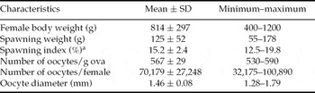
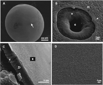
B. insignis fresh sperm possessed a mean of 23.5 × 10⁹ ± 1.5 × 10⁹ spermatozoa/ml and 95% motile sperm upon activation. Recently stripped oocytes were spherical, non-adhesive and greenish-brown. Spawning weight of 125 g, spawning index of 15.2%, 567 oocytes/g ova, 70179 oocytes/female and a mean oocyte diameter of 1.46 mm were observed (Table 1). The germinal vesicle position was central in 2% of stripped oocytes, peripheric in 63%, and breakdown in 35%. Under SEM observation, oocyte surface, which corresponds to the chorion or zona radiata, possessed a single micropyle and pore-canals (Fig. 1A). The micropyle was characterized by a conical vestibule and a narrow micropylar canal (Fig. 1B). At the animal pole, diameter and number of pore-canals increased towards the micropyle (Fig. 1B,C) whereas at the vegetative pole, the pore-canals showed a similar diameter and were uniformly distributed (Fig. 1D).
Female body weight and oocytes characteristics (n = 8 females) of tiete tetra Brycon insignis after hormone treatment

aSpawning index: spawning weight × 100/body weight.
Scanning electron micrographs of the oocyte surface of tiete tetra Brycon insignis. (A) Single micropyle (arrow) at the oocyte surface. (B) Details of micropyle (m) with vestibule (v), micropylar canal (c) and pore-canals (arrow) at the animal pole. (C) Fractured zona radiata (Zr) with pore-canals (arrow) at the oocyte surface(s). (D) Regular distribution of pore-canals at the vegetative pole.

Post-fertilization events
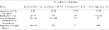
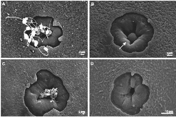
Some morphological post-fertilization events observed under SEM are depicted in Fig. 2A–D and Table 2. Several spermatozoa were observed at the entrance of the micropyle canal when sperm was added to oocytes before the addition of water (time 0; Fig. 2A) up to 20 s post-fertilization. The fertilization cone was characterized as a spherical structure blocking the entrance of micropyle and was observed 20 to 40 s post-fertilization (Fig. 2B). Agglutinated supernumerary spermatozoa expelled from the micropylar canal (Fig. 2C) and the beginning of micropyle closure were observed 100–180 s post-fertilization (Fig. 2D).
The moment when some post-fertilization events were first observed in species of the genus Brycon

ND, not determined.
aThe present study; bMaria (Reference Maria2008); cGaneco et al. (Reference Ganeco, Franceschini-Vicentini and Nakaghi2009); dNeumann et al. (Reference Neumann, Mendes and Nakaghi2007).
Scanning electron micrographs of some post-fertilization events in tiete tetra Brycon insignis. (A) Arrival of spermatozoa at the micropylar canal, 0–20 s post-fertilization. (B) Fertilization cone (arrow), 20–40 s post-fertilization. (C) Supernumerary spermatozoa expelled from the micropyle, 100–180 s post-fertilization. (D) Reduced micropylar canal diameter 100–180 s post-fertilization.

Embryonic development
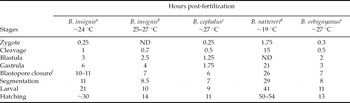
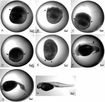
The embryonic development from fertilization to hatching lasted approximately 30 h at ~24 °C (~720 h-degree; Table 3). Under stereomicroscopy analysis, the following embryonic development stages were identified: zygote, cleavage, blastula, gastrula (blastopore closure), segmentation, larval and hatching (Fig. 3 and Table 3).
The moment when the embryonic development stages were first observed in species of the genus Brycon

ND, not determined.
aThe present study; bAndrade–Talmelli et al. (2001a); cAlexandre et al. (Reference Alexandre, Ninhaus-Silveira, Veríssimo-Silveira, Buzollo, Senhorini and Chaguri2010); dMaria (Reference Maria2008); eGaneco (Reference Ganeco2003).
fOr 90% of epiboly.
Stages of embryogenesis of tiete tetra B. insignis, observed under stereomicroscopy. (A) Zygote with formation of blastodisc (arrowhead; 0.25 h post-fertilization). (B) Cleavage with some blastomeres (arrow; 1.5 h post-fertilization). (C) Blastula (arrowheads; 3 h post-fertilization). (D) Beginning of gastrula, 30% epiboly – migration of blastodisc cells (arrows; 6 h post-fertilization). (E) End of gastrula, blastopore closure (arrowheads; 10–11 h post-fertilization). (F) Segmentation, optical vesicle (ov), somites (arrow head) and attached tail (arrow) (16 h post-fertilization). (G) Embryo at larval stage – free tail (arrow; 21 h post-fertilization). (H) Recently hatched larvae (~30 h post-fertilization).

The zygote stage was identified at 0.25 h and lasted up to ~1 h post-fertilization (Fig. 3A). This stage were characterized by: (a) the formation of the blastodisc on the animal pole comprising the active cytoplasm and nucleus; (b) the formation of the vegetative pole composed of yolk; and (c) the egg hydration characterized by the increase of the egg diameter from 1.46 to ~4 mm post-fertilization.
The cleavage stage (from 1 to ~3 h; Fig. 3B) was characterized by the beginning of cell division into 2, 4, 8, 16, 32 and 64 blastomeres. The cleavage followed meroblastic pattern and blastomeres of distinct sizes were observed. The cleavage pattern was observed as follows: the first cleavage plane was vertical, giving rise to two blastomeres; the second plane was vertical and perpendicular to the first one, giving rise to four blastomeres; the third cleavage was vertical and parallel to the first one, giving rise to eight blastomeres in a 4 × 2 arrangement; the fourth was vertical and parallel to the second cleavage, giving 16 blastomeres in a 4 × 4 display; the fifth plane was vertical and parallel to the first cleavage, giving 32 blastomeres in a 4 × 8 formation; and the sixth cleavage plane was horizontal, giving rise to two cell layers, with a total of 64 blastomeres. Between 1 and 3 h, embryos in different development stages (zygote, cleavage with different number of blastomeres, and blastula) were identified in a given sampling time. From 3 h onwards, embryos developed more uniformly.
During the blastula stage (from ~3 to ~6 h; Fig. 3C), blastomeres reorganized and blastoderm possessed a half-moon shape. The blastomeres underwent continuous divisions, but their pattern was undetermined.
The gastrula stage (from ~6 to ~11 h; Fig. 3D) was characterized by the migration of blastodisc cells into the vegetative pole through epiboly movement, with gradual cell expansion around the yolk. Two embryonic layers were formed at this stage: the epiblast and hypoblast. Epiboly movement culminated with blastopore closure when the yolk was completely surrounded by the blastoderm forming the yolk sac (Fig. 3E). At this moment, a fertilization rate of 26% was calculated.
The segmentation stage (from ~11 to ~21 h; Fig. 3F) began with the differentiation of embryonic germ layers. The somites, notochord, neural tube, early delimitation of the intestine and elongation of the embryo (mainly at the head–tail axis) were observed at this stage. Approximately 16 h post-fertilization, the embryos possessed about 18 somites, optic, otic and Kupffer's vesicles at the caudal region and an attached tail.
The larval stage began at ~21 h of the development and was characterized by the presence of a free tail and more than 26 somites. The notochord extended from the cephalic region up to the tail and a well defined primitive posterior intestine could be observed (Fig. 3G).
Vigorous muscle contractions of both the tail and body lead to chorion rupture and hatching of larvae ~30 h post-fertilization. Hatched larvae possessed a transparent and elongated body (Fig. 3H).
Discussion
Stripped oocytes of Brycon insignis were spherical, non-adhesive, greenish-brown, 1.46 mm of diameter and the germinal vesicle position was peripheric in 63% of the oocytes. These characteristics were similar to those reported for other species of the genus Brycon (Eckmann, Reference Eckmann1984; Vazzoler, Reference Vazzoler1996; Andrade-Talmelli et al., Reference Andrade-Talmelli, Kavamoto, Romagosa and Fenerich-Verani2001; Romagosa et al., Reference Romagosa, Narahara and Fenerich-Verani2001; Reynalte-Tataje et al., Reference Reynalte-Tataje, Zaniboni-Filho and Esquivel2004; Ganeco et al., Reference Ganeco, Franceschini-Vicentini and Nakaghi2009; Alexandre et al., Reference Alexandre, Ninhaus-Silveira, Veríssimo-Silveira, Buzollo, Senhorini and Chaguri2010). The oocyte surface of B. insignis possessed a single funnel-like shaped micropyle and pore-canals, similarly to other species of Characiformes (Rizzo et al., Reference Rizzo, Sato, Barreto and Godinho2002; Ganeco & Nakaghi, Reference Ganeco and Nakaghi2003; Ninhaus-Silveira et al., Reference Ninhaus-Silveira, Foresti and Azevedo2006; Neumann et al., Reference Neumann, Mendes and Nakaghi2007; Maria, Reference Maria2008; Ganeco et al., Reference Ganeco, Franceschini-Vicentini and Nakaghi2009; Alexandre et al., Reference Alexandre, Ninhaus-Silveira, Veríssimo-Silveira, Buzollo, Senhorini and Chaguri2010). The micropyle is a concave region located at the oocyte surface, composed of a continuous vestibule, with an internal canal that narrows towards the plasmatic membrane of the egg (Ganeco & Nakaghi, Reference Ganeco and Nakaghi2003). According to previous reports in teleost species, the internal canal opening allows the entrance of a single spermatozoon (Kobayashi & Yamamoto, Reference Kobayashi and Yamamoto1981; Rizzo & Bazzoli, Reference Rizzo and Bazzoli1993; Ganeco & Nakaghi, Reference Ganeco and Nakaghi2003; Alexandre et al., Reference Alexandre, Ninhaus-Silveira, Veríssimo-Silveira, Buzollo, Senhorini and Chaguri2010) as a mechanism to avoid polyspermy.
In the present study, spermatozoa were first observed in the entrance of the micropyle immediately after the mixture of sperm and oocytes even before the addition of water (time 0), similar to the Characiformes B. orbignyanus (Ganeco et al., Reference Ganeco, Franceschini-Vicentini and Nakaghi2009) and the Cypriniformes common carp Cyprinus carpio (Kudo, Reference Kudo1980). A fast spermatozoa arrival (from 10 to 30 s) was reported for B. nattereri and Brycon spp. (Table 2). Soon after the penetration of the fertilizing spermatozoa to the oocyte and the membrane fusion of both gametes, membrane flow occurs at the region of sperm penetration, and progresses toward the micropylar vestibule plugging the inner opening to block the supernumerary sperm from attaching and/or to penetrating into the space between egg envelope and oocyte plasma membrane forming a structure named fertilization cone (Kudo, Reference Kudo1980; Iwamatsu et al., Reference Iwamatsu, Onitake, Yoshimoto and Hiramoto1991; Brasil et al., Reference Brasil, Nakaghi, Santos, Grassiotto and Foresti2002; Murata, Reference Murata2003). In this study, the fertilization cone was spherical and was first observed 20 to 40 s post-fertilization, similarly to other species of genus Brycon (Table 2). The beginning of micropyle closure was observed 100 s post-fertilization, similarly to B. nattereri (Table 2). The micropyle shape and these post-fertilization events act as mechanisms to prevent polyspermy intercepting the entry of supernumerary spermatozoa (Iwamatsu et al., Reference Iwamatsu, Ishijima and Nakashima1993; Ganeco, Reference Ganeco2003; Murata, Reference Murata2003; Ganeco et al., Reference Ganeco, Franceschini-Vicentini and Nakaghi2009).
A relatively short embryonic development period is characteristic of neotropical rheophilic fish species, as water temperature during spawning season is warm (Sato et al., Reference Sato, Fenerich-Verani, Godinho, Godinho and Godinho2003). Incubation temperature influences the duration of development as well as the overall survival rate (Sato et al. Reference Sato, Fenerich-Verani, Verani, Vieira and Godinho2000; Hansen & Falk-Petersen, Reference Hansen and Falk-Petersen2002). In this study, hatching occurred ~30 h post-fertilization at a water temperature of ~24 °C. In a previous study using the same fish species (Andrade-Talmelli et al., Reference Andrade-Talmelli, Kavamoto, Romagosa and Fenerich-Verani2001), hatching occurred 14 h post-fertilization at 25–27 °C, similar to B. cephalus and B. orbignyanus (11 to 13 h at ~27 °C; Table 3). The B. insignis broodfish used in both studies were selected from the same Aquaculture Station at CESP, but 13 years apart. Currently, the incubation period of B. insignis eggs carried out by the CESP technicians is similar to what we have observed during our fertilization trial. It is possible that broodfish and their progenies underwent domestication during all these years and somehow incubation period became longer.
It is interesting to point out that the incubation period of B. nattereri eggs is very long (50 to 54 h) compared with other species of this genus. However, unlike most of the neotropical fish species for who spawning occurs during the rainy season, B. nattereri spawns after the rainy season up to the end of the dry season (Lima et al., Reference Lima, Albrecht, Pavanelli and Vono2007). Thus, water temperature is frequently below 20 °C and this delays embryonic development.
The embryonic development events observed in B. insignis are in accordance with previous observations described by Andrade-Talmelli et al. (Reference Andrade-Talmelli, Kavamoto, Romagosa and Fenerich-Verani2001) and were similar to those reported for other species of Characiformes (Lopes et al., Reference Lopes, Senhorini and Soares1995; Romagosa et al., Reference Romagosa, Narahara and Fenerich-Verani2001; Reynalte-Tataje et al., Reference Reynalte-Tataje, Zaniboni-Filho and Esquivel2004; Ninhaus-Silveira et al., Reference Ninhaus-Silveira, Foresti and Azevedo2006; Gomes et al., Reference Gomes, Scarpelli, Arantes, Sato, Bazzoli and Rizzo2007; Maria, Reference Maria2008; Ganeco et al., Reference Ganeco, Franceschini-Vicentini and Nakaghi2009; Alexandre et al., Reference Alexandre, Ninhaus-Silveira, Veríssimo-Silveira, Buzollo, Senhorini and Chaguri2010), as well as Siluriformes (Godinho et al., Reference Godinho, Fenerich, De and Narahara1978; Cardoso et al., Reference Cardoso, Alves, Ferreira and Godinho1995; Faustino et al., Reference Faustino, Nakaghi, Marques, Makino and Senhorini2007; Marques et al., Reference Marques, Nakaghi, Faustino, Ganeco and Senhorini2008; Amorim et al., Reference Amorim, Gomes, Martins, Sato, Rizzo and Bazzoli2009; Perini et al. Reference Perini, Sato, Rizzo and Bazzoli2010). During the first 3 h, B. insignis embryos in different stages (zygote, cleavage and blastula stage) in a given sampling time were identified. It has been reported that the variations in the velocity of embryonic development are related to the breeders’ age and the temperature of incubation (Morrison et al., Reference Morrison, Miyake and Wright2001). However, even within spawns fertilized and incubated under the same conditions, asynchrony of embryonic development has been observed in Characiformes such as B. cephalus (Alexandre et al., Reference Alexandre, Ninhaus-Silveira, Veríssimo-Silveira, Buzollo, Senhorini and Chaguri2010), B. nattereri (Maria, Reference Maria2008) and P. lineatus (Ninhaus-Silveira et al., Reference Ninhaus-Silveira, Foresti and Azevedo2006) as well as in Perciformes such as the Nile tilapia Oreochromis niloticus (Morrison et al., Reference Morrison, Miyake and Wright2001). From 3 h post-fertilization onwards, embryos developed more uniformly within each stage.
The pattern of egg cleavage in vertebrates depends on the amount and distribution of yolk and its proportion in relation to the cytoplasm that composes the zygote (Gilbert, Reference Gilbert1991). B. insignis eggs can be classified as macrolecithal (because of the large amount of yolk) or telolecithal (because yolk is concentrated at the vegetative pole) (Devlin & Nagahama, Reference Devlin and Nagahama2002). The cleavage of B. insignis followed a meroblastic or partial pattern, restricted to the animal pole as commonly observed in teleosts (Lagler et al., Reference Lagler, Bardach, Miller and Passino1977; Leme dos Santos & Azoubel, Reference Leme dos Santos and Azoubel1996; Ninhaus-Silveira et al., Reference Ninhaus-Silveira, Foresti and Azevedo2006). During the cleavage stage, the number of cells increased while their size decreased, as previously reported in teleost embryos (Castellani et al., Reference Castellani, Tse, Leme dos Santos and Faria1994; Ganeco, Reference Ganeco2003; Ninhaus-Silveira et al., Reference Ninhaus-Silveira, Foresti and Azevedo2006; Maria, Reference Maria2008; Alexandre et al., Reference Alexandre, Ninhaus-Silveira, Veríssimo-Silveira, Buzollo, Senhorini and Chaguri2010).
At blastula stage, the blastoderm presented a half-moon shape and irregular spaces between the blastomeres (blastocoele) with formation of two main regions (external cells of the blastodisc and the beginning of yolk syncytial layer) were observed. At gastrula stage, epiboly movements began, which extended up to the blastopore closure by the blastoderm cells and formation of the tail button. This event indicates that fertilization was successful (Perini et al., Reference Perini, Sato, Rizzo and Bazzoli2010). The differentiation of embryonic germ layers and elongation of the embryo at the head–tail axis were developed at segmentation stage. The release of the tail and the formation of more than 26 somites characterized the beginning at larval stage. During this stage, muscle contractions were observed. At hatching, muscle contractions are intensifies which culminates with chorion rupture. The blastula, gastrula, segmentation, larval and hatching stages were similar to the description of the embryonic development not only of other Characiformes (Castellani et al., Reference Castellani, Tse, Leme dos Santos and Faria1994; Andrade-Talmelli et al., Reference Andrade-Talmelli, Kavamoto, Romagosa and Fenerich-Verani2001; Ganeco, Reference Ganeco2003; Ninhaus-Silveira et al., Reference Ninhaus-Silveira, Foresti and Azevedo2006; Maria, Reference Maria2008; Alexandre et al., Reference Alexandre, Ninhaus-Silveira, Veríssimo-Silveira, Buzollo, Senhorini and Chaguri2010), but also of Siluriformes (Marques et al., Reference Marques, Nakaghi, Faustino, Ganeco and Senhorini2008; Perini et al., Reference Perini, Sato, Rizzo and Bazzoli2010) and Cypriniformes (Kimmel & Law, Reference Kimmel and Law1985; Trinkaus, Reference Trinkaus1984).
The morphology of oocyte, post-fertilization events and embryonic development stages here observed are in accordance with similar studies previously carried out in B. insignis, in other species of Characiformes, as well as Cypriniformes, Perciformes and Siluriformes. Such knowledge is important for a better understanding of reproductive features of a species, helpful to elucidate issues related to fish rearing at this stage, useful to further taxonomic, ecological and conservational studies in the B. insignis.
Acknowledgements
This research was supported by FAPEMIG (CVZ 1609–06; APQ 2578–5–04–07; CAEG APQ 02715–02), CNPq (556495/2008–0; 141748/2008–7) and ANEEL P&D CESP (0061–017/2006), and is part of the first author's PhD project. The authors warmly thank D. Caneppele and L.H.C. Oliveira of the Hydrobiology and Aquaculture Station of CESP, Paraibuna/SP for assistance with experiments.