Introduction
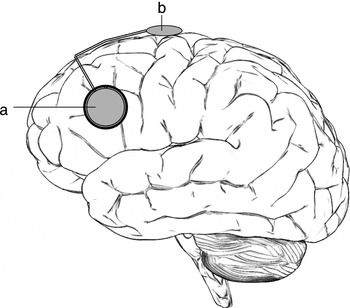
The infiltrative nature of high-grade gliomas makes complete surgical resection difficult, leading to local recurrence and treatment failure in the majority of cases. At the time of recurrence, treatment options include re-operation, chemotherapy, and various radiation therapy modalities. One such modality, the GliaSite radiation therapy system (RTS) (Figure 1), delivers iodine-125 (I-125) brachytherapy through a balloon applicator placed in the resection cavity at the time of surgery.
The GliaSite Radiation Therapy System (RTS). A distal balloon segment (a) is attached by a catheter to a subcutaneous injection port (b).

The GliaSite procedure is generally well tolerated, with only rare cases of infection and symptomatic radiation necrosis reported.Reference Tatter, Shaw and Rosenblum1,Reference Chan, Weingart and Parisi2 Hemorrhagic complications have not been described. We describe a case of intracranial hemorrhage (ICH) in a patient undergoing GliaSite RTS manipulation while receiving anticoagulation for a previously diagnosed pulmonary embolism. After reviewing the relevant literature, we propose measures to decrease such complications.
Case report
A 58-year-old man was diagnosed with a right parietal glioblastoma multiforme in June 2004. He underwent gross total resection, placement of Gliadel (BCNU, carmustine) wafers, and external beam radiation therapy (total 60 Gy) with daily concomitant temozolomide. This was followed by six cycles of monthly adjuvant temozolomide. During the first treatment cycle, he developed symptomatic, bilateral pulmonary emboli. He was anticoagulated with low molecular weight heparin (LMWH) followed by warfarin.
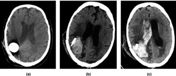
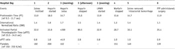
In April 2005, a routine follow-up magnetic resonance imaging scan demonstrated local tumor recurrence. In May 2005, he underwent a second resection and placement of GliaSite RTS. Approximately 6 weeks later, he was admitted to the inpatient oncology service for radioisotope loading, with a planned dose of 45 Gy to a depth of 0.5 cm. On hospital day 1, 15 ml of an aqueous solution of I-125 (Iotrex) was placed using a non-coring needle in the GliaSite subcutaneous port after a CT scan (Figure 2a) confirmed proper GliaSite placement. At the time, the patient remained on his outpatient warfarin dose, though he had a subtherapeutic international normalized ratio of 1.4 (typical therapeutic range 2–3) (Table 1).
Non-contrast head CT scans performed during hospitalization. (a) The GliaSite planning CT (Hospital Day 1) shows no hemorrhage. (b) Approximately 8 hours after Iotrex removal from the GliaSite RTS (Hospital Day 5), there is a 4.7 × 3.2 cm right parietal intracavitary hematoma outside the confines of the GliaSite balloon. (c) Approximately 13 hours after Iotrex removal from GliaSite RTS (Hospital Day 6), the right parietal hematoma has increased in size to 5.6 × 3.6 cm and dissected into the right ventricular system.

Clinical events and laboratory data during hospitalization

FFP, fresh frozen plasma; LMWH, low molecular weight heparin.
The patient reported a mild headache over the next 3 days, but had no other neurologic symptoms. In response to subtherapeutic anticoagulation with warfarin, an infusion of intravenous unfractionated heparin was started without bolus on hospital day 2. The follow-up activated partial thromboplastin time (aPTT) was subtherapeutic, and a heparin bolus was administered and the infusion rate increased. This led to a transiently supratherapeutic aPTT ratio (Table 1). Later that day, owing to lack of intravenous access, the unfractionated heparin infusion was discontinued and weight-based LMWH (enoxaparin 90 mg subcutaneously twice daily) was started.
On hospital day 4, warfarin, which had been administered since admission, was discontinued. On hospital day 5, the patient received his morning enoxaparin dose. Approximately 4 hours later, the Iotrex aqueous I-125 solution was removed from the GliaSite RTS using a non-coring needle in the subcutaneous port. Shortly after this procedure, the patient reported worsening headaches. These persisted despite analgesics, and a non-contrast head computed tomography was performed (Figure 2b). This demonstrated a right parietal intraparenchymal hematoma. At the time, the patient's international normalized ratio was 1.4, and the aPTT was normal (Table 1). The patient was transferred to the neurosurgical intensive care unit, where he received fresh frozen plasma.
On hospital day 6, the patient deteriorated clinically and was intubated. A repeat non-contrast head computed tomography (Figure 2c), performed approximately 5 hours after the previous scan, revealed dissection of the hematoma into the right ventricular system. The patient was taken to the operating room, where he underwent craniotomy, hematoma evacuation, removal of the GliaSite RTS, and intraventricular catheter placement. The platelet count, which had been normal throughout the hospitalization and was 149,000/ml at the time of hemorrhage, fell to 66,000/ml after surgery. Platelets were transfused. The patient did not recover and died on hospital day 14.
Discussion
Radiation therapy options are often limited in the treatment of recurrent gliomas. Because 80–90% of these tumors recur less than 2 cm from the resection bed,Reference Lee, Fraass and Marsh3,Reference Sneed, Gutin and Larson4 an area within the original radiation port, the risk of damage to normal brain tissue may preclude the use of further external beam radiation therapy. As a result, local radiotherapy techniques such as GliaSite RTS have been developed. Treatment with this radiotherapy device has been well tolerated. Although up to 40% of patients experience nausea and headache, these symptoms are typically self-limited.Reference Chan, Weingart and Parisi2 Less common but more serious adverse effects include wound infection, transient expressive aphasia, pseudomeningocele, infectious and chemical meningitis, and symptomatic radiation necrosis.Reference Tatter, Shaw and Rosenblum1,Reference Chan, Weingart and Parisi2 To date, there have been no reports of hemorrhagic complications, and the use of anticoagulation has not been commented upon in these studies.
Because venous thromboembolism occurs in up to 30% of individuals with brain tumors,Reference Ruff and Posner5,Reference Gerber, Grossman and Streiff7 the issue of anticoagulation arises frequently in this patient population. Despite the risk of ICH, anticoagulation is generally considered safe and more effective than mechanical treatment (such as vena cava filters) in these patients.Reference Ruff and Posner5–Reference Gerber, Grossman and Streiff7 However, the use of anticoagulation near the time of neurosurgical procedures may increase hemorrhagic complications. In a study using prophylactic LMWH started at the time of anesthesia induction, 5 of 46 patients (11%) suffered ICH.Reference Dickinson, Miller, Patel and Gupta8 ICH has also occurred in patients receiving anticoagulation after placement of Ommaya reservoirs.Reference Sandberg, Bilsky, Souweidane, Bzdil and Gutin9
In contrast to the invasiveness of neurosurgical procedures, the instillation and removal of Iotrex from the GliaSite RTS require only accessing a subcutaneous port with a small needle, a procedure performed in similar devices (such as intravascular mediports) in anticoagulated patients without difficulty. However, in the GliaSite RTS these actions may lead to a change in balloon contact with surrounding brain tissue. This is suggested by animal studies, in which the inflated GliaSite RTS balloon is highly conformal to the margin of the resection cavity, with up to 98% parenchymal contact on Magnetic resonance images.Reference Stubbs, Strickland, Frank, Simon, McMillan and Williams10 In the patient described here, Iotrex removal and balloon deflation occurred during a period of full anticoagulation with LMWH, which may have disrupted fibrin bands that had formed during the period of balloon inflation and initiated the bleeding event. However, as the patient received multiple forms of anticoagulation throughout the hospital course, the degree to which unfractionated heparin or warfarin may have also contributed to the hemorrhage cannot be determined.
These events suggest that the manipulation of the GliaSite balloon may create a risk of hemorrhage in anticoagulated patients, although the magnitude of this risk is not addressed by this case report. Depending upon clinical circumstances, precautions such as correcting thrombocytopenia and withholding anticoagulation (up to 6 hours for unfractionated heparin, up to 24 hours for LMWH, and up to several days for warfarin) before GliaSite manipulation should be considered. Particular care should be taken with patients receiving LMWH; because these agents do not affect routine coagulation parameters, assessing anticoagulation status requires a detailed review of medication administration.