Introduction
Acute invasive fungal sinusitis is a rapidly progressive angio-invasive infection of the paranasal sinuses with a propensity to spread to the surrounding vital structures, often resulting in a fatal outcome. Based on the classification by deShazo and colleagues,Reference deShazo, O'Brien, Chapin, Soto-Aguilar, Gardner and Swain1 acute invasive fungal sinusitis forms a distinct subset under the spectrum of invasive fungal sinusitis, and is the most common type. Acute invasive fungal sinusitis is caused most commonly by filamentous fungi of the order Mucorales and class zygomycetes. A lower proportion of cases (less than 30 per cent) are caused by Aspergillus species, which belong to the class Ascomycetes.Reference Bakhshaee, Bojdi, Allahyari, Majidi, Tavakol and Najafzadeh2,Reference Malleshappa, Rupa, Varghese and Kurien3 These fungi are ubiquitous in the environment; they can enter the human body and in the right circumstances cause invasive mycosis.
Acute invasive fungal sinusitis affects patients with uncontrolled diabetes mellitus (especially when associated with metabolic acidosis), profound and prolonged neutropenia, and haematological malignancies, patients who have undergone bone marrow or solid organ transplantation,Reference Roden, Zaoutis, Buchanan, Knudsen, Sarkisova and Schaufele4 those with iron overload,Reference Spellberg, Edwards and Ibrahim5 and patients receiving deferoxamine therapy.Reference Boelaert, de Locht, Van Cutsem, Kerrels, Cantinieaux and Verdonck6 The long-term prognosis is often dictated by the severity of immunosuppression and the degree of correction of various predisposing factors.
The presentation of acute invasive fungal sinusitis can be quite variable depending upon the primary area of involvement, along with the extent of disease at presentation. In the initial stages, symptoms may be very mild and similar to sinusitis. The subtle presenting symptoms compounded with the lack of collaborative findings on imaging often preclude early diagnosis. Lack of conspicuous sinonasal symptoms when overshadowed by prominent orbital and neurological symptoms can result in delayed diagnosis and initiation of treatment, affecting overall survival.
A high clinical suspicion is warranted in immunocompromised individuals presenting with acute sino-orbital symptoms.Reference Turner, Soudry, Nayak and Hwang7 Frozen section examination of tissues aids early and accurate diagnosis. Timely diagnosis helps initiate prompt and aggressive treatment in the form of urgent surgical debridement followed by antifungal therapy, which is the ‘gold standard’ treatment. When combined with simultaneous correction of patient-related immunosuppressive factors, overall survival and prognosis tend to improve.
Unfortunately, in spite of rapid improvements in surgical techniques and medical treatment, not only has the incidence of the disease increased considerably in the last decade,Reference Bitar, Van Cauteren, Lanternier, Dannaoui, Che and Dromer8 overall survival has also remained stagnant, at around 50–80 per cent.Reference Bakhshaee, Bojdi, Allahyari, Majidi, Tavakol and Najafzadeh2,Reference Malleshappa, Rupa, Varghese and Kurien3,Reference Monroe, McLean, Sautter, Wax, Andersen and Smith9
The recognition of prognostic factors early in the course of the disease can have a profound effect on the outcome. Conditions such as diabetes mellitusReference Turner, Soudry, Nayak and Hwang7 and acute myeloid leukaemiaReference Chen, Sheng, Cheng, Chen, Tsay and Tang10 are reported to have the worst prognosis. The causative fungi also have an effect on outcome, with Mucorales being the more virulent.Reference Hirabayashi, Idowu, Kalin-Hajdu, Oldenburg, Brodie and Kersten11 Patients with involvement of the hard palate, pterygopalatine fossa,Reference Zuniga and Turner12 brain and orbit are also reported to have poorer outcomes. Recent studies have additionally noted that increasing levels of C-reactive protein is a poor prognostic factor.Reference Cho, Jang, Hong, Chung, Kim and Dhong13
The present study aimed to analyse the various prognostic factors of acute invasive fungal sinusitis, primarily comparing patients with diabetes mellitus and those with underlying haematological conditions, using a large cohort from a single institution.
Materials and methods
Study design and participants
An observational study was conducted in a cohort of patients diagnosed with acute invasive fungal sinusitis at an ENT department of a tertiary care hospital in South India. The study period was from August 2014 to December 2019.
Inclusion criteria
All patients diagnosed with acute invasive fungal sinusitis based on histopathological evidence were included in the study.
Data collection
Details regarding demography, co-morbidities, prior treatment history, clinical features, diagnostic endoscopic findings, fungal smear and culture results, histopathological reports (including frozen section when performed), radiological findings, clinicoradiological staging, surgical procedures performed, antifungal therapy, complications during treatment, and outcome at the time of discharge and at last follow up, were collected.
Patient investigation
A detailed history and clinical examination were carried out for all patients with suspected acute invasive fungal sinusitis. The laterality, extent and stage of disease were ascertained by endoscopic examination and imaging. Endoscopic biopsy was performed in all patients at first presentation or intra-operatively. The biopsy was taken from the obviously diseased site, or from the middle turbinate if all areas looked grossly normal. Frozen section analysis was available only during the daytime and was hence performed only for those patients in whom the specimen was available for processing during that period. All patients underwent contrast-enhanced computed tomography of the paranasal sinuses. Additional magnetic resonance imaging of the brain and face was conducted for patients with suspected extra-paranasal sinus involvement.
From 2017 onwards, the surgical protocol followed was slightly different based on the findings from our previously published study.Reference Malleshappa, Rupa, Varghese and Kurien3 All patients who underwent surgery had endoscopic debridement of all sinuses bilaterally, irrespective of clinicoradiological evidence of disease. All sinuses were sampled separately and examined for the presence of disease. Patients were then given antifungal therapy depending on the organism identified. Emphasis was also given to management of the underlying conditions and rehabilitation of the patients.
Regular endoscopic inspection of the operative cavity was performed. Patients were declared disease-free once clinical findings were normal and repeat biopsy results were negative. Deaths during the course of treatment were determined, whether associated with the disease or other causes.
The histopathological reports of surgical specimens were analysed in order to group patients into those with anterior disease (sinuses draining into middle meatus) or posterior disease (posterior ethmoid and sphenoid sinuses). The final biopsy report was used to divide the patients into unilateral disease and bilateral disease groups.
Definitions, criteria and staging
Acute invasive fungal sinusitis was diagnosed by the presence of invasion of mucosa, submucosa, bone and/or blood vessels on histopathological examination of the biopsy specimen using haematoxylin and eosin as well as fungal stains.
Mixed fungal sinusitis was defined as two or more types of fungal sinusitis identified in the same specimen. Fungal co-infection was defined as two or more fungal species seen in the same patient on fungal culture or histopathology.
Staging of disease was performed to plan treatment. The clinicoradiological staging used in this study has been described previously.Reference Rupa, Maheswaran, Ebenezer and Mathews14 Broadly, stage 1 referred to disease confined to the nose and paranasal sinuses. Stage 2 referred to disease extending beyond the nose and paranasal sinuses to areas such as the anterior orbit, subcutaneous tissue, pterygopalatine fossa or palate, which was resectable. Finally, stage 3 referred to disease that had extended beyond the nose and paranasal sinuses and was unresectable (typically with intracranial, deep skull base or orbital apex involvement).
Follow-up details after discharge from hospital were obtained from the patients’ medical records or telephonically if the patient had not reported to the out-patient section for follow up.
A disease-free status was defined as the absence of initial symptoms, along with biopsy proven and/or radiological evidence of resolved disease. Patient outcome was defined as survival or mortality at last contact of follow up. Mortality was assessed as all-cause mortality during the course of the disease.
Statistical analysis
Descriptive analysis was used for the data description. Means, standard deviations and ranges were calculated for continuous variables, and numbers and percentages for categorical variables. Penalised logistic regression was used for univariate and multivariate analysis, to examine the relationship between the study variables and mortality. The results are presented as odds ratios with 95 per cent confidence limits. A p-value of less than 0.05 was considered as significant.
Ethical approval
Approval for the conduct of the study was obtained from the institutional review board (approval number: 10632).
Results
Demography
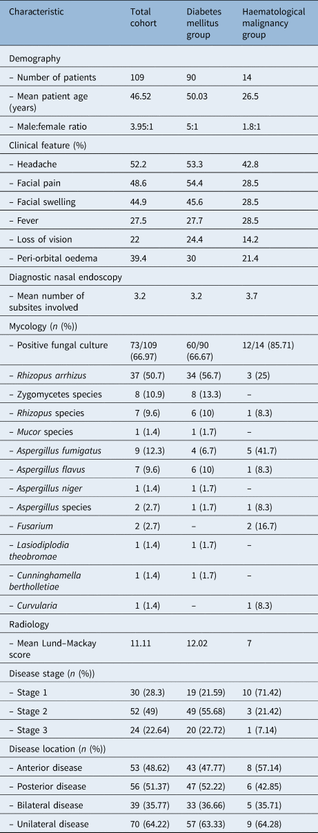
Based on the inclusion criteria, a total of 109 patients were included in the study, with a male to female ratio of 3.95:1 (Table 1). The male predominance was more pronounced in the diabetic subgroup. Patients’ ages ranged from 3 to 78 years (mean, 46.5 years), with 66.9 per cent of patients being in the 31 to 60-year age group. Patients in the haematological malignancy subgroup were younger, with a mean age of 26.5 years.
Patient characteristics

Ninety patients (82.6 per cent) had diabetes mellitus and 14 patients (12.8 per cent) had haematological malignancy. However, five patients had no underlying conditions. Among the diabetics, other associated major co-morbidities included 27 patients with hypertension, 7 with chronic kidney disease and 4 with co-existing human immunodeficiency virus (HIV) infection. The various haematological conditions seen were: acute lymphocytic leukaemia, in four patients; acute myeloid leukaemia, in three patients; and biphenotypic leukaemia, aplastic anaemia, plasma cell dyscrasia, auto-immune lymphoproliferative disorder, central nervous system lymphoma, B-cell lymphoma and thalassaemia major (post stem cell transplant), in one patient each.
Clinical features
Facial pain (54.4 per cent), headache (53.3 per cent) and facial swelling (45.6 per cent) were the most common symptoms (in descending order) among diabetic patients. In contrast, among those with haematological malignancy, headache (42.8 per cent) was more prevalent than other common symptoms such as facial swelling (28.5 per cent), fever (28.5 per cent) and facial pain (28.5 per cent). Nasal symptoms were comparatively less common, with only 24.4 per cent of diabetic patients and 21.4 per cent of those with haematological malignancy having nasal obstruction or nasal discharge. Peri-orbital swelling and loss of vision were the most encountered orbital symptoms in both groups.
Endoscopy
On diagnostic rigid nasal endoscopy, the middle meatus and middle turbinate were seen as pale or slough-covered areas in 46.3 per cent of patients. Fourteen patients (12.8 per cent) in our study had a normal appearance of mucosa on diagnostic endoscopy. The diabetic subgroup showed involvement of two or three nasal subsites, while only one subsite was involved in the majority (one-third) of patients with haematological malignancy.
Fungal smear and culture
Fungal samples of 73 of the 109 patients showed a positive fungal culture. The two main groups of fungi isolated on culture in the cohort were zygomycetes in 53 (72.6 per cent) and Aspergillus in 19 (26 per cent) culture-proven cases. The most frequently recorded species was Rhizopus arrhizus, in 37 of the 73 positive samples (50.68 per cent), while in 8 patients (10.9 per cent) zygomycetes remained unspecified. Among the patients who grew Aspergillus in their culture, the commonest species were Aspergillus fumigatus, in nine patients, and Aspergillus flavus, in seven patients. Atypical fungi like Fusarium were seen in two patients; Curvularia, Cunninghamella bertholletiae and Lasiodiplodia theobromae were seen in one patient each. Fungal co-infections were seen in five patients.
Zygomycetes was the aetiological agent in 81.67 per cent of the culture-positive diabetic patients, whereas in the haematological malignancy subgroup it accounted for only 33.33 per cent of patients. In contrast, Aspergillus species was the causative agent in 58.33 per cent of the haematological group, and was the aetiological agent in 20 per cent of diabetics.
Imaging
The most frequent sites of involvement were the maxillary (87.7 per cent) and ethmoid (87.2 per cent) sinuses, followed by the sphenoid sinus (77.35 per cent). Though maxillary and ethmoid sinuses were almost equally involved in diabetics, patients with haematological malignancy showed a predominant ethmoid sinus involvement. Involvement of the orbit was seen in 16 patients (15 per cent) and intracranial extension was noted in 22 patients (20.7 per cent). A comparison of both groups using Lund–Mackay scoring revealed that the diabetic group had much higher Lund–Mackay scores compared to the haematology group (score of 12 vs 7).
Histopathology
Frozen section analysis was conducted for 78 patients (71.5 per cent). Of these, 55 patients (70.5 per cent) had positive results. Based on histopathology, the maxillary sinus was involved in most patients (69 patients, 63.3 per cent). Anterior and posterior ethmoid sinuses were involved in 52 patients (47.7 per cent) and 44 patients (40.4 per cent) respectively. A positive biopsy finding was obtained for the frontal sinus in only nine patients.
Based on the histopathological reports, 20 patients (18.3 per cent) had mixed pathology. In addition to acute invasive fungal sinusitis, 10 patients had chronic invasive fungal sinusitis, 6 had chronic granulomatous fungal sinusitis, 2 had both chronic invasive fungal sinusitis and chronic granulomatous fungal sinusitis, 1 had co-existing chronic invasive fungal sinusitis and a fungal ball, and 1 had a fungal ball alone. Of the 20 patients with mixed infection, all except 1 patient were diabetic.
Ten diabetic patients (11.36 per cent) and four haematological disorder patients (30.76 per cent) showed a positive biopsy report from a radiologically normal sinus.
Disease stage
Most of the patients in the cohort had stage 2 disease. This was true for the diabetic subgroup also. However, 71.4 per cent of patients with haematological malignancy presented with stage 1 disease. Disease staging could not be carried out in three patients as the images were not available.
Disease location
Based on the final biopsy, the posterior sinuses (posterior ethmoid and sphenoid sinuses) were marginally more involved than anterior sinuses in diabetics, whereas 57.14 per cent of cases with underlying haematological malignancy showed anterior sinus involvement. In most patients (70 patients, 64.22 per cent), the disease was confined to one side. Extra-paranasal sinus extension to the infratemporal fossa was seen in 64.15 per cent of the total cohort, 72.72 per cent of the diabetic patients and 28.57 per cent of the haematological disorder patients.
Treatment
All patients except one underwent surgery, which varied depending on the extent of the disease. Surgery was endoscopic alone or performed in combination with an open approach, and was followed by systemic antifungal treatment. Almost all of our patients received amphotericin B initially. Some patients were switched from amphotericin B to voriconazole later on, based on culture results. Only 81 patients (74.3 per cent) completed the antifungal therapy; the rest discontinued at different stages for various reasons.
Outcome
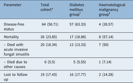
At the end of treatment, 64 patients (58.71 per cent) were disease-free. Nineteen patients (17.43 per cent) were lost to follow up. Twenty-six patients (23.85 per cent) died either because of acute invasive fungal sinusitis or due to other causes during the course of treatment. Of the 90 diabetic patients, 57 (63.33 per cent) survived, whereas only 4 of the 14 patients with underlying haematological conditions (28.57 per cent) had a favourable outcome (Table 2).
Patient outcome

Data represent numbers (and percentages). *n = 109; †n = 90; ‡n = 14
In the Mucorales group, 12 patients died of acute invasive fungal sinusitis or other causes, and 14 patients were lost to follow up. Hence, the survival rate was 50.94 per cent (27 out of 53). In the Aspergillus group, three patients were lost to follow up; nine patients showed a disease-free status, giving a survival rate of 47.36 per cent (9 out of 19).
Follow up
A total of 61 patients had a minimum of three months’ follow up post treatment. Of these, 18 patients had 3–6 months of follow up, whereas 6 patients had 6–12 months of follow up. Thirty-seven patients had more than one year of follow up. The longest follow-up duration was 72 months.
Logistic regression analysis for mortality
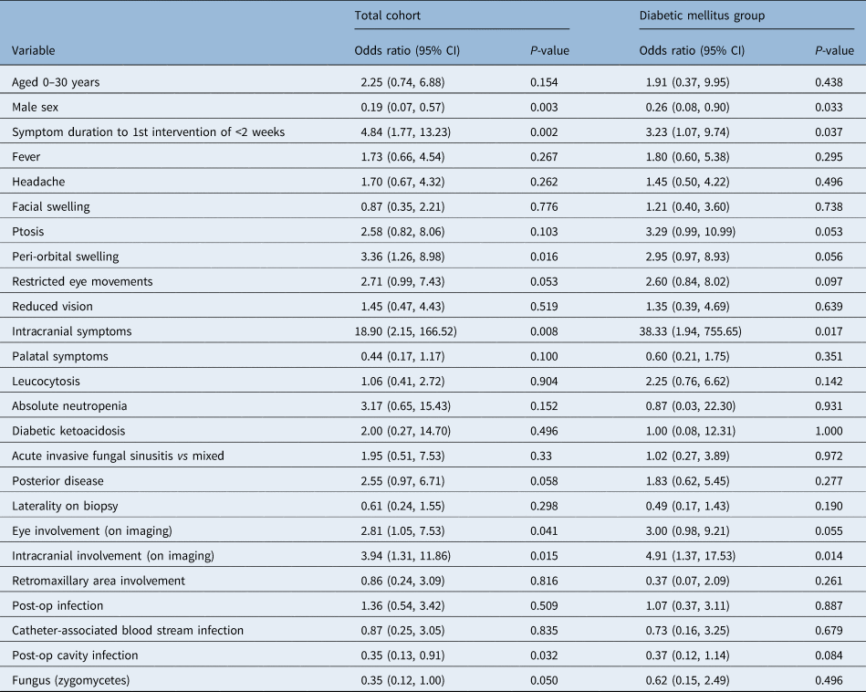
On univariate analysis of all patients except those lost to follow up, factors associated with increased mortality rate in our cohort were: female sex (p = 0.003), ocular findings like peri-orbital swelling (p = 0.016), restricted ocular movements (p = 0.053) and intracranial symptoms (p = 0.008). Other factors significantly associated with death in the cohort were: duration of less than two weeks between symptoms and first intervention (p = 0.002), posterior disease (p = 0.058), eye involvement on imaging (p = 0.041), intracranial involvement on imaging (p = 0.015), causative fungus being zygomycetes (p = 0.050), and any post-operative sinus cavity infection (p = 0.032).
Symptoms including fever, headache, facial swelling, ptosis, decreased vision and palatal involvement were not significantly associated with death from acute invasive fungal sinusitis. In addition, bilateral disease, retromaxillary area involvement and mixed infection failed to show significant association with adverse outcome from acute invasive fungal sinusitis. There was no significant relationship between the variables of leukocytosis, absolute neutropenia, diabetic ketoacidosis, catheter-associated blood stream infection and mortality.
In the diabetic cohort, however, ptosis showed a significant association (p = 0.053) with mortality (Table 3).
Univariate penalised logistic regression analysis for mortality

CI = confidence interval; post-op = post-operative
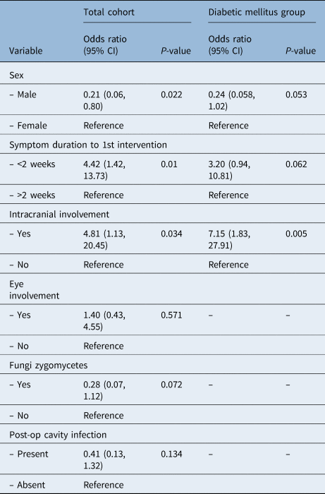
Multivariate analysis was performed using variables selected based on their significance in the univariate analysis. Female sex and intracranial involvement were significant factors for the whole cohort and the diabetic group too. A less than two-week duration between initial symptoms and first intervention was an independent significant factor for the full cohort; however, the association only neared significance (p = 0.062) for the diabetic cohort. Other factors that were strongly associated with poor prognosis on univariate analysis were not found to be significant on multivariate analysis (Table 4).
Multivariate penalised logistic regression for mortality

CI = confidence interval; post-op = post-operative
Discussion
The present study investigated clinical variables in acute invasive fungal sinusitis that affect patients’ overall survival. Female sex, a duration of less than two weeks between symptoms and first intervention, and intracranial involvement were independent predictors of poor survival. Other risk factors significantly associated with increased mortality were: clinical symptoms including peri-orbital swelling, restricted ocular movements and intracranial symptoms, posterior sinus involvement, radiological signs of eye involvement, causative fungus being zygomycetes, and any post-operative sinus cavity infection.
Acute invasive fungal sinusitis is mostly thought of as a disease affecting immunocompromised and immunosuppressed individuals; it is very uncommon in immunocompetent individuals.Reference DelGaudio, Swain, Kingdom, Muller and Hudgins15 A review of literature on acute invasive fungal sinusitis showed varied underlying conditions predominating the cohorts; in some articles, most patients had haematological malignancies,Reference Monroe, McLean, Sautter, Wax, Andersen and Smith9,Reference Gode, Turhal, Ozturk, Aysel, Midilli and Karci16–Reference Wandell, Miller, Rathor, Wai, Guyer and Schmidt19 while in others most of the cohort were diabetic.Reference Turner, Soudry, Nayak and Hwang7,Reference Cho, Jang, Hong, Chung, Kim and Dhong13 Our cohort had a predominance of diabetic patients (82.6 per cent), with some patients presenting with diabetic ketoacidosis or newly diagnosed diabetes mellitus. Five patients in the cohort had no co-morbidities.
There has been a reported increase in incidence of acute invasive fungal sinusitis over the last decade. This could be attributed to the increased prevalence of diabetes mellitus and HIV infection, an increase in the number of patients undergoing bone marrow and solid organ transplants, and the availability of better diagnostic methods.
The clinical course of the disease is known to be very aggressive, and published mortality rates in the literature vary from 20 to 80 per cent, with haematological disorder patients having a higher mortality rate.Reference Turner, Soudry, Nayak and Hwang7,Reference Cho, Jang, Hong, Chung, Kim and Dhong13,Reference Wandell, Miller, Rathor, Wai, Guyer and Schmidt19 Our cohort showed a survival rate of 58.71 per cent at the end of treatment. After excluding patients lost to follow up, 64 patients survived, equating to a survival rate of 71.1 per cent. A significant difference was seen in survival between diabetics (77 per cent) and patients with haematological conditions (33.33 per cent), which is similar to other reports in literature.Reference Bitar, Van Cauteren, Lanternier, Dannaoui, Che and Dromer8,Reference Cho, Jang, Hong, Chung, Kim and Dhong13,Reference Wandell, Miller, Rathor, Wai, Guyer and Schmidt19 This difference could be attributed to underlying predisposing factors in diabetic patients – such as ketoacidosis and hyperglycaemia – which are easier to reverse. Wandell et al. showed the presence of high haemoglobin A1c to be a good prognostic factor correlating with increased survival presumably because of its higher reversibility,Reference Wandell, Miller, Rathor, Wai, Guyer and Schmidt19 although this was not seen in our study. In addition, the presence of more resectable lesions in this population could explain the difference in survival rates. The immunocompromised state of an acute invasive fungal sinusitis patient makes them susceptible to other life-threatening infections as well during the course of the treatment, sometimes causing mortality. Hence, determination of disease-specific survival is difficult.
Although advanced age and male gender have been shown, in various cohorts,Reference Monroe, McLean, Sautter, Wax, Andersen and Smith9,Reference Payne, Mitzner, Kunchala, Roland and McGinn18 to worsen survival,Reference Turner, Soudry, Nayak and Hwang7 the absence of significant effects of age and sex has also been reported.Reference Gode, Turhal, Ozturk, Aysel, Midilli and Karci16 Older age is associated with poor functional status and decreased physiological reserves, which may lead to poor outcomes. Although the cohort was predominantly male, contrary to many reports, female gender was an independent variable associated with a fatal outcome in our study. The diabetic cohort also showed the same association. Early and more aggressive intervention should be attempted in female patients, to lessen the prognostic discrepancy between the two sexes.
The presenting symptoms were often quite varied, with a conspicuous lack of nasal symptoms. Headache, followed by facial pain and facial swelling, were the most common symptoms in our study, similar to the findings of Wandell et al.Reference Wandell, Miller, Rathor, Wai, Guyer and Schmidt19 In a study by Payne et al., on a cohort predominantly comprising haematological malignancy patients, fever was the most common presenting symptom;Reference Payne, Mitzner, Kunchala, Roland and McGinn18 in our study, fever was seen only in 27.5 per cent of patients. Headache, cranial nerve palsy, visual loss and orbital pain were the most common presenting symptoms in a study by Cho et al. that comprised a greater number of diabetics (51 per cent),Reference Cho, Jang, Hong, Chung, Kim and Dhong13 similar to our study. In the only published systematic review on acute invasive fungal sinusitis, by Turner et al., most patients presented with facial swelling, fever and nasal congestion.Reference Turner, Soudry, Nayak and Hwang7
A duration of less than two weeks between symptom onset and first intervention was independently associated with increased mortality (odds ratio = 4.42, 95 per cent confidence interval = 1.42–13.73, p = 0.01), indicating the aggressive nature of the disease. Similar results were shown by Pirochamai et al.Reference Piromchai and Thanaviratananich20 Cho et al.Reference Cho, Jang, Hong, Chung, Kim and Dhong13 showed negative survival (mortality) with a shorter duration of symptoms, indicating the crucial role of early diagnosis and prompt intervention. However, DelGaudio et al. showed that rapid diagnosis and treatment improved only morbidity, and may not have a similar impact on mortality.Reference DelGaudio and Clemson21 Conversely, one report showed the absence of any significant impact of prompt intervention on survival with a mean delay in treatment of 16.6 ± 7.7 days.Reference Saedi, Sadeghi and Seilani22 Surprisingly, Wandell et al. showed that a longer duration between symptom onset and the initiation of treatment improved survival.Reference Wandell, Miller, Rathor, Wai, Guyer and Schmidt19 This was explained by the presence of more indolent disease with very subtle symptoms in the subset of patients for whom it took a longer time to diagnose and initiate treatment, compared to the patients with more aggressive disease in whom treatment was initiated urgently.
Zygomycetes (48.62 per cent) and Aspergillus (17.43 per cent) were the two main fungi isolated on culture in the cohort, which is similar to findings reported in recent literature.Reference Turner, Soudry, Nayak and Hwang7,Reference Monroe, McLean, Sautter, Wax, Andersen and Smith9,Reference Cho, Jang, Hong, Chung, Kim and Dhong13,Reference Gode, Turhal, Ozturk, Aysel, Midilli and Karci16,Reference Payne, Mitzner, Kunchala, Roland and McGinn18 Although many studies have not related a particular fungus with a poor prognosis,Reference Valera, do Lago, Tamashiro, Yassuda, Silveira and Anselmo-Lima23 a few have reported the contrary. Burton et al. reported increased mortality associated with Mucorales species owing to the increased presence of iron chelators depleting the host iron stores.Reference Burton, Jafari, Asmerom, Swisher, Gabriel and DeConde24 This was compounded in the presence of neutropenia and was found to worsen the prognosis.Reference Fernandez, Crocetta, Demattè, Farneti, Stanzani and Lewis25 In addition, the incidence of orbital and neurological involvement was found to be more prevalent with Mucorales infection.Reference Ingley, Parikh and DelGaudio26 However, Cho et al. found no significant differences between Aspergillus and Mucor in patients with diabetes or haematological malignancies.Reference Cho, Jang, Hong, Chung, Kim and Dhong13 Atypical fungi are known to have a very poor prognosis. Fusarium is said to be resistant to amphotericin and is associated with 80 per cent mortality. In our study, the two patients with haematological malignancy whose culture grew Fusarium both died due to rapid progression of disease. Mixed infections were observed in 18.3 per cent of the study cohort, which was much higher than reported.Reference Wandell, Miller, Rathor, Wai, Guyer and Schmidt19
The angio-invasive character of the disease predisposes rapid spread to extra-paranasal sites, often carrying a grave prognosis. In our study, patients with intracranial or intra-orbital involvement had a poor prognosis, both in the entire cohort and in the diabetic subgroup. In the literature, intracranial involvement and associated cranial neuropathy have been associated with shortened survival.Reference Turner, Soudry, Nayak and Hwang7,Reference Monroe, McLean, Sautter, Wax, Andersen and Smith9 However, Gode et al. have reported otherwise.Reference Gode, Turhal, Ozturk, Aysel, Midilli and Karci16 The impact of orbital involvement on survival is also debatable, with some reports showing decreased survival in patients with visual loss and ophthalmoplegia.Reference Monroe, McLean, Sautter, Wax, Andersen and Smith9,Reference Bhansali, Bhadada, Sharma, Suresh, Gupta and Singh27 On the contrary, some studies have reported an absence of a significant impact on survival in patients who underwent orbital exenteration,Reference Hargrove, Wesley, Klippenstein, Fleming and Haik28 and a lack of poor prognosis associated with orbital involvement.Reference Turner, Soudry, Nayak and Hwang7
• Orbital or facial symptoms in immunocompromised patients should warrant a high index of suspicion of acute invasive fungal sinusitis
• Sinonasal symptoms and radiological findings are often subtle
• Female sex, intracranial involvement and less than two weeks between symptom onset and treatment initiation are independent factors associated with increased mortality
• Orbital or intracranial symptoms, posterior sinus disease, and causative organism being zygomycetes are poor prognostic factors
• Early debridement and antifungal therapy improve overall survival, even in patients with bilateral, palatal or retromaxillary involvement
• The association between post-operative sinus cavity infection and mortality highlights the importance of regular nasal and sinus cavity cleaning and surveillance
Nasal septalReference Saedi, Sadeghi and Seilani22,Reference Foshee, Luminais, Casey, Farag, Prestipino and Iloreta29 and palatalReference Gode, Turhal, Ozturk, Aysel, Midilli and Karci16 involvement have been reported to increase mortality. This, however, was not seen in our study, probably because of the feasibility of a complete excision. Involvement of the pterygopalatine fossa, an early sign of acute invasive fungal sinusitis detected by loss of fat stranding on imaging is another reservoir of fungal burden.Reference Gamba, Woodruff, Djang and Yeates30 Debridement of this area has been shown to improve survival.Reference Hosseini and Borghei31 Retromaxillary area involvement, though seen in 64.15 per cent of patients, was not associated with any increased adverse outcome in our study.
Acute invasive fungal sinusitis is mainly seen in immunocompromised patients,Reference DelGaudio, Swain, Kingdom, Muller and Hudgins15 primarily owing to the poor neutrophilic response. In haematological malignancies, neutropenia (absolute neutrophil count of less than 1000 per microlitre of blood) is a known risk factor.Reference DelGaudio and Clemson21 However, prolonged neutropenia was not reported as an actual negative prognostic factor in some reports.Reference Turner, Soudry, Nayak and Hwang7 Neutropenia, leucocytosis and diabetic ketoacidosis failed to have any significant impact on mortality in the present study cohort. However, patients who developed post-operative cavity infection had a significant risk of mortality (p = 0.032). Meticulous post-operative follow up, with saline nasal douching and cavity clearance, is required to reduce the risk of post-operative cavity infection, thus improving the overall outcome.
Conclusion
Acute invasive fungal sinusitis requires a high index of suspicion in immunocompromised patients presenting with predominantly facial and orbital symptoms but subtle sinonasal symptoms. The recognition of prognostic factors helps to stratify and counsel patients, and aids planning of early medical and surgical treatment. Female sex, a duration of less than two weeks between symptoms and first intervention, the presence of any orbital or intracranial symptoms, posterior sinus disease, and the causative organism being zygomycetes are poor prognostic factors warranting urgent and more aggressive intervention. Early debridement followed by the initiation of antifungal medication will improve overall survival, even in patients with bilateral, palatal or retromaxillary involvement. The significant association between post-operative sinus cavity infection and mortality highlights the importance of nasal and sinus cavity care after debridement.
Competing interests
None declared