Case report
A 1-year-old male patient who had undergone a Norwood Stage 1 operation with Sano modification for hypoplastic left heart syndrome was admitted to the paediatric cardiology department for regular follow-up prior to a Norwood Stage 2 operation. The patient was asymptomatic except for mild cyanosis. On examination, a large ascending aortic pseudoaneurysm was detected via echocardiography and this finding was confirmed with conventional angiography and thorax CT angiography.
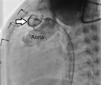
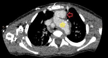
Conventional angiography and thorax CT angiography revealed that the pseudoaneurysm was 18.5 × 13.3 mm in diameter and arising from the anterior surface of the ascending aorta before the branching of the innominate artery (Figs 1–2). There was no compression of neighbouring structures. Since the sudden rupture of an aortic pseudoaneurysm might be life-threatening, aneurysmectomy with Norwood Stage 2 operation was immediately performed after obtaining informed consent.
Conventional angiography of the patient; arrow indicates the pseudoaneurysm in ascending aorta.

CT angiographic image of the patient; red arrow shows the pseudoaneurysm in the ascending aorta; The asteriks represents the transverse aorta.

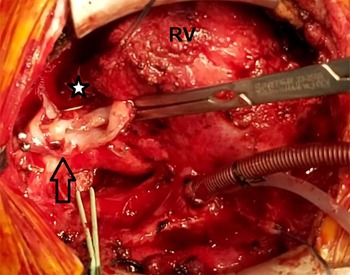
The patient was cannulated from the right common carotid artery and the right internal jugular vein. Cardiopulmonary bypass was initiated. After an uneventful sternal re-entry, right atrium was directly cannulated. The right pulmonary artery was looped and the patient was cooled to 26°C.The sac was clamped and incised, however, the intact aortic endothelium could not be reached and complete resection of pseudoaneurysm was failed (Fig 3). It was thought that primary suture repair was not possible and it was decided to perform an open repair.
Intraoperative image of the patient; RV; right ventricle, arrow indicates the resected pseudoaneurysm sac. Asterisk shows Sano shunt.

Then, the patient was cooled to 20°C and deep hypothermic circulatory arrest with antegrade selective cerebral circulation was established. The clamp under the sac was removed and the pseudoaneurysm was opened with ascending aorta. The pseudoaneurysm was originated from the anterior suture line of the transverse arcus patch, and there was no infectious finding in the pseudoaneurysm. The pseudoaneurysm was completely resected and the incision was extended to the distal arcus aorta. It was observed that the prior xenograft was completely calcified and native aortic tissue around the patch had become fragile. The defect was repaired with a bovine pericardium. Following aneurysmectomy, closure of Sano shunt combined with unilateral (right) bidirectional cavopulmonary anastomosis was performed during rewarming. The patient was disconnected from bypass with minor inotropic support and transferred to the intensive care unit.
Tissue culture was negative and the post-operative period of the patient was unremarkable. The patient was discharged on post-operative day 10.
Discussion
Pseudoaneurysms of the ascending aorta are uncommon but life-threatening due to the risk of spontaneous rupture. The incidence is less than 0.5% of all patients undergoing cardiac operations. It is detected much less frequently in the paediatric population, however, the exact incidence is unknown. There are only few paediatric case reports published in the English literature in which surgical treatment of ascending aortic pseudoaneurysm and Norwood Stage 2 palliation concurrently performed.
Immunological deficiencies, aortic wall or leaflet defects, trauma, congenital heart diseases, aortic dissection, tissue necrosis due to biological glue, hypertension, sepsis, aortic cannulation and cardioplegia sites, suture lines related to previous surgery are the known predisposing factors for the development of aortic pseudoaneurysm.
Ascending aortic pseudoaneurysms are either asymptomatic or have non-specific symptoms due to their mass effects such as dyspnoea, haemoptysis, chest pain, sepsis, respiratory failure, cardiogenic shock and thrombus.Reference Kanter, Mahle, Vincent and Berg1,Reference Masri, Majdalani and Bitar2 The diagnosis is based significantly on a high index of suspicion or mediastinal imaging for symptoms.Reference Atiyah, Mohsin, Al Faraidi, Al-Hawri, Al Otay and Al Najashi3
The timing and mode of treatment of ascending aortic pseudoaneurysms are controversial. Due to the risk of spontaneous rupture, regardless of size or site, some authors prefer carrying out the surgical or percutaneous treatment as soon as the diagnosis is established.Reference Malec, Mroczek, Gorecki and Kordon4,Reference Venkataraman, Thangavelu and Muthukumar5 Others prefer pre-treatment with antibiotics and delay the operation in order to avoid an infected operation field. They suggest that the inflamed and fragile nature of the infected aortic tissue may cause aortic graft re-infection and weakness of suture lines. The surgical treatment of aortic pseudoaneurysm is a high-risk procedure and the mortality ranges from 29 to 46 %. Massive haemorrhage due to the rupture of a pseudoaneurysm during sternal reentry may lead to sudden death especially when the pseudo-aneurysm is larger than 55 mm in diameter in adults.Reference Li, Zhou and Zhang6 The most important part of successful surgical treatment is the opening of the chest without cardiac injury and preventing exsanguination to avoid brain injury. The use of various perfusion techniques during sternal re-entry has been reported in paediatric patients.Reference Masri, Majdalani and Bitar2,Reference Atiyah, Mohsin, Al Faraidi, Al-Hawri, Al Otay and Al Najashi3,Reference Mustafa, Neijenhuis, Furci and Tsang7
We cannulated the common carotid artery and the internal jugular vein prior to opening the chest in our patient. Neck cannulation is a well- known and useful technique in paediatric patients with small femoral arteries and veins and is effective by inserting a large enough cannula to initiate cardiopulmonary bypass.Reference Mustafa, Neijenhuis, Furci and Tsang7 With the support of cardiopulmonary bypass, the heart and the ascending aorta became decompressed. The sternal re-entry was uneventful with careful dissection of surrounding tissue of the ascending aorta and pseudoaneurysm.
Surgical repair of an ascending aortic aneurysm is the conventional management approach and considered the gold standard treatment. However, in certain patients with an extremely high risk of surgical mortality, such as immunocompromised heart transplant recipients, these patients may be managed by catheter based interventions, such as Amplatzer device occlusion, stents, vascular plugs, coil embolisation and thrombin injection of the pseudoaneurysm. It has been advised that these devices should not be used in patients with an ongoing infectious process. Due to the fact that this technique requires extreme expertise to perform, it is considered as an alternative technique in patients with a high risk of surgical mortality.Reference Kanter, Mahle, Vincent and Berg1,Reference Masri, Majdalani and Bitar2
In our patient, we opted for surgical repair of the pseudoaneurysm because the patient was already scheduled for Norwood Stage 2 operation.
During the operation, we noted that the pseudoaneurysm had arisen from the anterior suture line of the xenograft bovine patch which was anastomosed to the diminutive transverse arcus aorta. The resected tissue was very fragile and the remaining aortic patch material was heavily calcified. The development of the pseudoaneurysm may have been related to an improper suture technique or abnormal elastic properties of either the calcified xenograft material or the remaining native aortic wall. It has been suggested that when the aortic tissue is reconstructed with non-viable material it is more exposed to the negative effects of shear forces than other aortic tissues.Reference Masri, Majdalani and Bitar2 Moreover, in a histological study, elastic fragmentation and fibrotic changes were more commonly noted in the heart walls of patients with hypoplastic left heart syndrome when compared to normal controls.Reference Jiang, Xu, Tang and Xia8 This histological abnormality inherent to hypoplastic left heart syndrome may have caused the development of the post-operative pseudoaneurysm.
There are few case reports regarding concurrent surgical treatment of ascending aortic pseudoaneurysm and Norwood Stage 2 operation. We believe that this case report will contribute to the literature on this rare association.
Acknowledgements
None.
Financial support
The clinicians received no grants from any funding agency and no support from any commercial or not-for-profit sector.
Conflicts of interest
None.
Ethical standards
The authors assert that this work complies with the ethical standards of the relevant national guidelines on human experimentation and with the Helsinki Declaration of 1975, as revised in 2008. This case was approved by the patient’s family.