Implications
The oxidative damage of broilers exposed to reactive oxygen species (ROS) can reduce the growth performance, which hinders the development of poultry industry. The present study showed that oxidative stress induced by hydrogen peroxide (H2O2) had a negative effect on histomorphology and redox status in the liver of broilers while underlying oxidative damage caused a decline in growth performance. This may attribute to apoptosis and autophagy processes triggered by excessive ROS that suppress the nuclear factor-κB (NF-κB) signaling pathway. The results will provide important evidence for our ultimate understanding of alleviating broiler oxidative stress so as to establish strategies for scientific management and nutrition regulation in the broiler industry.
Introduction
The oxidative stress was first defined as ‘a disturbance in the pro-oxidant-antioxidant balance in favor of the former.” It is regarded as an event of increased levels of ROS in the cell. The oxidative stress may lead to oxidative damage when the excessive ROS attack almost all the cellular macromolecules, which impairs the development, growth and reproduction of animals (Cichoż-Lach and Michalak, Reference Cichoż-Lach and Michalak2014; Sies et al., Reference Sies, Berndt and Jones2017). Poultry is more susceptible to oxidative stress in the context of the negative impact of genetic selection and intensive breeding (Zhang et al., Reference Zhang, Xiao, Lee and Ahn2011; Sihvo et al., Reference Sihvo, Immonen and Puolanne2013). Previous studies showed that oxidative stress had a deleterious effect on growth performance of broilers (Zhang et al., Reference Zhang, Hu, Lu, Yang, Zhang and Wang2015 and Reference Zheng, Wu, Song, Zhang, Zhang, Zhang, Zhang, Wang and Wang2016). Oxidative stress could cause systemic hypoxemia to reduce individual growth, which indicated that the growth performance may be associated with oxidative processes in broilers (Kassahn et al., Reference Kassahn, Crozier, Pörtner and Caley2009). In an organism, the liver is demonstrated to be a primary target organ attacked by ROS (Sanchez-Valle et al., Reference Sanchez-Valle, Chavez-Tapia, Uribe and Mendez-Sanchez2012). Yet, the molecular mechanism of oxidative damage in the liver of broilers remains unclear. Thus, investigating the mechanism of oxidative damage in the liver of broilers could help in assessing the relationship between oxidative stress and final growth performance.
The oxidative study has always been particularly topical in poultry science. However, little information is available on oxidative stress induced by increased ROS levels directly. Hydrogen peroxide is highly reactive and results in the generation of other ROS (e.g. hydroxyl radicals), which is well suited for redox sensing and redox signaling (Weidinger and Kozlov, Reference Weidinger and Kozlov2015; Sies et al., Reference Sies, Berndt and Jones2017). Thus, it has been reported that the administration of H2O2 could directly induce oxidative stress in broilers. Oxidative stress is demonstrated to induce cell death and the ROS are also responsible for the rebuilding of the liver (Cichoż-Lach and Michalak, Reference Cichoż-Lach and Michalak2014). Moreover, apoptosis can regulate the cell number, which is induced by ROS in numerous cells (Simon et al., Reference Simon, Haj-Yehia and Levi-Schaffer2000). On the other hand, autophagy is demonstrated to play a major role in hepatocyte homeostasis affected by high levels of metabolic stress (Ueno and Komatsu, Reference Ueno and Komatsu2017). In addition, the key factors of NF-κB signal pathway are involved in system-wide oxidative stress response and play an important role in apoptosis and autophagy (Zhong et al., Reference Zhong, Umemura, Sanchez-Lopez, Liang, Shalapour, Wong, He, Boassa, Perkins, Ali, McGeough, Ellisman, Seki, Gustafsson, Hoffman, Diaz-Meco, Moscat and Karin2016; Kondylis et al., Reference Kondylis, Kumari, Vlantis and Pasparakis2017; Sies et al., Reference Sies, Berndt and Jones2017). Recently, It has been confirmed that oxidative stress induced by aflatoxin B2 triggered apoptosis and autophagy in broiler hepatocytes, and the initiation of autophagy was mediated by NF-κB signal pathway in the intestine of piglets exposed to H2O2 (Yin et al., Reference Yin, Duan, Cui, Ren, Li and Yin2015; Chen et al., Reference Chen, Li, Guo, Xie and Chen2016). However, little information about the occurrence of cellular apoptosis and autophagy through the ROS generation in NF-κB signal pathway was reported in the liver of broilers exposed to H2O2. In the current study, we investigate the change of growth performance of broilers exposed to H2O2. We further explored the occurrence of apoptosis and autophagy, as well as the expression of NF-κB in these signaling pathways, by increasing body ROS levels. Our results provided experimental evidence for possible molecular mechanisms of oxidative damage in the liver of broilers.
Material and methods
Preparation of hydrogen peroxide solution
The various concentrations (2.5%, 5.0% and 10.0%) of H2O2 solution were obtained by serial dilutions of H2O2 solution (30.0%; Sigma-Aldrich Co. Ltd, Shanghai, China) with 0.75% sodium chloride buffer (saline). The H2O2 solutions were protected from light in a dry place after preparation. The concentration was determined using commercially available assay kits (A064; Nanjing Jiancheng Bioengineering Institute, Nanjing, China).
Experimental design, animals, and management
The experimental design and procedures were approved by the Animal Care and Use Committee of Nanjing Agricultural University, Nanjing, China. A total of 320 1-day-old male Arbor Acres broiler chickens were obtained from a commercial hatchery (Hewei Agricultural Development Co. Ltd, Xuancheng, China) with an average weight of 50.12±0.05 g. Chicks were weighed and randomly distributed to five treatments. In total, 40 cages were provided for the five treatment chicks, and there were eight replicated cages per treatment with eight broilers per cage (110×60×50 cm). All birds were raised on a basal diet formulated according to the nutrient requirements of Arbor Acres broilers for 1 to 21 days starter diet and 22 to 42 days grower diet. The composition and nutrition levels of the basal diet are given in the Supplementary Table S1. The control treatment received no injection; birds in saline treatment received intraperitoneal injection of physiological saline (0.75%, 1.0 ml/kg BW); birds in H2O2 treatments (H2O2(0.74), H2O2(1.48) and H2O2(2.96)) received an intraperitoneal injection of H2O2 with 0.74, 1.48 and 2.96 mM/kg BW, respectively. All birds in saline and H2O2 treatments received an injection at days 16 and 37 during the experimental period. Birds were allowed ad libitum access to mash feed and water in three-layer cages in an environmentally controlled room. All birds were enjoyed continuous fluorescent illumination with 23 h light and 1 h dark throughout the 42 days experiment. The temperature was maintained at 32°C to 34°C from day 1 to 3, and then gradually reduced to a final temperature of 20°C at the rate of 2°C to 3°C per week. Birds were weighed after feed deprivation for 12 h, and feed intake were recorded by replicate at the days 21 and 42 for the calculation of feed/gain ratio during the experiment.
Sample collection and preparation
At the end of the experiment, one bird with a BW close to the mean BW of the replicate (cage) (total eight birds per treatment) was selected and weighed. Then the bird was euthanized by cervical dislocation and necropsied immediately for liver tissue collection. Each sample from the left lobe of the liver was sliced into different sections. The main slice of the liver was used for ROS detection, histology evaluation, and detection of the apoptotic cells. Another slice was frozen in liquid nitrogen immediately and stored at −80°C until further analysis.
Detection of reactive oxygen species
A sensitive fluorescent dichlorofluorescein-diacetate probe was used to detect the intracellular ROS concentrations by the ROS assay kit (E004; Nanjing Jiancheng Bioengineering Institute) according to the manufacturer’s instruction. The formation of the fluorescent-oxidized derivative of dichlorofluorescein was monitored using FACS Calibur flow cytometer (FC500 MCL/MPL; Beckman Coulter Inc., Los Angeles, CA, USA) with excitation at 488 nm and emission at 525 nm. The ROS generation was quantified by the mean fluorescence intensity over that of the control.
Hematoxylin–eosin staining
Hepatic samples were fixed in paraformaldehyde solution (4.0%) and tissue sections were embedded in paraffin embedding medium, then serially sectioned at 6 μm. The dewaxing sections were stained with hematoxylin–eosin and evaluated under a conventional light microscope (Olympus BX50 microscope; Olympus Corporation, Tokyo, Japan). Images were captured with Evolution LC color light camera (Media Cybernetics Inc., Rockville, MD, USA).
Measurement of tissue oxidative stress parameters
The total antioxidant capacity, the activities of total superoxide dismutase and glutathione peroxidase were determined using commercially available assay kits (A015, A001, and A005, respectively; Nanjing Jiancheng Bioengineering Institute), and following the manufacturer’s instructions. The content of protein carbonyl was estimated by the reaction between the substrate with 2,4-dinitrophenylhydrazine using the commercial kit A087 (Nanjing Jiancheng Bioengineering Institute). The concentrations of 8-hydroxy-2'-deoxyguanosine and advanced oxidation protein products were measured by commercial ELISA kits (Nanjing Angle Gene Biotechnology Institute, Nanjing, China) as the manufacturer’s instructions. The content of malondialdehyde was quantified using the commercial kit A003 (Nanjing Jiancheng Bioengineering Institute). The content of total hepatic protein was determined by the method of Coomassie brilliant blue using the commercial kit A045-2 (Nanjing Jiancheng Bioengineering Institute).
Terminal deoxynucleotide-transferase-mediated deoxyuridine triphosphate nick end labeling assay
Hepatic samples were fixed in paraformaldehyde solution (4.0%) and tissue sections were embedded in paraffin, then serially sectioned at 6 μm. To detect DNA fragmentation, a terminal deoxynucleotide-transferase-mediated deoxyuridine triphosphate nick end labeling assay was performed using an in situ cell death detection kit (Vazyme Biotech Co., Ltd, Nanjing, China) according to the manufacturer’s instructions. After mounting terminal deoxynucleotide-transferase-mediated deoxyuridine triphosphate nick end labeling-positive cells, the nuclei were counterstained with DAPI, and the sections were observed at 400× magnification under a conventional light microscope (Olympus BX50 microscope). The nuclear color of normal cells was blue, whereas the nuclear color of apoptotic cells was nigger-brown. Images were captured with Evolution LC color light camera. The positive cells were quantified using Image-Pro Plus 5.1 (Media Cybernetics, Inc.). Five slices were measured in each treatment and five randomly chosen microscopic areas were measured in each slice. Then the numbers were counted and averaged according to Mozdziak et al. (Reference Mozdziak, Evans and McCoy2002). The rate of apoptotic to hepatic cells were calculated, with an average of the ratios as the representative value for replicate.
RNA extraction and real-time quantitative PCR
Total RNA was isolated using Trizol reagent (TaKaRa Biotechnology Co. Ltd, Dalian, China). The purity and quantity of total RNA were assessed by a NanoDrop 1000 spectrophotometer (Thermo Scientific, Wilmington, DE, USA). The total RNA was treated with DNase I (TaKaRa Biotechnology Co. Ltd) to remove DNA and reverse transcribed to complementary DNA with Veriti 96-Well Thermal Cycler (Model 9902; Applied Biosystems, Singapore City, Singapore) using a Prime Script RT Master Mix kit (TaKaRa Biotechnology Co. Ltd). Real-time quantitative PCR was performed using QuanStudio5 RT-qPCR detection system (Applied Biosystems Co. Ltd, Foster City, CA, USA) and SYBR Premix Ex Taq Kits (TaKaRa Biotechnology Co. Ltd). The PCR program used was as follows: 95°C for 30 s, followed by 40 cycles of 95°C for 5 s and 60°C for 30 s, then 95°C for 15 s, 60°C for 1 min, 95°C for 15 s. Each sample was measured in triplicate and the gene expression was calculated using the
 $$2^{{{\minus}\Delta \Delta C_{t} }} $$
method. Details of primers were as follows: CAGCCAACTGGGAGGTGTAT (forward) and GCTGCTTGGAGCTTTCATTC (reverse) for NF-κB subunit 1 (p50); TCTCCCAGCCCATCTATGAC (forward) and CCTCGATGTCCTCTTTCTGG (reverse) for v-rel reticuloendotheliosis viral oncogene homolog A (RelA); GACAACGGAGGATGGGATG (forward) and CAGGCTCAGGATGGTCTTCA (reverse) for B-cell CLL/lymphoma 2 (Bcl-2); CAACAGGAAGAACACGCTGA (forward) and TCAGTCTCGGCCCACTATCT (reverse) for Bcl-2 associated X (Bax); ACAGCAAGCGAAGCAGTTTT (forward) and TCACCTCTGAAAAGGCTGGT (reverse) for caspase-3; CAGCAAACCTACACCAACCA (forward) and CGCAACTCCTCTTCTCTGATG (reverse) for caspase-6; TCTCCTACAGAAGCCCAAGC (forward) and TCTTGCTGCTCACCTCTTGA (reverse) for caspase-8; ATTCCTTTCCAGGCTCCATC (forward) and CACTCACCTTGTCCCTCCAG (reverse) for caspase-9; CTTGGACTGTGTGCAGCAAT (forward) and ACCACCACTGCCACCTGTAT (reverse) for autophagy-related gene 6 (Beclin1); GCTGGACAAGACCAAGTTCC (forward) and TTCACCAGCAGGAAGAAAGC (reverse) for microtubule-associated protein 1 light chain 3 (LC3)-I; ATCCGAGATCAGCATCCAAC (forward) and GTGATCTGGCACCAGGAACT (reverse) for LC3-II; GAGGGTAGTGAAGGCTGCTG (forward) and CATCAAAGGTGGAGGAATGG (reverse) for GAPDH.
Western blot analysis
Frozen samples of the liver were powdered in liquid nitrogen with a mortar and pestle, and the homogenates were centrifuged for collecting supernatants. The protein concentrations were determined by using the BCA protein assay kit (Sangon Biotech Co. Ltd, Shanghai, China). Total cellular protein (60 μg) was electrophoresed with a SDS-polyacrylamide gel and transferred to a polyvinylidene difluoride membrane. Membrane blocking was conducted with 5.0% fat-free milk for 1 h. After washing, the membrane was incubated with primary antibodies overnight at 4°C with gentle shaking. Then the membrane was incubated with a second antibody of horseradish peroxidase-conjugated (1 : 3000; Cell Signaling Technology Inc., Beverly, MA, USA) at room temperature for 3 h. The primary antibodies for total caspase-3 (1 : 500; product number: 9662S), total LC3-II (1 : 1000; product number: 2775S), total NF-κB (1 : 1000; product number: 12540S) and β-actin (1 : 1500; product number: 3700S) were from Cell Signaling Technology Inc. The membrane was developed using Super Signal West Pico chemiluminescent substrate (Thermo-Scientific Co. Ltd, Waltham, MA, USA), and semi-quantitative analysis of protein was determined by band intensity using Kodak 1D Scientific Imaging Systems (v.3.6.2; Kodak, New Haven, CT, USA). The densities of the bands were quantified using Scion Image software (Scion Corporation, Frederick, MD, USA). The intensity of specific band was normalized to β-actin.
Statistical analyses
The data on growth performance using the replicate (each replicate included eight broilers) as the experimental unit (n=8). The data on the concentration of ROS, the antioxidants activity and content of oxidative products, the mRNA expression of apoptosis response, autophagy response and NF-κB signal pathway, and the protein expression of caspase-3, LC3-II and NF-κB were analyzed using the individual broiler as the experimental unit (n=8). Differences between the mean values of each treatment were evaluated by the one-way ANOVA (SAS 9.2) to test the homogeneity of variances via Levene’s test and followed with Tukey’s multiple comparison test. Differences among treatment means were considered to be significant at P<0.05.
Results
Growth performance
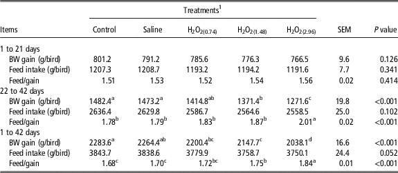
No significant difference (P>0.05) was observed in growth performance among treatments in 1 to 21 days (Table 1). Broilers exposed to 1.48 and 2.96 mM/kg BW H2O2 treatments showed lower (P<0.05) BW gain than broilers in control and saline treatments in 22 to 42 days. Broilers in H2O2(2.96) exhibited higher (P<0.05) feed/gain ratio than broilers in control and saline treatments in 22 to 42 days. Broilers in H2O2(1.48) and H2O2(2.96) treatments exhibited lower (P<0.05) BW gain, and higher (P<0.05) feed/gain ratio than broilers in the control and saline treatments in 1 to 42 days. The data about death number and mortality of broilers is given in the Supplementary Table S2.
Effects of oxidative stress induced by hydrogen peroxide (H2O2) on growth performance of broilers

The results are presented by mean values and the SEM. Data are the mean of eight replicates with eight broilers each (n=8).
a,b,c,dMeans within a row with no common superscripts differ significantly (P<0.05).
1 Control is the non-injection treatment. Saline is 1 ml/kg BW physiological saline (0.75%) injection treatment. H2O2 treatments (H2O2(0.74), H2O2(1.48) and H2O2(2.96)) received an intraperitoneal injection of H2O2 with 0.74, 1.48 and 2.96 mM/kg BW per bird.
Generation of intracellular reactive oxygen species
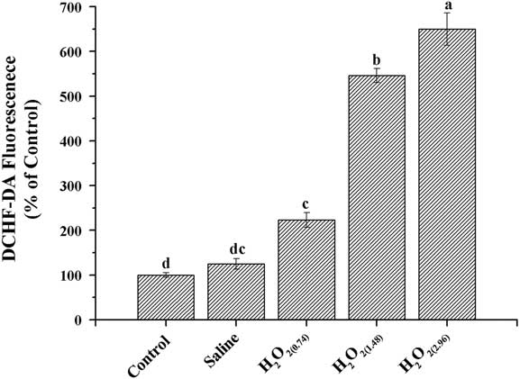
To evaluate the direct effect of H2O2 on the ROS concentration, we detected ROS production using 2',7'-Dichlorodihydrofluorescein diacetate probe after exposure to H2O2 (Figure 1). Broilers in H2O2(1.48) and H2O2(2.96) treatments exhibited higher (P<0.05) ROS formation in the liver as compared with those of the control and saline treatments, and the highest (P<0.05) level of mean fluorescence intensity was detected in broilers exposed to 2.96 mM/kg BW H2O2.
The concentration of reactive oxygen species in the liver of broilers exposed to hydrogen peroxide (H2O2) at 42 days. Control is the non-injection treatment. Saline is 1 ml/kg BW physiological saline (0.75%) injection treatment. H2O2 treatments (H2O2(0.74), H2O2(1.48) and H2O2(2.96)) received an intraperitoneal injection of H2O2 with 0.74, 1.48 and 2.96 mM/kg BW per bird. All data were represented as the mean value±SE of eight sample birds per treatment (n=8). a,b,c,dDifferent letters within the same time points indicate significant differences (P<0.05).

Histological evaluation
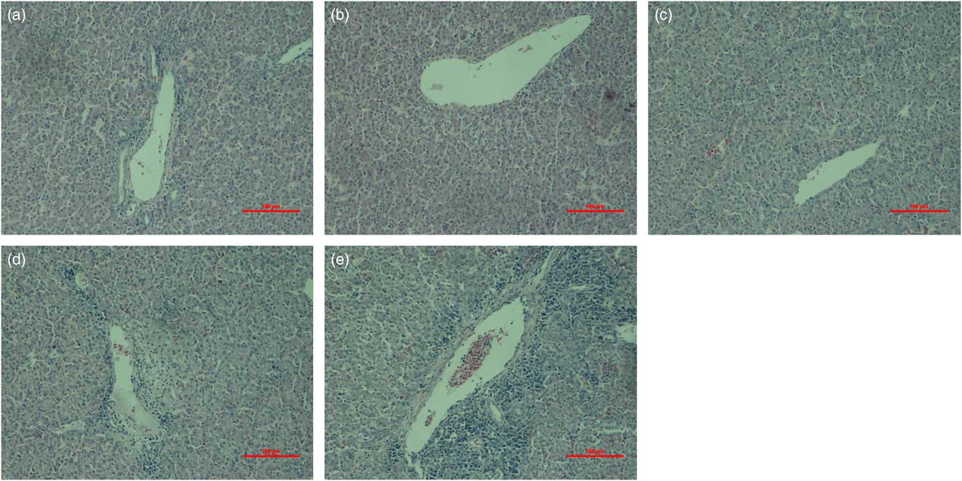
The histological evaluation of the liver sections revealed that the hepatocyte was aligned and the hepatic tissue was almost non-necrotic in the control, saline, and H2O2(0.74) treatments. On the contrary, hepatocellular necrosis and accumulation of mixed inflammatory cells around the necrotic area were observed in the livers of broilers subjected to 1.48 and 2.96 mM/kg BW H2O2. Especially in broilers subjected to 2.96 mM/kg BW H2O2, the hepatocyte was misaligned, and untight and hemorrhagic spots and lymphocytes infiltration were identified (Figure 2).
(color online) Hematoxylin–eosin staining of liver sections. Broilers were exposed to hydrogen peroxide (H2O2). Liver morphology was normal in birds of control, saline and H2O2(0.74) (a, b and c), whereas, all of H2O2(1.48) and H2O2(2.96) demonstrated hepatocellular necrosis and accumulation of mixed inflammatory cells around the necrotic area in the livers (d and e). Scale bar: 100 μm. (a) Control; (b) Saline; (c) H2O2(0.74); (d) H2O2(1.48); (e) H2O2(2.96).

Antioxidants activity and content of oxidative products
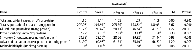
The activities of total superoxide dismutase and glutathione peroxidase were lower (P<0.05), and the contents of protein carbonyl, 8-hydroxy-2'-deoxyguanosine, advanced oxidation protein products and malondialdehyde were higher (P<0.05) in the liver of broilers exposed to 2.96 mM/kg BW H2O2 than broilers in control and saline treatments (Table 2). The contents of protein carbonyl and malondialdehyde were higher (P<0.05) in the liver of broilers exposed to 1.48 mM/kg BW H2O2 than broilers in control and saline treatments.
Effects of oxidative stress induced by hydrogen peroxide (H2O2) on antioxidants activity and content of oxidative products in the liver of broilers

The results are presented by mean values and the SEM. Data are the mean of eight replicates (n=8).
a,b,cMeans within a row with no common superscripts differ significantly (P<0.05).
1 Control is the non-injection treatment. Saline is 1 ml/kg BW physiological saline (0.75%) injection treatment. H2O2 treatments (H2O2(0.74), H2O2(1.48) and H2O2(2.96)) received an intraperitoneal injection of H2O2 with 0.74, 1.48 and 2.96 mM/kg BW per bird.
Apoptosis and autophagy response
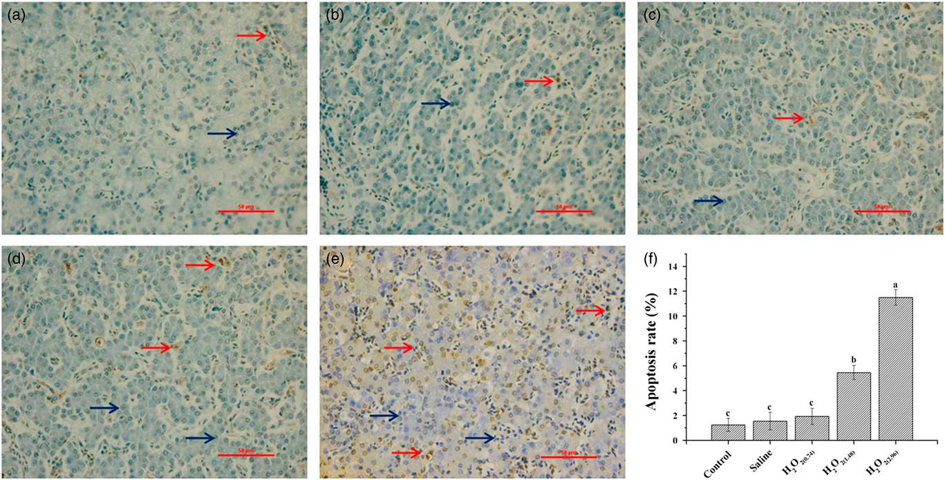
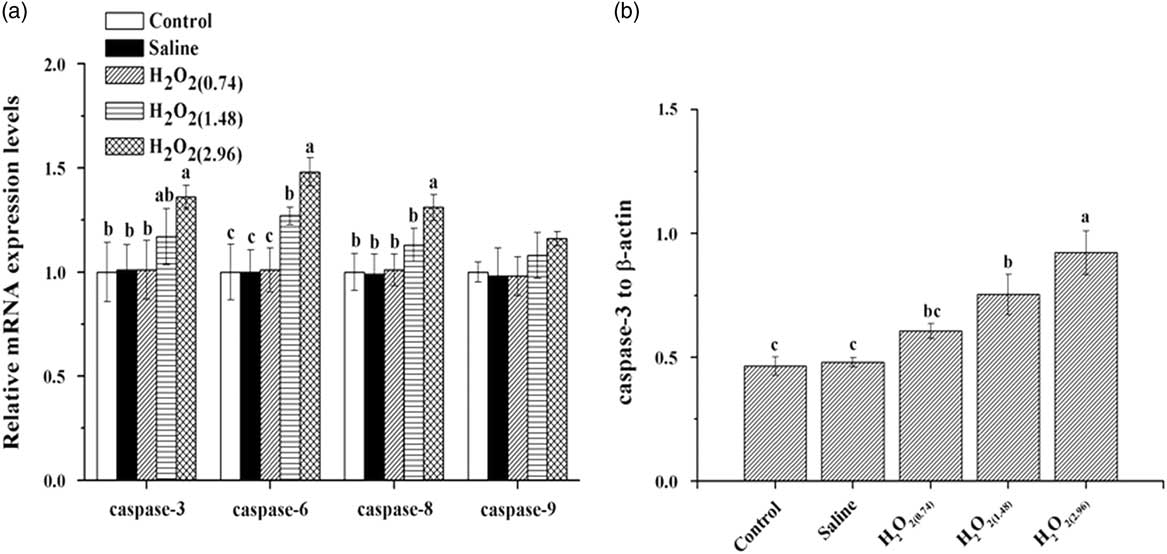
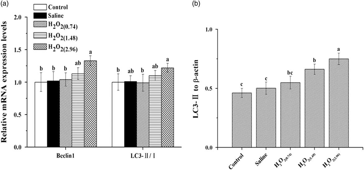
The terminal deoxynucleotide-transferase-mediated deoxyuridine triphosphate nick end labeling assay was performed to detect DNA strand breaks that occurred in preference to nuclear fragmentation. The result revealed significant more (P<0.05) apoptotic cells in the liver of broilers in H2O2(1.48) and H2O2(2.96) in comparison with that of the control and saline treatments (Figure 3). Broilers exposed to 2.96 mM/kg BW H2O2 had higher (P<0.05) mRNA expressions of caspase-3, 6 and 8, and higher (P<0.05) protein expression of total caspase-3 in comparison with broilers in control and saline treatments (Figure 4). Increased mRNA expression of caspase-6 and protein expression of total caspase-3 were also observed in broilers exposed to 1.48 mM/kg BW H2O2 (P<0.05). The full membranes with the molecular weight ladder used for western blot analysis of β-actin and caspase-3 are given in the Supplementary Figures S1 and S2. As shown in Figure 5, the administration of 2.96 mM/kg BW H2O2 up-regulated (P<0.05) mRNA expressions of beclin1 and LC3-II/LC3-I, and increased the protein expression of total LC3-II in the liver of broilers as a comparison with those in control and saline treatments. The similar effect on the protein expression of total LC3-II was also observed in the liver of broilers exposed to 1.48 mM/kg BW H2O2. The full membranes with the molecular weight ladder used for western blot analysis of β-actin and LC3-II is given in the Supplementary Figures S3 and S4.
(color online) Terminal deoxynucleotide-transferase-mediated deoxyuridine triphosphate nick end labeling assay of liver sections. Broilers were exposed to hydrogen peroxide (H2O2). The normal hepatocytes (blue arrow) and terminal deoxynucleotide-transferase-mediated deoxyuridine triphosphate nick end labeling-positive hepatocytes (red arrow) were shown. Scale bar: 50 μm. (a) Control; (b) Saline; (c) H2O2(0.74); (d) H2O2(1.48); (e) H2O2(2.96). The apoptosis rate in the liver of broilers exposed to H2O2 at 42 days are shown (f). All data were represented as the mean value±SE of eight sample birds per treatment (n=8). a,b,cDifferent letters within the same time points indicate significant differences (P<0.05).

Apoptosis response was measured via real-time quantitative PCR (a) and western blot (b) analyses in the liver of broilers exposed to hydrogen peroxide (H2O2) at 42 days. Control is the non-injection treatment. Saline is 1 ml/kg BW physiological saline (0.75%) injected treatment. H2O2 treatments (H2O2(0.74), H2O2(1.48) and H2O2(2.96)) received an intraperitoneal injection of H2O2 with 0.74, 1.48 and 2.96 mM/kg BW per bird. All data were represented as the mean value±SE of eight sample birds per treatment (n=8). a,b,cDifferent letters within the same time points indicate significant differences (P<0.05).

Autophagy response was measured via real-time quantitative PCR (a) and western blot (b) analyses in the liver of broilers exposed to hydrogen peroxide (H2O2) at 42 days. Control is the non-injection treatment. Saline is 1 ml/kg BW physiological saline (0.75%) injection treatment. H2O2 treatments (H2O2(0.74), H2O2(1.48) and H2O2(2.96)) received an intraperitoneal injection of H2O2 with 0.74, 1.48 and 2.96 mM/kg BW per bird. All data were represented as the mean value±SE of eight sample birds per treatment (n=8). Beclin1=autophagy-related gene 6; LC3=microtubule-associated protein 1 light chain 3. a,b,cDifferent letters within the same time points indicate significant differences (P<0.05).

The nuclear factor-κB signal pathway in the liver of broiler
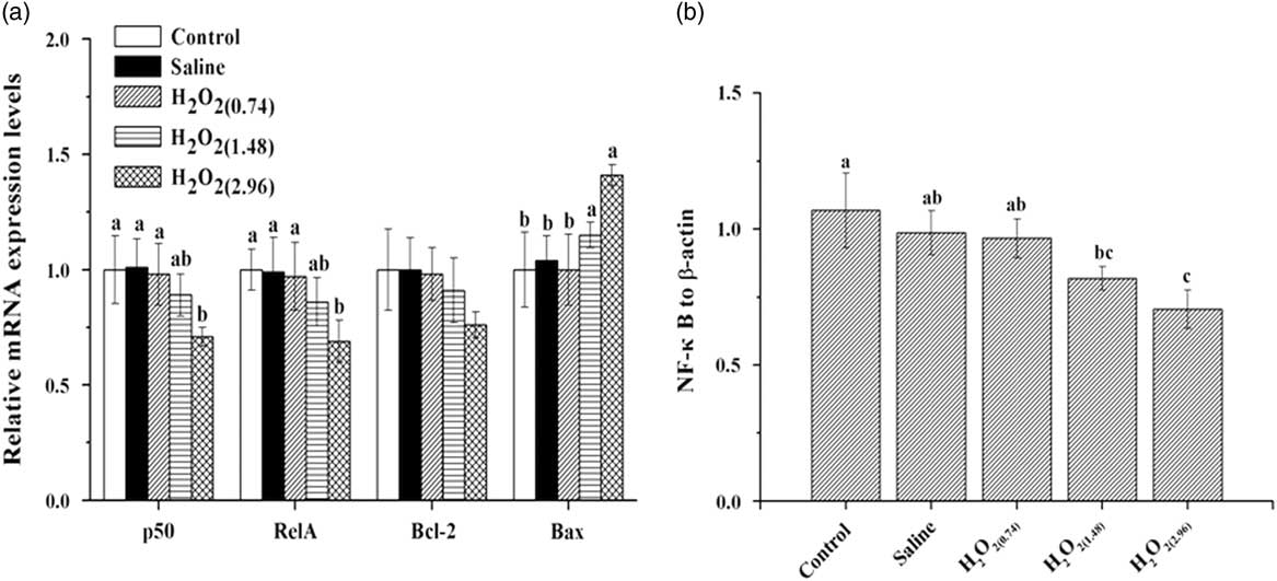
Decreased (P<0.05) mRNA expressions of p50, RelA and protein expression of total NF-κB were detected in the liver of broilers exposed to 2.96 mM/kg BW H2O2, whereas the mRNA expression of Bax was increased (P<0.05) in the liver of broiler exposed to 2.96 mM/kg BW H2O2 as comparison with those of control and saline treatments (Figure 6). In addition, increased (P<0.05) mRNA expression of Bax occurred in the liver of broilers exposed to 1.48 mM/kg BW H2O2 in comparison with the broilers of control and saline treatments. The full membranes with the molecular weight ladder used for western blot analysis of β-actin and NF-κB is given in the Supplementary Figures S5 and S6.
The nuclear factor-κB signal path was measured via real-time quantitative PCR (a) and western blot (b) analyses in the liver of broilers exposed to hydrogen peroxide (H2O2) at 42 days. Control is the non-injection treatment. Saline is 1 ml/kg BW physiological saline (0.75%) injection treatment. H2O2 treatments (H2O2(0.74), H2O2(1.48) and H2O2(2.96)) received an intraperitoneal injection of H2O2 with 0.74, 1.48 and 2.96 mM/kg BW per bird. All data were represented as the mean value±SE of eight sample birds per treatment (n=8). p50=nuclear factor kappa B subunit 1; RelA=v-rel reticuloendotheliosis viral oncogene homolog A; Bcl-2=B-cell CLL/lymphoma 2; Bax=Bcl-2 associated X. a,b,cDifferent letters within the same time points indicate significant differences (P<0.05).

Discussion
Oxidative stress contributes significantly to the impairment of poultry growth (Fellenberg and Speisky, Reference Fellenberg and Speisky2006). The present study showed a significant decrease in BW gain and a significant increase in feed/gain ratio of broilers exposed to H2O2 (2.96 mM/kg BW) at 22 to 42 days and 1 to 42 days. Similarly, oxidative stress induced by lipopolysaccharide (LPS, the concentration of 250 μg/kg BW) decreased the average daily weight gain, the average daily feed intake and the feed conversion ratio of broilers (Zheng et al., Reference Zheng, Wu, Song, Zhang, Zhang, Zhang, Zhang, Wang and Wang2016). The decreased BW gain and gain/feed ratio of broilers might also result from oxidative stress induced by heat stress (Zhang et al., Reference Zhang, Hu, Lu, Yang, Zhang and Wang2015). However, our findings were different from the result from broilers exposed to oxidative stress induced by tert-butyl hydroperoxide (0.2 mM/kg BW, Boostani et al., Reference Boostani, Sadeghi, Mousavi, Chamani and Kashan2015). The difference in growth performance may be attributed to the distinct characteristic or processing technology of inducers. Oxidative stress could cause systemic hypoxemia to reduce individual growth (Kassahn et al., Reference Kassahn, Crozier, Pörtner and Caley2009). Therefore, the dramatic reduction in BW gain in our study might mainly result from the promotion of oxidative processes in broilers exposed to H2O2.
In an organism, the liver is demonstrated to be a primary target attacked by ROS (Sanchez-Valle et al., Reference Sanchez-Valle, Chavez-Tapia, Uribe and Mendez-Sanchez2012). In the current study, H2O2 injection generated elevated ROS levels in the liver of broilers. This result supported the hypothesis that the administration of H2O2 could directly induce oxidative stress. The exceed ROS can destroy the balance between pro-oxidant and antioxidant systems. Antioxidant enzymes such as glutathione peroxidase and total superoxide dismutase, are the major defense system against excessive ROS levels. In the present study, the activities of glutathione peroxidase and total superoxide dismutase were significantly decreased in the liver of broilers exposed to 2.96 mM/kg BW H2O2. Similarly, a decrease of antioxidant enzymes activities were reported in the stomach of rat exposed to 15.0% H2O2 and in the liver of broilers exposed to LPS (Mohamadin et al., Reference Mohamadin, Ashour, El-Sherbeny, Alahdal, Morsy and Abdel-Naim2009; Zheng et al., Reference Zheng, Wu, Song, Zhang, Zhang, Zhang, Zhang, Wang and Wang2016). Meanwhile, higher contents of protein carbonyl, 8-hydroxy-2'-deoxyguanosine, advanced oxidation protein products and malondialdehyde were observed in the liver of broilers exposed to a relative high concentration of H2O2. These results were consistent to that reported by Wan et al. (Reference Wan, Zhang, He, Bai, Zhang and Wang2017) in the liver of broilers reared under high ambient temperature. It is well known that malondialdehyde is commonly used to monitor the level of lipid oxidation and the extent of damage, and protein carbonyl and advanced oxidation protein products are representative biomarkers for protein oxidation. 8-hydroxy-2'-deoxyguanosine as an accurate marker of DNA oxidation is associated with oxidative damage of tissue. Oxidative stress could alter irretrievably lipids, proteins and DNA contents to trigger hepatic damage (Li et al., Reference Li, Tan, Wang, Zhang, Lao, Wong and Feng2015). In addition, previous research indicated that the oxidative process caused structural and functional abnormalities in the liver (Cichoż-Lach and Michalak, Reference Cichoż-Lach and Michalak2014). Therefore, we hypothesized that oxidative stress induced by H2O2 might result in the initiation and progression of oxidative damage of the liver in broilers.
Apoptosis, a mechanism that regulates the cell number, is crucial for the processes of animal growth (Salvesen and Dixit, 1999). The ROS, especially H2O2, can induce apoptosis in numerous cells (Simon et al., Reference Simon, Haj-Yehia and Levi-Schaffer2000). The prior studies reported that apoptosis could be triggered by oxidative stress, caused by H2O2 in human normal hepatocyte cells and 3, 5-diethoxycarbonyl-1, 4-dihydrocollidine in the liver of mouse (Tao et al., Reference Tao, Lehwald, Jang, Baek, Xu, Omary and Sylvester2013; Tang et al., Reference Tang, Cao, Li, Wang, Jia, Liu, Chen, Cai, Shang and Zhao2016). Accordingly, the present study showed a significant number of cells undergoing apoptosis through terminal deoxynucleotide-transferase-mediated deoxyuridine triphosphate nick end labeling assay, and significant increase of caspases expression at mRNA and protein levels in the liver of broilers exposed to H2O2. The caspase family participates in the events that lead to apoptosis of animal cells, and the distinct members such as caspase-3, 6, 8 are involved in both the initiation and execution phases of cell death (Salvesen and Dixit, Reference Salvesen and Dixit1999). Moreover, caspase-3 plays a central role in different apoptotic pathways. Thus, the administration of H2O2 could induce apoptosis of hepatocytes in broilers, which attributed to the increase of ROS levels acting as signaling molecules. On the other hand, the liver is rich in lysosomes which is originally discovered in the liver of rat, and owns high levels of metabolic-stress-induced autophagy (Ueno and Komatsu, Reference Ueno and Komatsu2017). Previous studies reported that the aging-associated decline in autophagic activity is associated with the increased levels of oxidative stress which result in protein oxidation, and the lipotoxic effects of oxidative stress can suppress autophagic activity (Donati et al., Reference Donati, Cavallini, Paradiso, Vittorini, Pollera, Gori and Bergamini2001; Uddin et al., Reference Uddin, Nishio, Ito, Suzuki and Isobe2012; Ueno and Komatsu, Reference Ueno and Komatsu2017). Consistent with prior reports, the autophagy was triggered in hepatocytes of broilers duo to the oxidative damage caused by administration of H2O2 in the present study. The LC3 is most abundant in autophagosomal membranes and monitor the autophagosome and autophagic activity, and Beclin1 contributes to the formation of the autophagosome. Both are widely used as markers of autophagy (Yang and Klionsky, Reference Yang and Klionsky2009). Therefore, we expected that the initiation of autophagy might result from the beginning of autophagosome promoted by the increased production of ROS as similar with the report of Kara et al. (Reference Kara, Gedikli, Sengul, Gelen and Ozkanlar2016). It has been reported that ROS induced autophagy, and the autophagy could be used to reduce the oxidative damage in turn (Scherz-Shouval and Elazar, Reference Scherz-Shouval and Elazar2011). Thus, it is reasonable to conclude that autophagy plays an important role in protecting the liver of broilers from oxidative damage. Moreover, oxidative stress induced by aflatoxin B2 could trigger apoptosis and autophagy in hepatocytes of broilers (Chen et al., Reference Chen, Li, Guo, Xie and Chen2016). In this way, the elevated levels of ROS caused by H2O2 could increase both apoptosis and autophagy in the liver of broilers, simultaneously.
The key factors of NF-κB signal pathway are known as molecular redox switches for system-wide oxidative stress response (Sies et al., Reference Sies, Berndt and Jones2017). As we are known, the heterodimer that includes p50 and RelA, regularly addressed as NF-κB. In the current study, the administration of 2.96 mM/kg BW H2O2 decreased the expression of NF-κB and increased the expression of Bax mRNA. The NF-κB signal pathway is demonstrated to control cell survival (Kondylis et al., Reference Kondylis, Kumari, Vlantis and Pasparakis2017). The NF-κB is subject to multiple regulations by redox changes to participate in anti-apoptosis, and the Bcl-2/Bax ratio can regulate cell death by stimulation of external factors. Previous studies have reported that apoptosis was regulated by NF-κB signal pathway through ROS, caused by microcystins in the liver of rat and deoxyelephantopin in human hepatocellular carcinoma G2 cells (Chen et al., Reference Chen, Li, Guo, Xie and Chen2016; Mehmood et al., Reference Mehmood, Maryam, Zhang, Li, Khan and Ma2017). Similarly, the present study indicated that oxidative stress induced by H2O2 initiated apoptosis via inhibiting NF-κB activation and diminishing the Bcl-2/Bax ratio in the liver of broilers. Yet, our findings are different from the results in T-cells treated with H2O2 (Dumont et al., Reference Dumont, Hehner, Hofmann, Ueffing, Droge and Schmitz1999). This difference of NF-κB signal regulation in apoptosis may be related to stimulating factors and test objects, as well as the degree of the NF-κB activation signal (Barkett and Gilmore, Reference Barkett and Gilmore1999). Meanwhile, the recent studies suggested the interplay of the NF-κB and autophagy (Criollo et al., Reference Criollo, Senovilla, Authier, Maiuri, Morselli, Vitale, Kepp, Tasdemir, Galluzzi, Shen, Tailler, Delahaye, Tesniere, De Stefano, Younes, Harper, Pierron, Lavandero, Zitvogel, Israel, Baud and Kroemer2010; Zhong et al., Reference Zhong, Umemura, Sanchez-Lopez, Liang, Shalapour, Wong, He, Boassa, Perkins, Ali, McGeough, Ellisman, Seki, Gustafsson, Hoffman, Diaz-Meco, Moscat and Karin2016). Hence, we attempted to elucidate the relationship between ROS and autophagy via the NF-κB signal, which defined the impact of oxidative stress on autophagy in the liver of broilers. Prior studies showed that the accumulation of ROS in transcription factor α-treated cells and in dihydroartemisinin-treated cancer cells, initiated autophagy by suppressing NF-κB activation (Djavaheri-Mergny et al., Reference Djavaheri-Mergny, Amelotti, Mathieu, Besançon, Bauvy and Codogno2007; Hu et al., Reference Hu, Chen, Zhang, Lou and Zhou2014). A similar effect was observed on the regulation of NF-κB in autophagy by the increase of ROS in the present study. Yet, it was noteworthy that different results were reported about the modification of ROS levels in in vitro related researches (He et al., Reference He, Zhu, Li, Zhong, Tan and Wang2017; Nopparat et al., Reference Nopparat, Sinjanakhom and Govitrapong2017). The possibility of the difference may attribute to the source of intracellular ROS or the test object. We previously found that in breast muscle of broilers, a high concentration of ROS might suppress NF-κB signal to trigger both apoptosis and autophagy after oxidative damage (Chen et al., Reference Chen, Zhang, Li, Gao and Zhou2017). This result suggested the same effect of elevated ROS levels on hepatic apoptosis and autophagy by regulation of NF-κB signal.
In summary, our findings indicated that oxidative stress induced by H2O2 had a negative effect on histomorphology and redox status in the liver of broilers, underlying a decline in growth performance of broilers. This could attribute to apoptosis and autophagy processes triggered by excessive reactive oxygen species that suppressed the NF-κB signaling pathway. Most importantly, these observations provided additional evidence to explain a possible molecular mechanism of oxidative damage in broilers.
Acknowledgments
This work was supported by the National Key Research and Development Program of China (No. 2016YFD0500501); and the Three Agricultural Projects of Jiangsu Province (SXGC2017281).
Declaration of interest
The authors declare no conflict of interest.
Ethics statement
This study was approved by the Institutional Animal Care and Use Committee of Nanjing Agricultural University, Nanjing, China.
Software and data repository resources
None of the data were deposited in an official repository.
Supplementary material
To view supplementary material for this article, please visit https://doi.org/10.1017/S1751731118000903