Introduction
The genus Blarneya was described by Hawksworth et al. (Reference Hawksworth, Coppins and James1979) for a sterile, corticolous lichen with a pinkish white thallus and conspicuous white sporodochia. The thalli were considered to be initially lichenicolous on different species of Roccellaceae and on Belonia caudata, eventually becoming autonomous after killing the host mycobiont and taking over its photobiont (Trentepohlia). The species was described from Ireland and later discovered in SW England and Wales, the Western Pyrenees (France and Spain) and Macaronesia (Canary Islands and Madeira) (Etayo Reference Etayo1989, Reference Etayo1992, Reference Etayo1998; Chambers & Purvis Reference Chambers, Purvis, Smith, Aptroot, Coppins, Fletcher, Gilbert, James and Wolseley2009).
Recent field work by the authors has led to the discovery of additional localities of Blarneya in the Pyrenees, the Canary Islands, continental Africa (Democratic Republic of Congo), the USA (Florida and Hawaiian Islands) and the Galapagos Islands (Ecuador). Several saxicolous populations were discovered, differing slightly from the corticolous ones, suggesting that more than one species might be involved, implying that Blarneya could no longer be accepted as a monotypic genus. Among the material examined, we also encountered specimens that were apparently fertile with Tylophoron-like ascomata. This observation led us to wonder if the sporodochial thalli called Blarneya might be an anamorph of Tylophoron, or if these specimens were just sterile Blarneya overgrowing fertile thalli of Tylophoron. The genus Tylophoron has recently been shown to be a calicioid member of the Arthoniales using molecular data (Lumbsch et al. Reference Lumbsch, Lücking and Tibell2009), while Blarneya is considered as an anamorphic ascomycete of uncertain position referred by some authors to the Arthoniales based on the chemistry and the method of conidiogenesis (e.g., Chambers & Purvis Reference Chambers, Purvis, Smith, Aptroot, Coppins, Fletcher, Gilbert, James and Wolseley2009).
Therefore, we carefully studied all available specimens both macroscopically and microscopically; we analyzed their chemistry by TLC or, in the case of small thalli, by spot test reactions. In several cases we were able to culture specimens from conidia of recently collected material and we were able to sequence LSU rDNA from eight specimens, including four saxicolous ones. The results conclusively demonstrate that Blarneya is an anamorph of Tylophoron and, further, allow us to describe two new Tylophoron species with Blarneya-type sporodochia.
Material and Methods
Morphology
Sections were investigated microscopically on material mounted in water and 5% KOH. Measurements of asci and ascospores all refer to material examined in water; conidia were measured in KOH, which dissolves surrounding crystals. Ascospores and conidia measurements are indicated as (minimum–) x-σx − x+σx (–maximum) followed by the number of measurements (n); the length/breadth ratios of the ascospores and conidia are indicated as l/b. For other characters, the minimum and the maximum values are given. The thallus colour always refers to herbarium specimens.
Thin-layer chromatography (TLC) of acetone extracts of almost all specimens was performed in solvent systems C, EA and G (Orange et al. Reference Orange, James and White2001).
Taxon sampling and cultures
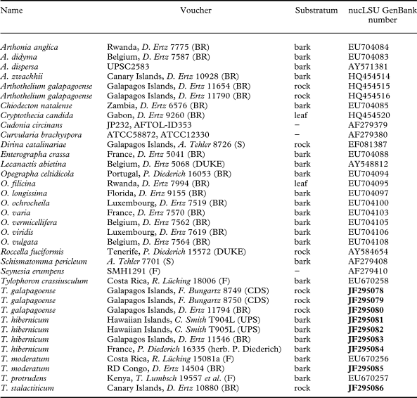
Cultures were isolated from ascospores or conidia of freshly collected material on malt-yeast extract medium following Yoshimura et al. (Reference Yoshimura, Yamamoto, Nakano, Finnie, Kranner, Beckett and Varma2002). We obtained cultures from three specimens of Blarneya for this study (Ertz 10880, 11546, 11794). All cultures were obtained from conidia. When cultures were not available, well-preserved and freshly collected lichen specimens lacking any visible symptoms of fungal infection were used for DNA isolation. We obtained 9 new sequences of Arthoniales for this study from France, the Democratic Republic of Congo, the Galapagos Islands (Ecuador), the Hawaiian Islands (USA) and the Canary Islands (Spain). For the phylogenetic analyses, 27 sequences were retrieved from GenBank in addition to our own sequences (Table 1). Three outgroup species were chosen based on Lutzoni et al. (Reference Lutzoni, Kauff, Cox, McLaughlin, Celio, Dentinger, Padamsee, Hibbett, James and Baloch2004): Curvularia brachyspora (Dothideomycetes), Seynesia erumpens (Sordariomycetes) and Cudonia circinans (Leotiomycetes).
Specimens and DNA sequences used in this study, with their respective voucher information. GenBank accession numbers in bold refer to sequences (9) generated by this project. All other sequences (27 GenBank accession numbers) were obtained directly from GenBank

Molecular data
Genomic DNA was isolated from mycobiont cultures or from lichen specimens using the Puregene Genomic DNA Purification Kit (GENTRA Systems, Minnesota), following the manufacturer's Plant Tissue extraction protocol. Amplification reactions were prepared for a 50 µl final volume containing 5 µl 10x Taq Buffer (Roche), 2·5 µl of each of the 20 µM primers, 1 µl of 10 mg ml−1 bovin serum albumin (Ambion # 2616), 1 µl of 25 mM MgCl2, 1·25 U Taq DNA polymerase (Roche) and 1 µl of template genomic DNA. PCR was performed on Peltier Thermal Cyclers PTC-100 or PTC-150 (MJ Research). A targeted fragment of about 1 kb at the 5′end of the nuclear LSU rDNA was amplified using primers LR0R (Rehner & Samuels Reference Rehner and Samuels1994), LIC15R (Miądlikowska et al. Reference Miądlikowska, McCune and Lutzoni2002) with LR5 or LR6 (Vilgalys & Hester Reference Vilgalys and Hester1990). PCR products were purified using the QIAquick PCR Purification Kit (Qiagen). The yield of the PCRs was verified by running the products on a 1% agarose gel using ethidium bromide. Both strands were sequenced directly using BigDye terminators (Applied Biosystems) and the amplification primers. Additional primers for sequencing were used: LR3R and LR3 (Vilgalys & Hester Reference Vilgalys and Hester1990; Vilgalys' website, http://www.botany.duke.edu/fungi/mycolab). Sequence fragments were assembled with Sequencher version 4.6 (Gene Codes Corporation, Ann Arbor, Michigan). Sequences were subjected to BLAST searches to verify their closest relatives and to detect potential contaminations.
Phylogenetic analyses
NucLSU sequences were aligned manually using MacClade 4.05 (Maddison & Maddison Reference Maddison and Maddison2002). Ambiguous aligned regions and introns representing a total of 224 bp were delimited manually and excluded from the analyses.
Maximum Parsimony (MP) analysis was performed in PAUP*4.0b10 (Swofford Reference Swofford2002) on the nucLSU matrix of 778 unambiguously aligned characters including 296 variable characters, of which 211 are parsimony-informative. The nucLSU matrix was subjected to a symmetric step matrix constructed with the STMatrix 2.2 MacOS Program (François Lutzoni & Stefan Zoller, Department of Biology, Duke University). Heuristic searches were used with 1000 random addition sequence replicates, MaxTrees set to autoincrease, tree-bisection-reconnection (TBR) branch swapping, MulTrees option in effect and gaps treated as a fifth character state. The MP analysis yielded one most parsimonious tree (1436·05 steps, CI = 0·5413, RI =0·7213). Bootstrap values (MP-bs) were obtained from 1000 replicates with 3 random addition sequences (all other parameters identical to the original MP search).
The best-fit model of DNA evolution was chosen using the Akaike information criterion (AIC) as implemented in Modeltest v. 3.06 (Posada & Crandall Reference Posada and Crandall1998). Bayesian analyses were carried out using the Metropolis-coupled Markov chain Monte Carlo method (MCMCMC) in MrBayes v. 3.1.2 (Huelsenbeck & Ronquist Reference Huelsenbeck and Ronquist2001; Ronquist & Huelsenbeck Reference Ronquist and Huelsenbeck2003). Analyses were run under the GTR model of nucleotide substitution including a proportion of invariable sites and a discrete gamma distribution with six rate categories. Two parallel MCMCMC runs were performed each using four independent chains and 10 000 000 generations, sampling trees every 1000th generation. The proportion of burn-in trees sampled before reaching equilibrium was estimated using TRACER v.1.5 (Rambaut & Drummond Reference Rambaut and Drummond2007) by plotting the log-likelihood values of the sample points against generation time. Posterior probabilities (PP) were determined by calculating a majority-rule consensus tree generated from the last 9000 of the 10 000 trees sampled.
The Maximum Likelihood (ML) analysis was performed using GARLI (Zwickl Reference Zwickl2006, v.0.951 for OS X) with default settings, and a single most likely tree was produced (–lnL = 5048·749961). Maximum Likelihood bootstrap values (ML-bs) were derived from 1000 bootstrap replicates using default settings.
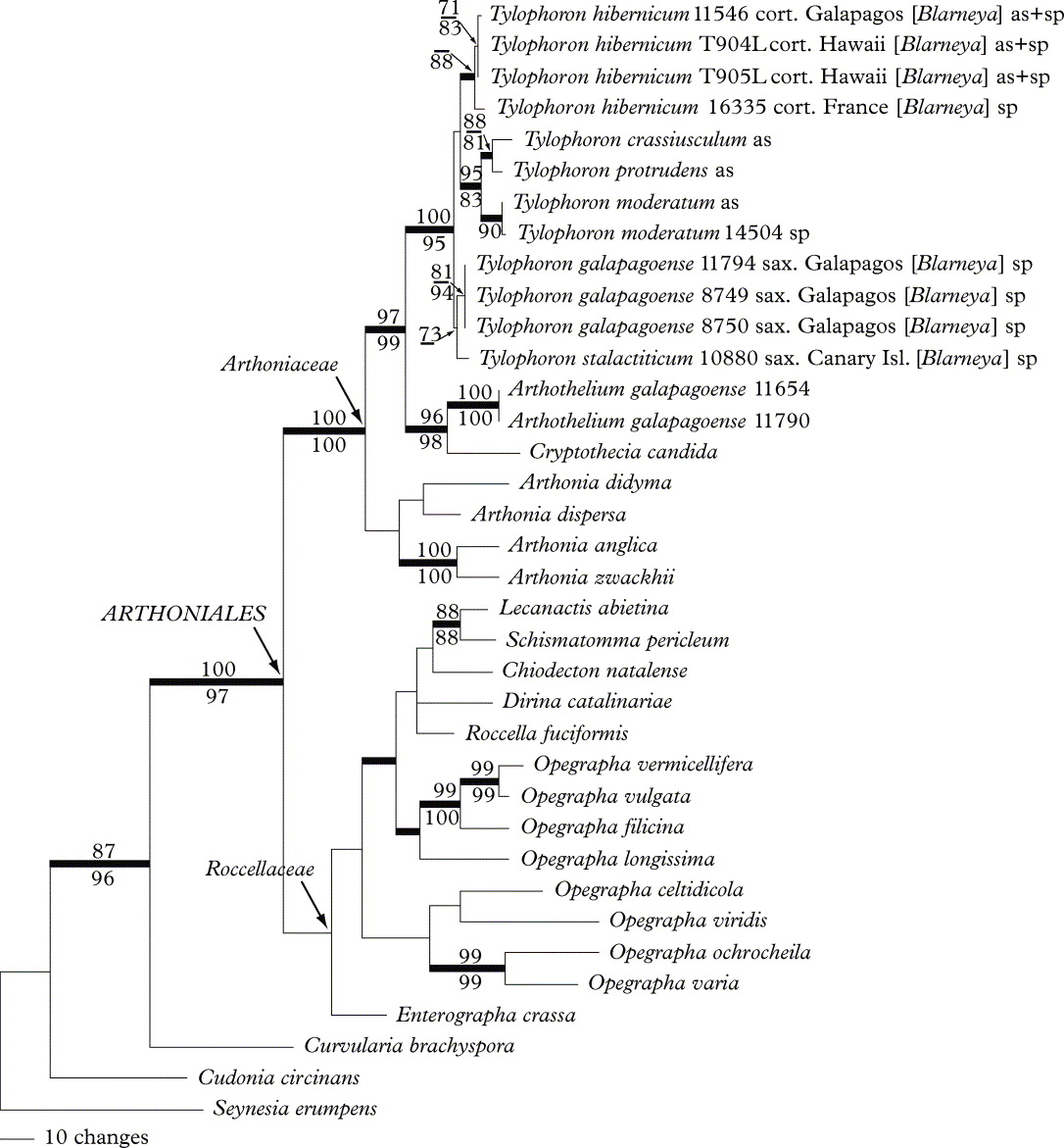
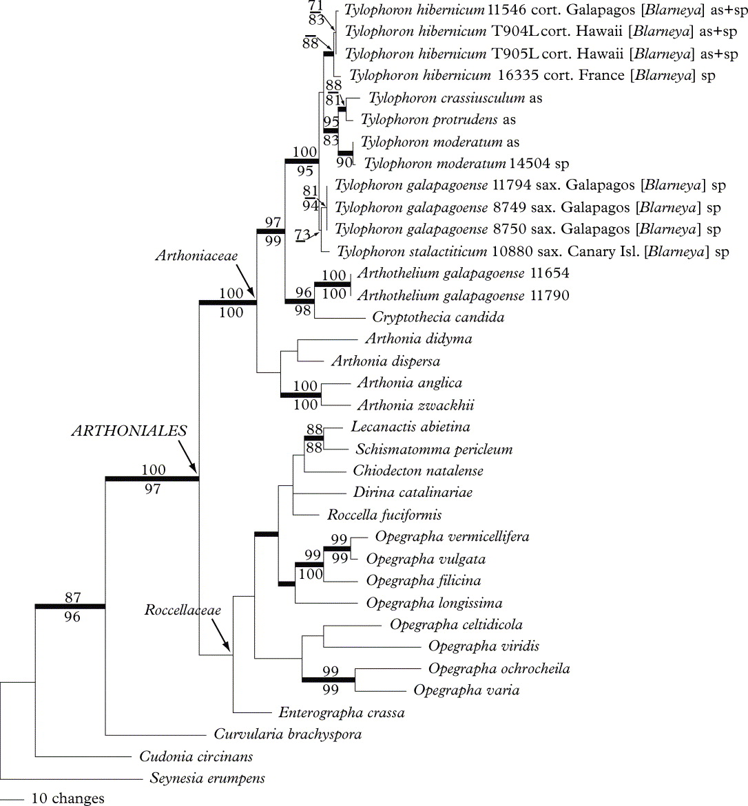
The MP tree and the ML tree did not contradict the Bayesian tree topology for the strongly supported branches and hence only the majority rule consensus tree of the Bayesian analysis is shown here, with the branch supports of the two other analyses (Fig. 1). PP ≥ 95%, ML-bs ≥ 70% and MP-bs ≥ 70% were considered to be significant.
One locus (nucLSU) 50% majority-rule consensus tree produced by the Bayesian analysis and representing the proposed phylogenetic relationships among 33 specimens of Arthoniales. Internal branches with a PP ≥ 95% are considered strongly supported and represented by thicker lines. MP-bs values ≥ 70 are shown above, and ML-bs values ≥ 70 are shown below, internal branches. Collecting numbers of authors following the species names act as specimen and sequence identifiers. Tylophoron specimens with Blarneya-like sporodochia are followed by the name Blarneya within brackets, those with ascomata are followed by ‘as', those with sporodochia are followed by ‘sp’, those with ascomata and sporodochia are followed by ‘as+sp’.
Results and Discussion
The most striking result of the phylogenetic analysis is the position of Blarneya within the Arthoniaceae and its very close relationship with Tylophoron in its usual delimitation (Lumbsch et al. Reference Lumbsch, Lücking and Tibell2009). The discovery of Tylophoron-like ascomata emerging from the surface of thalli with Blarneya-like sporodochia further supports this result (Fig. 2D & E). As Blarneya is considered to be initially a lichenicolous fungus, eventually lichenized (Hawksworth et al. Reference Hawksworth, Coppins and James1979), it is futile to argue, on morphological observations alone, that these ascomata were not produced by Tylophoron thalli invaded by Blarneya. However, all sequences of Blarneya used here for the phylogenetic analysis were obtained directly from Blarneya-like sporodochia. Moreover, three sequences are based on culture isolates from Blarneya-like conidia: specimens Ertz 10880 from Tenerife, and Ertz 11546 and 11794 from the Galapagos Islands. In addition, one sequence comes from the Pyrenees (Diederich 16335) and one from Tenerife (Ertz 10880), where the genus Tylophoron is not known. For all these reasons, we can exclude any contamination with Tylophoron for all sequences obtained from the ‘Blarneya’ material studied here. Instead we can conclude that the ascomata observed on the sporodochial thalli belong to these thalli.
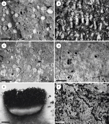
Tylophoron species with Blarneya-type sporodochia. A, T. galapagoense, thallus and sporodochia (Bungartz 8750); B, T. stalactiticum, thallus with sporodochia present at the tip of the hanging stipes (Ertz 10880 & Diederich); C–E, T. hibernicum; C, thallus and sporodochia (Diederich 16335); D, thallus with ascomata and sporodochia (Smith T904); E, cross section through an ascoma (Smith T904); F, Tylophoron moderatum, anamorphic thallus from the Galapagos Islands (Bungartz 7109), the pale white thallus is covered by an irregular felt of brownish sporodochia. Scales: A–D = 2 mm, E = 0·1 mm, F = 5 mm.

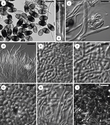
Regarding their very close phylogenetic relationship, we consider Blarneya a synonym of Tylophoron and therefore combine B. hibernica into Tylophoron (see taxonomy below). Ascomata and ascospores are morphologically and anatomically identical, which also supports this new synonymy. Several morphological differences nevertheless exist between the anamorphic states of Tylophoron hibernicum and the previously accepted Tylophoron species, in particular T. moderatum. In T. hibernicum the sporodochia are always pale and usually form well-delimited cushions (Fig. 2C & D), whereas the anamorph of T. moderatum is characterized by a dark brown felt of sporodochia that are not distinctly delimited and often fuse to form irregular aggregates (Fig. 2F). The conidia in T. hibernicum are hyaline, simple or 1-septate (Fig. 3E & F); they are dark brown and always aseptate in T. moderatum (Fig. 3I). These distinct morphological and anatomical differences of the anamorphic thalli would suggest distinguishing two different genera with almost identical ascomata and ascospores. However, molecular evidence clearly does not support this split. All sequences obtained from thalli with Blarneya-type sporodochia are included within the same, strongly supported clade as Tylophoron. They do not form a separate monophyletic group. The molecular data support a single genus, and the differences between anamorphs must therefore be regarded as significant only at species level.
Tylophoron species with sporodochia (A–C in water; D–I in KOH). A–F, T. hibernicum (A, Ertz 11546; B–E, Smith T904; F, Diederich 16335); A, brown, ellipsoid, one-septate ascospores, the spores are typically slightly pointed and more strongly pigmented around the septum; B–C, asci and paraphyses; D, pycnoconidia; E–F, sporodochial conidia; G, T. galapagoense (Bungartz 8750), sporodochial conidia; H, T. stalactiticum, sporodochial conidia (holotype); I, T. moderatum, sporodochial conidia (Ertz 14504). Scales: A–I = 10 µm.

In this context it is particularly interesting that thalli with brown, felty sporodochia have so far been confirmed with certainty only for Tylophoron moderatum. It appears that this species forms two distinct types of thalli: either exclusively with apothecia (the teleomorph), or only producing conidia arranged in brown, irregularly delimited, felty sporodochia (the anamorph, Fig. 2F). Unlike the Tylophoron species that form Blarneya-type sporodochia (T. hibernicum and T. galapagoense), the brown anamorphs of T. moderatum have never been found with ascomata. These exclusively anamorphic thalli have been ascribed to T. moderatum based on similarities in their thallus structure, chemistry, ecology and features of axenic cultures of the mycobiont obtained from ascospores and conidia, respectively (Tibell Reference Tibell1996). Field observations also suggested that both types of thalli exist in Tylophoron crassiusculum, but no sporodochial thalli are known from T. protrudens (but see notes under the description of T. hibernicum). Here, we have sequenced one anamorphic thallus of Tylophoron from Africa producing dark brown and aseptate conidia (Ertz 14504). Our phylogenetic tree shows that this thallus clearly belongs to Tylophoron moderatum, thus providing evidence for the existence of an anamorphic thallus characterized by a brown, diffusely delimited sporodochial felt.
In our phylogenetic tree (Fig. 1), the clade including Tylophoron is strongly supported but poorly resolved. The nucLSU sequences alone are certainly not sufficient for resolving the species circumscription, as it is too conserved. However, it must be noted that the corticolous specimens of Tylophoron with Blarneya-like sporodochia group together with strong support (PP=100, ML-bs=88 but MP-bs=68), just like the saxicolous ones, but here with weak support (PP=78, ML-bs=68 and MP-bs=73). Interestingly, the corticolous specimen from the Galapagos Islands groups with the other corticolous specimens from Hawaii and the Pyrenees, but not with the saxicolous specimens from the Galapagos Islands as would have been expected with a single species. In Galapagos, the saxicolous specimens generally grow in sheltered, shaded sites, under rock overhangs, but they are generally found in dry forests, whereas the corticolous specimen has been collected in humid forests in the highlands. Based on morphological, chemical, ecological and molecular data, we decided to describe two new species and to restrict the name Tylophoron hibernicum to the corticolous specimens.
Taxonomy
Key to all currently accepted species of Tylophoron
1 Thallus with sporodochia, sometimes also with ascomata ... 2
Thallus lacking sporodochia, with numerous ascomata ... 5
2(1) Thallus with an irregular, not distinctly delimited felt of dark brown sporodochia, lacking ascomata ... T. moderatum
Thallus with cushions of clearly delimited, white to cream, rarely pinkish, stipitate or sessile sporodochia, sometimes also with ascomata ... 3
3(2) Thallus thick, compact, of densely packed hyphae, with a smooth surface, distinctly creamy white to beige, thick, C+ strongly red; saxicolous ... T. galapagoense
Thallus thin, of loosely interwoven hyphae with a felty surface, bright to pale greyish or bluish white, rarely cream coloured (prolonged herbarium storage?), C− or C+ weakly red ... 4
4(3) Thallus C−, with hanging, stipitate sporodochia; saxicolous ... T. stalactiticum
Thallus C+ weakly red, with sessile sporodochia; corticolous ... T. hibernicum
5(1) Thallus C+ red; ascomata 0·6–1·2 mm diam. ... T. protrudens
Thallus C−; ascomata 0·3–0·8 mm diam. ... 6
6(5) Thallus corticate, excipulum laterally 160–180 µm thick ... T. gibsonii
Thallus ecorticate ... 7
7(6) Lateral part of excipulum 55–100 µm thick, of richly branched and interwoven hyphae ... T. crassiusculum
Lateral part of excipulum 15–25 µm thick, of sparsely branched, periclinally arranged hyphae ... T. moderatum
The Taxa
Tylophoron Nyl. ex Stizenb
Ber. Thät. St. Gallen Naturwiss. Ges. 1861–1862: 177 (1862); type: Tylophoron protrudens Nyl.
Syn. nov. Blarneya D. Hawksw., Coppins & P. James, Bot. J. Linn. Soc. 79: 358 (1979); type: Blarneya hibernica D. Hawksw., Coppins & P. James. Originally monotypic.
Tylophoron galapagoense Bungartz, Ertz, Diederich & Tibell sp. nov
MycoBank No: MB 519736
A Tylophorone hibernico thallo leviore et crassiore cremeo differt.
Typus: Ecuador, Galapagos Islands, Isabela, Volcán Darwin, south-western slope, above Tagus Cove, 724 m, transition zone, SW-exposed lava flow of weathered AA-lava with scarce vegetation (Macraea laricifolia, Dodonaea viscosa, …), on rock, overhang, 2007, D. Ertz 11794 (CDS 37153—holotypus; BR, hb. Diederich—isotypi).
Thallus crustose, superficial, more or less smooth, creamy white to pale brownish, ecorticate, thin, up to 0·5 mm thick; hyphae irregularly branched, hyaline, mainly 2–2·5 µm diam., forming numerous convex to subglobose sporodochia 0·5–1 mm diam., which are similar in colour, but paler than the thallus. Sporodochial conidia hyaline, smooth to slightly roughened, mostly simple, more rarely 1-septate, with rounded or truncate apices, spherical, ellipsoid or rarely oblong, simple conidia (4·5–)5–6·5(–9) ×(3–)3·5–4·5(–5·5) µm, l/b ratio 1·1–1·9 (n = 60), 1-septate conidia (7–)8–11(–15) ×(3–)3–4·5(–6) µm, l/b ratio 2–3·2 (n = 70). Prothallus whitish cream, byssoid, 0·5–1 mm wide. Photobiont Trentepohlia.
Ascomata densely covering the thallus of one specimen (Aptroot 65477), but all mazaedia composed of degenerated ascospores and brownish hyphae, no intact ascospores found.
Pycnidia unknown.
Chemistry. Thallus C+ red, K−, KC+ red, P−, UV−. Sporodochia C+ red, K+ yellow, KC+ red, P−, UV−. TLC: lecanoric acid in all specimens examined. In addition, a pale orange substance in daylight after heating in Ertz 11794.
Notes. The new species is distinguished from T. hibernicum by a smoother and thicker, more compact thallus, which is always distinctly cream coloured. Furthermore, the C+ red reaction of the thallus is always stronger than in T. hibernicum. In some specimens of T. galapagoense (e.g., Bungartz 8749 and 8750) the majority of conidia are simple (Fig. 3G), in contrast to T. hibernicum in which one-septate conidia usually dominate (Fig. 3F). However, in other specimens of both species, a mixture of one-septate and simple conidia occurs. Overall, even within sporodochia that predominantly produce one-septate conidia, a few simple conidia can typically be found, and vice versa. Because one-septate conidia easily fracture, it is difficult to assess if the formation of simple vs. one-septate ones follows a different conidiogenesis. Specimens that produce mostly simple conidia generally have conidia with a more spherical shape, whereas simple conidia derived secondarily from septate ones that broke apart, are typically more elongate, with an oval to ellipsoid, not spherical shape. Overall we could not find any clear correlation between these differences and a particular species, and the predominance of simple vs. one-septate, spherical vs. elongate conidia may perhaps be related more to environmental factors than to genetic factors.
Tylophoron galapagoense is currently known only from the Galapagos, on five different islands, where all specimens have been found growing on rock below shaded overhangs or in crevices, from the coast through the dry lowland forests to the transition zone. However, in Santiago the species has also been found in the highlands, where it is known from hidden crevices of the dry, N- NE-exposed steep crater rim of Cerro Gavilan. On Volcán Darwin, where the type specimen was collected, the western slopes of the volcano lie within the rain-shadow of Fernandina. Here the vegetation is generally dry and the transition zone reaches unusually high altitudes. Whereas on the opposite sides of the island humid forests abound, the dry lava flows on the western slopes are characterized by low, transition zone shrubs. In contrast, the corticolous T. hibernicum, although occasionally found in the transition zone, appears more common in the humid forests of the Galapagos highlands.
Additional selected specimens examined. Ecuador: Galapagos Islands: Santa Cruz Island: along dirt road to Mina Granillo Rojo, off the main road to the channel, 583 m, transition zone, dry deciduous forest with basalt outcrops in between on N-slope of island, on sheltered rock face, 2007, D. Ertz 11576 (BR); ibid., F. Bungartz 7113 (CDS 37598); Mina Granillo Rojo, on the N-side of the island, above the mine, 633 m, transition zone, lava outcrop in open forest, ENE-exposed overhang (70°–80°) of basalt boulder in rock outcrop, on volcanic rock, 2010, F. Bungartz 8749 (CDS 44629); ibid., F. Bungartz 8750 (CDS 44630). Isabela Island: Volcán Alcedo, along the trail going up the E-slope, basalt rubble field to the SE-side of the trail and the barranco, 530 m, dry zone, basalt rubble field with scattered vegetation, 2006, A. Aptroot 64943 (CDS 31522); Volcán Sierra Negra, Cerro Orchilla, c. 4 km W of Puerto Villamil, 56 m, dry zone, S exposed slope of hill, open Bursera graveolens forest, SE-exposed overhang of basalt outcrop, 2008, Bungartz 8449 (CDS 41095). Pinta Island, SW-part of the island, along trail going up the SW slope to Las Pampas on the western saddle, 316 m, transition zone, open forest, top of basalt boulder, 2008, Nugra 564 (CDS 38942). Santiago Island, summit of Cerro Gavilan, inner N- and NE-exposed crater rim, 840 m, humid zone N- and NE-exposed, steep basalt cliffs of crater rim with ferns growing in crevices, 2006, A. Aptroot 65760 (CDS 32352). Pinzón Island, along the trail going up from Playa Escondida, N- to W-facing cliff above a crater, 318 m, dry transition zone with Cordia lutea, Croton scouleri, and at the bottom of the cliff also Scalesia baurii ssp. baurii, 2006, A. Aptroot 64030 (CDS 30591).
Tylophoron hibernicum (D. Hawksw., Coppins & P. James) Ertz, Diederich, Bungartz & Tibell comb. nov
MycoBank No: MB 519738
Basionym: Blarneya hibernica D. Hawksw., Coppins & P. James, Bot. J. Linn. Soc. 79: 358 (1979); type: Ireland, Co. Kerry, Killarney, Eagles Nest, on Cresponea premnea in dry recess of ancient Quercus, 1966, P. W. James (K!—holotype).
Thallus crustose, superficial, felty, pale greyish or rarely creamy or bluish white, ecorticate, thin, up to 0·3 mm thick; hyphae irregularly branched, hyaline, mainly 1·5–2 µm diam., forming numerous convex to subglobose sporodochia 0·4–1 mm diam. which are white, cream or sometimes with a pinkish or orange tinge, usually fading to white in the herbarium. Sporodochial conidia hyaline, smooth to slightly roughened, 0–1-septate, with rounded or truncate apices, simple conidia ellipsoid to oblong, rarely spherical, (4–)4·5–6·5(–8) × (3–)3–4(–5), l/b ratio 1·3–1·9 (n = 60), 1–septate conidia often distinctly constricted at the septum, (6–)7–11·5(–15) × (3–)3·5–4·5(–5) µm, l/b ratio 1·9–2·9 (n = 70) (for a detailed description of the conidiogenesis see Hawksworth et al. Reference Hawksworth, Coppins and James1979). Prothallus brown, byssoid, 1–4(–5) mm wide. Photobiont Trentepohlia.
Ascomata very rare, sessile, short cylindrical to conical, 0·5–1 mm diam., 0·4–0·6 mm high (excluding the mass of ascospores), 1–1·5(–2) times as wide as high. Thalline margin well-developed, c. 50–60 µm wide, approx. half the height of the ascomata, densely inspersed with minute colourless crystals dissolving in K (observed in polarized light), of hyaline hyphae 1·5–2 µm diam. Excipulum 10–25 µm laterally, sometimes thinner or absent below, of dark brown, sparingly branched and interwoven hyphae, 1·5–2 µm diam. Mazaedium well-developed, black, sometimes only slightly projecting over the thalline margin but often extending further. Paraphyses branched, anastomosing, 1·5 µm wide. Asci dissolving at early stages, cylindrical, with eight uniseriate ascospores, 35–45 × 4–5 µm. Ascospores 1-septate, dark brown, with a heavily pigmented band around the central part, ellipsoid, sometimes constricted at the septum, occasionally with slightly pointed ends, wall thick, (9–)10·5–13·5(–17) × (5·5–)6·5–8·0(–9) µm, l/b ratio 1·4–1·8 (n = 110).
Pycnidia rare (only observed in Smith T904), present at the margin of the thallus, immersed, visible as brownish spots of c. 0·15–0·3 mm, surrounded by a white thalline margin of c. 50–80 µm thick; pycnidial wall very reduced; pycnoconidia filiform, straight or slightly curved, hyaline, 9–14 × 1 µm.
Chemistry. Thallus (incl. margin of ascomata) C+ weakly red, K−, KC+ red, P−, UV− or UV+ orange. Sporodochia C+ red, K+ yellow, KC+ red, P−, UV− or UV+ orange. TLC: lecanoric acid in all specimens examined; in addition, lichexanthone is present in Ertz 11546, 14915 and 14898, the last two having an obvious UV+ orange thallus; trace of a fatty acid in Diederich 16335. Hawksworth et al. (Reference Hawksworth, Coppins and James1979) did TLC of two specimens of Blarneya hibernica when describing that species: the holotype overgrowing Cresponea premnea (sub Lecanactis premnea) and a second specimen overgrowing Lecanactis abietina. They detected lecanoric and schizopeltic acids as major substances, and in addition a xanthone, a fatty acid, and unknown substances fluorescing ice blue and yellow in ultra-violet light. No schizopeltic acid could be detected in our specimens. The holotype of Blarneya hibernica is extremely reduced (3 sporodochia on Cresponea premnea) and therefore does not allow re-examination by TLC. As the second specimen examined by Hawksworth et al. (Reference Hawksworth, Coppins and James1979) grows on Lecanactis abietina, a lichen producing schizopeltic acid in large amounts, a contamination of schizopeltic acid from the host is very likely. Therefore, we conclude that the detection of schizopeltic acid inBlarneya hibernica is likely to be the result of contamination from other lichen thalli.
Notes. Hawksworth et al. (Reference Hawksworth, Coppins and James1979) suggested that Tylophoron hibernicum starts as a lichenicolous species overgrowing other lichens, taking over their algae, and eventually developing an independent thallus. All specimens collected by us have large thalli, and therefore we could not confirm that they started growth as a lichenicolous fungus. Tylophoron hibernicum differs from other known Tylophoron species by wider ascospores and, when ascomata are lacking, by a thin, bright to pale greyish or bluish white thallus with a felty surface.
It must be noted here that the New York Botanical Garden Herbarium has published on its website (http://sciweb.nybg.org/science2/hcol/lena/index.asp) a photograph of a specimen (Lendemer 15891) collected in Florida, USA, with Blarneya-like sporodochia (but without ascomata) under the name Tylophoron protrudens; the specimen obviously belongs to Tylophoron hibernicum according to the results of our study. It seems that the colleagues in NY had no doubt that their material belongs to Tylophoron but they did not make the ‘connection’ with what Europeans have called Blarneya.
Fertile specimens examined (with ascomata and sporodochia). Ecuador: Galapagos Islands: Santa Cruz Island, c. 1 km N of Bellavista along dirt road to Media Luna, 143 m, humid zone, farm area, coffee plantation with Cedrella odorata trees and one large Ceiba pentandra tree, on trunk of C. odorata, 2007, D. Ertz 11546 (BR).—USA: Hawaiian Islands: Kailua, east side of Olomana mauka op Kalanianaole Hwy, near Waimanalo Quarry, on dead Acacia confusa, 2008, C. Smith T904, T905 (UPS).
Sterile specimens examined (with sporodochia only). France: Pyrénées-Atlantiques: forêt de Sare between Sare and Col de Lizarrieta, on Quercus, 2006, P. Diederich 16335 (hb. Diederich).—Spain: Navarra: 20 km E of San Sebastian, col d'Ibardin, on Quercus in old forest in valley, 1991, P. Diederich 9761, 9762 & J. Etayo (hb. Diederich).—Democratic Republic of Congo: Equateur Prov.: Lisala, quelques km en aval de Lisala sur la rive droite du fleuve Congo, 350 m, forêt secondaire dense sur sol sec à hydromorphe, sur gros tronc, 2009, D. Ertz 14133 (BR). Orientale Prov.: Kisangani, réserve forestière de Masako située dans une boucle de la rivière Tshopo à environ 14 km au NNE de Kisangani, gros tronc de Gilbertiodendron, 2009, D. Ertz 14898 (BR); ibid., gros tronc près de la station de recherche, D. Ertz 14915 (BR).—USA: Florida: Everglades National Park, along the road from Florida city to Flamingo, Mahogany Hammock trail, 7 m, big trunk in a small patch of forest, 2005, D. Ertz 9074 (BR).—Ecuador: Galapagos Islands: Santa Cruz Island: abandoned farm along the northern part of the loop road from Bellavista to Garrapatero, 143 m, humid zone, overgrown farm area with introduced trees, trunk of Persea americana (c. 40 cm in diam.), 2006, F. Bungartz 3703 (CDS 27558); along the road from Bellavista to Baltra, between Bellavista and Santa Rosa, close to the road, 571 m, humid zone, agricultural area, farmland with an alley of large Cedrela odorata trees, trunk of Syzygium sp. (c. 65 cm diam.), 2006, F. Bungartz 3931 (CDS 27813); ibid., trunk of Cedrela, A. Aptroot 64494 (CDS 31066); on farm N of Bellavista, 250 m, humid zone, agricultural area, trunk of Psidium, 2005, A. Aptroot 63329 (CDS 30073); along road from Bellavista to El Garrapatero, c. 4 km W from the campsite of the National Park, 159 m, dry zone, deciduous dry lowland forest, trunk of Erythrina velutina (c. 25 cm diam.), 2006, F. Bungartz 3571 (CDS 27370). San Cristóbal Island, trail to Ochoa, at the border of the National Park, c. 1 km N of El Progreso, 297 m, transition zone, agricultural area, farm with Citrus sp., Inga schimpfii, Coffea arabica and Ciruela trees, trunk of Inga schimpfii, 2008, F. Bungartz 8638 (CDS 41284).
Tylophoron stalactiticum Ertz & Diederich sp. nov
MycoBank No: MB 519737
A Tylophorone hibernico sporodochiis stipitatis pendentibus et thallo C− differt.
Typus: Canary Islands, Tenerife, S of Garachico, W of San Juan del Reparo, 565 m, volcanic rock, 2007, D. Ertz 10880 & P. Diederich (BR—holotypus; hb. Diederich—isotypus).
Thallus crustose, superficial, felty, white, ecorticate; hyphae irregularly branched, hyaline, mainly 1·5–2 µm diam., forming numerous convex to subglobose, pale cream sporodochia 0·4–1 mm diam., produced on white, elongate, straight, simple or exceptionally bifurcate, hanging stipes with a rough surface, 0–1·5 mm long and 0·4–0·6 mm diam. Sporodochial conidia hyaline, smooth, 0–1-septate, with rounded or truncate apices, simple conidia (4·5–)5·5–7·5(–8·5) × (3–)3–4(–4·5) µm (n = 50), 1-septate conidia (7–)8·5–12·5(–15) × (3–)3–4(–4·5) µm (n = 60). Prothallus whitish, byssoid, 0·5–1 mm wide. Photobiont Trentepohlia.
Ascomata and pycnidia unknown.
Chemistry. Thallus and stipes of sporodochia C−, K−, KC−, P−, UV−. Sporodochia C+ red, K+ yellowish, KC+ red, P−, UV−. TLC: lecanoric acid, cf. roccellic acid and a colourless substance becoming pale olivaceous or brownish, UV+ green after heating.
Etymology. The stipitate, hanging sporodochia strongly resemble stalactites.
Notes. The new species is easily distinguished from all other known Tylophoron species with Blarneya-type sporodochia by the stipitate sporodochia and the white, felty C− thallus. It is known only from the type locality on underhangs of volcanic rocks in Tenerife (Canary Islands).
We particularly want to thank Emmanuël Sérusiaux for his critical and useful comments on the manuscript, as well as Paul Kirk and the keeper of the herbarium K for their effort to locate the holotype of Tylophoron hibernicum. The first author thanks the University of Kisangani, the Royal Museum for Central Africa and the Royal Belgian Institute of Natural Sciences for support during field work in the Democratic Republic of the Congo in 2009. We thank the Galapagos National Park, especially the technical director Washington Tapia for specimen export permits and continued support of the Galapagos Lichen Inventory. Frauke Ziemmeck (Charles Darwin Foundation) helped with collecting, data entry and curating of Galapagos material. André Aptroot contributed important Galapagos collections of ‘Blarneya’. This publication is contribution number 2033 of the Charles Darwin Foundation for the Galapagos Islands. The Galapagos Lichen Inventory received funding from the National Geographic Society (Grant #8162-2007), The Paul and Bay Foundations, Basler Stiftung, and the Erwin-Warth Stiftung. The first author received funding from the Fonds National de la Recherche Scientifique, Belgium (FNRS).