Highlights
-
Low-cost customized implants are a safe, effective and affordable option for low-income populations.
-
Customized locally printed implants are related to an increase in quality of life and are non-inferior compared with commercial implants.
-
Access to high-quality implants could decrease time to cranioplasty, which is related to functional outcome improvement.
Introduction
Cranioplasty is a neurosurgical procedure aimed to repair a skull defect secondary to a previous craniectomy by using a biocompatible implant that restores the original cranial contour.This implant can be manufactured and molded according to the skull defect’s dimensions and anatomical location in a very precise manner, using specialized software and the individualized anatomical features of the patient, which are obtained from data provided by imaging studies such as CT scans.Reference Czyżewski, Jachimczyk and Hoffman1 Recently, low-cost customized implants have become a widely used option for patients in low- and middle-income countries, since their efficacy and safety are non-inferior to commercially manufactured implants. They are a more affordable option for these patients, preventing them from catastrophic health expenditures related to neurosurgical care.
The role of customized cranioplasty in the improvement of motor and cognitive functions has been defined;Reference Zegers, Ter Laak-Poort, Koper, Lethaus and Kessler2–Reference Singh, Singh, Jain and Walia8 nonetheless, little is known about its impact on the quality of life (QoL) of patients and their performance in daily activities.Reference Giese, Antritter, Unterberg and Beynon9,Reference Worm, Finger, Ludwig do Nascimento, Rynkowski and Collares10 QoL is defined as a multidimensional construct that includes physical, psychological, social, and environmental health and well-being and is often used to evaluate the effectiveness of medical interventions. Understanding QoL is important for improving symptom relief, care and rehabilitation.Reference Haraldstad and Wahl11–Reference Shiraz, Rahtz, Bhui, Hutchison and Korszun15
Patients who undergo cranioplasty may have functional disabilities or experience ongoing issues related to pain or discomfort long after their treatment is completed, and these may be overlooked without a proper QoL assessment. It has been determined that significant neurological improvement after cranioplasty could occur within the first month.Reference Malcolm, Rindler and Chu16 Some previous studies have reported an improvement in QoL within the first 3 months after cranioplasty,Reference Worm, Finger, Ludwig do Nascimento, Rynkowski and Collares10 although the time between the procedure and the expected benefit in QoL in the context of customized implants is not yet precisely defined.
Further research is needed to achieve a better understanding of the impact of cranioplasty on patients’ QoL and develop realistic expectations of its benefits after the procedure, providing more comprehensive care with efficient support for their recovery and rehabilitation.
The aim of this study was to evaluate the long-term QoL improvement of patients who underwent customized-implant cranioplasty in our center.
Methods
Patient selection
We reviewed the clinical records of all consecutive patients who underwent cranioplasty in our academic urban training center between 2018 and 2023. For the primary analysis, only patients who achieved a proper follow-up after cranioplasty of a minimum of 12 months and completed QoL assessment by answering all the items in both the SF-36 and EuroQol-5D-3L tools were included. Patients with bone defects unsuitable for cranioplasty or incomplete clinical records were excluded. We retrospectively reviewed all patients’ medical records and radiological reports and recorded demographic features, such as sex, age and indication for craniectomy; pre- and postoperative data such as defect’s surface area, timing between craniectomy and cranioplasty, implant material, surgical time, length of hospitalization, surgical complications, reintervention, and implant removal rates were also recorded.
Ethical considerations
Approval was obtained from the Ethics in Research Committee of our institution. All procedures were performed using the ethical standards of the institutional research committee and meeting the 1964 Helsinki Declaration and its later amendments or comparable ethical standards. The committee approved a waiver of written informed consent. All respondents gave verbal consent before beginning the QoL survey. The data that support the findings of this study are available on reasonable request.
Customized implants modeling
Polymethyl methacrylate (PMMA) custom-made implants were used in most of the patients, through computer-aided design and manufacturing inside our institution using data extracted from preoperative CT scans. A 3D model was then generated using open-source image-editing software (like InVesalius, Blender, MeshLab and MeshMixer). After the borders were adjusted to the patient’s cranial defect, the implant’s mold was modeled and exported to a stereolithography format for printing using polylactic acid and then sterilized through hydrogen peroxide gas plasma for use in the implant’s modeling and customization. The cost of these personalized implants is less than 5% of a commercial implant. Specialized desktop printers used for the molds’ manufacture were Formlabs Form 3B and Ultimaker 2+. All 3D-printed custom cranial implants were made using the same technique. The manufacturing process has been reviewed and approved by the ethics committee of our institution and the regulating bodies of our university (Universidad Autónoma de Nuevo León), as by state industrial quality and intellectual property organisms.
Implants made from other biocompatible materials were used in some patients when a PMMA prosthesis was not feasible. These included titanium mesh, autologous bone graft and hand-modeled PMMA. In all cases where PMMA custom implants were used, the decision was taken by both the patient and physician.
Cranioplasty technique
Following the prior incision, the scalp was dissected and reflected in order to expose the cranial defect. Cranioplasty was performed in the standard fashion, securing the implant to the skull with self-tapping titanium screws and miniplates. A Jackson-Pratt wound drain was placed in the subgaleal plane in all cases. When an autologous bone flap was used, the graft was stored in a specialized cryopreservation device in our institution until the cranioplasty was performed.
All patients received preoperative antibiotics, and a postoperative CT scan was obtained, usually on the 1st postoperative day. Brain edema, active infection in the surgical site and hydrocephalus were considered contraindications for the procedure.
Assessment of quality of life
Quality of life was evaluated quantitatively by using two validated scoring systems: the Short Form 36 Health Survey (SF-36)Reference Hays, Sherbourne and Mazel17 and the EuroQol EQ-5D-3L questionnaire.Reference Brooks18 The former is an internationally accepted health-related survey of 36 questions summarized in an 8-scale profile that includes physical, emotional, social and self-perception domains, each of them ranked in a 0–100 individual score, while the latter is a standardized, non-disease-specific, 5 dimensions instrument for describing and valuing health-related quality of life.
The questionnaire application was made by a phone call survey or during the patient’s last follow-up visit. The assessment was made in an ambispective longitudinal fashion, with the self-reported QoL before cranioplasty taken as a reference and compared with self-reported QoL at 3, 6 and 12 months after cranioplasty. This approach allowed the researchers to track QoL changes over time and evaluate the long-term impact of cranioplasty on patients’ daily activities and overall well-being.
Statistical analysis
An initial descriptive analysis was performed using absolute frequencies and percentages to represent qualitative data, while quantitative continuous data were presented with their median and interquartile range (IQR) due to the small sample size. A repeated measures ANOVA was used to evaluate the differences between the patient’s SF-36 domains and EuroQol-5D-3L scores at 3, 6 and 12 months. An exploratory univariate analysis was made to search for predictors of QoL score measured with SF-36 at 12 months, using Student’s t-test for comparing mean QoL scores between socio-demographic (categorical) features, while the correlation between QoL score and continuous variables, such as age, cranioplasty timing and surface area of the cranial defect were tested through the Spearman ρ test. This exploratory analysis also included the test of demographic and surgical features in the repeated measures linear model in order to search for possible modifiers of the QoL improvement through time. The Friedman test was used to compare the proportion of EuroQol-5D-3L self-reported QoL outcomes in each of its five domains through the pre- and post-cranioplasty periods, with the latter including the measurements at 3, 6 and 12 months post-intervention. A post hoc analysis was also conducted with a Bonferroni correction to assess any significant difference between pairs of evaluated periods in both EuroQol-5D-3L and SF-36 assessments. The whole statistical analysis was conducted using SPSS IBM ® version 27.
Results
Demographic and surgical data
During the study period, a total of 72 patients who underwent cranioplasty in our center were initially screened; from these, only 28 patients continued their follow-up for a minimum of 12 months, without any missing data in their clinical records, and answered the telephonic survey. These were included in the final sample and analysis (see Table 1).
Baseline characteristics of the patient population included in the primary analysis (n = 28)

Table 1 Long description
A table with 13 rows and 2 columns summarizing baseline characteristics of 28 patients who underwent cranioplasty. The columns are labeled 'n' and '%'. The rows include various characteristics such as sex, median age, diagnosis, laterality, hemicraniectomy, median defect's surface area, median timing of cranioplasty, implant material, median surgical time, postoperative complications, and median time of hospitalization. Each row provides the count and percentage for each characteristic. Notable trends include a higher percentage of male patients (82.1 percent), traumatic brain injury as the most common diagnosis (79.2 percent), and PMMA-3D as the most used implant material (76.9 percent). Postoperative complications are reported in 28.6 percent of patients, with hematoma and seizures each occurring in 14.3 percent of cases.
TBI = traumatic brain injury; ICH = intracerebral hemorrhage; PMMA = polymethyl methacrylate; IQR = interquartile range.
The median age (IQR) of the study population was 30 (20–52) years, and 22 (82%) were male. Indications for primary craniectomy were traumatic brain injury (TBI) (79.2%), stroke (8.3%), spontaneous intracerebral hemorrhage (ICH) (8.3%) and infection (4.2%). Altogether, the median cranioplasty timing was 14 months. The median surgical time and days of hospitalization were 3 hours and 7 days, respectively. Most of the patients received a biocompatible custom-made 3D-modeled PMMA (n = 20; 76.9%), whereas the remaining received a biocompatible non-custom-made or autologous bone graft implant.
There were a total of 12 surgical complications in 8 cases (28.5%), with seizures (n = 4; 14.2%) and hematoma (n = 4; 14.2%) as the most common. Surgical revision was made in five cases (17.9%) due to surgical complications, and implant removal became necessary in three cases (10.7%).
Evaluation of quality of life
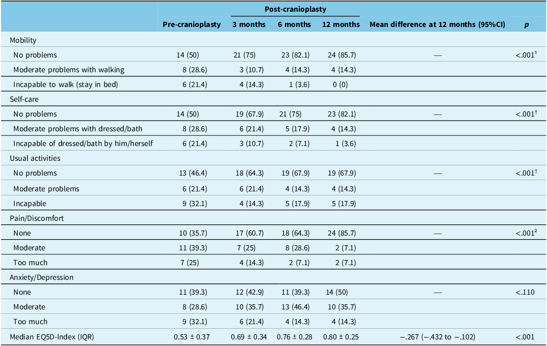
Assessment by EuroQol-5D-3L (see Table 2) revealed an overall significant improvement (p = <0.001) in almost every QoL dimension, except for “Anxiety/Depression” (see Figure 1, A–E). This improvement was most significant when comparing baseline with 12 months after cranioplasty in the dimensions of “Mobility” (“No problems” in 50% vs 85.7% patients, respectively; p = 0.016 at post hoc analysis) and “Pain/Discomfort” (“None” in 35.7% vs 85.7% patients, respectively; p = 0.005 at post hoc analysis). The overall mean EQ5D index showed a significant improvement at 12 months (mean difference −0.267, 95% CI −0.432 to −0.102; p = <0.001).
EQ-5D-3L quality-of-life evaluation before and after cranioplasty in (A) mobility, (B) self-care, (C) usual activities, (D) anxiety/depression and (E) pain/discomfort.

Contrast in quality-of-life score assessed by EuroQol-5D-3L scale pre- and post-cranioplasty

Table 2 Long description
A table comparing quality-of-life scores pre- and post-cranioplasty across various dimensions. The table has 5 rows and 6 columns. Column headers are Pre-cranioplasty, 3 months, 6 months, 12 months, Mean difference at 12 months (95% CI), and p. Row labels are Mobility, Self-care, Usual activities, Pain/Discomfort, Anxiety/Depression, and Median EQ5D-Index (IQR). Row 1: Mobility. No problems: 14 (50), 21 (75), 23 (82.1), 24 (85.7), --, <0.001. Moderate problems with walking: 8 (28.6), 3 (10.7), 4 (14.3), 4 (14.3), --, <0.001. Incapable to walk (stay in bed): 6 (21.4), 4 (14.3), 1 (3.6), 0 (0), --, <0.001. Row 2: Self-care. No problems: 14 (50), 19 (67.9), 21 (75), 23 (82.1), --, <0.001. Moderate problems with dressed/bath: 8 (28.6), 6 (21.4), 5 (17.9), 4 (14.3), --, <0.001. Incapable of dressed/bath by him/herself: 6 (21.4), 3 (10.7), 2 (7.1), 1 (3.6), --, <0.001. Row 3: Usual activities. No problems: 13 (46.4), 18 (64.3), 19 (67.9), 19 (67.9), --, <0.001. Moderate problems: 6 (21.4), 6 (21.4), 4 (14.3), 4 (14.3), --, <0.001. Incapable: 9 (32.1), 4 (14.3), 5 (17.9), 5 (17.9), --, <0.001. Row 4: Pain/Discomfort. None: 10 (35.7), 17 (60.7), 18 (64.3), 24 (85.7), --, <0.001. Moderate: 11 (39.3), 7 (25), 8 (28.6), 2 (7.1), --, <0.001. Too much: 7 (25), 4 (14.3), 2 (7.1), 2 (7.1), --, <0.001. Row 5: Anxiety/Depression. None: 11 (39.3), 12 (42.9), 11 (39.3), 14 (50), --, <0.110. Moderate: 8 (28.6), 10 (35.7), 13 (46.4), 10 (35.7), --, <0.110. Too much: 9 (32.1), 6 (21.4), 4 (14.3), 4 (14.3), --, <0.110. Row 6: Median EQ5D-Index (IQR). 0.53 ± 0.37, 0.69 ± 0.34, 0.76 ± 0.28, 0.80 ± 0.25, -0.267 (-0.432 to -0.102), <0.001.
Pre-cranioplasty versus:
¹ 6 months (p = 0.050) and 12 months (p = 0.016).
² 12 months (p = 0.005).
† No significant differences between time periods.
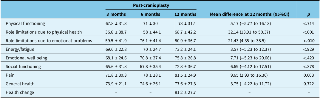
Simultaneously, the QoL estimation of the post-cranioplasty period by using SF-36 (see Table 3) showed a significant long-term improvement (baseline vs 12 months of follow-up) in the domains of “Role limitations due to physical health” (mean difference −32.14, 95% CI −50.37 to −13.91; p = <0.001), “Role limitations due to emotional problems” (mean difference −21.43, 95% CI −38.5 to −4.35; p = 0.010) and “Pain” (mean difference −9.65, 95% CI −16.36 to −2.93; p = 0.003) (see Figure 2, A–I). The mean score in the “Health change” domain, which takes the overall health perception of the patient 1 year ago as a reference and compares it with the actual health perception, showed a mean score of 81.2 ± 27.7.
SF-36 quality-of-life evaluation before and after cranioplasty in (A) a general overview and in each subscale: (B) physical functioning, (C) role limitations due to physical health, (D) role limitations due to emotional problems, (E) energy/fatigue, (F) emotional well-being, (G) social functioning, (H) pain and (I) general health. PF = physical functioning; RLPH = role limitations due to physical health; RLEP = role limitations due to emotional problems; EF = energy/fatigue; EWb = emotional well-being; SF = social functioning; P = pain; GH = general health.

Figure 2 Long description
Panel A: A radar chart shows a general overview of quality-of-life evaluations before and after cranioplasty. The chart includes axes for physical functioning, role limitations due to physical health, role limitations due to emotional problems, energy/fatigue, emotional well-being, social functioning, pain, and general health. Panel B: A vertical bar graph compares physical functioning at 3 months and 12 months, showing similar values for both time points. Panel C: A vertical bar graph illustrates role limitations due to physical health, with a significant improvement from 3 months to 12 months (p<0.001). Panel D: A vertical bar graph shows role limitations due to emotional problems, indicating a notable improvement from 3 months to 12 months (p=0.010). Panel E: A vertical bar graph depicts energy/fatigue levels, with similar values at 3 months and 12 months. Panel F: A vertical bar graph presents emotional well-being, showing similar values for both time points. Panel G: A vertical bar graph illustrates social functioning, with similar values at 3 months and 12 months. Panel H: A vertical bar graph shows pain levels, with a significant reduction from 3 months to 12 months (p=0.003). Panel I: A vertical bar graph depicts general health, showing similar values for both time points.
Quality-of-life assessment by SF-36 scale during follow-up post-cranioplasty, stratified by subscale scores; mean ± SD

Table 3 Long description
The table presents quality-of-life assessment data using the SF-36 scale during follow-up post-cranioplasty, stratified by subscale scores. It includes mean and standard deviation values for various domains at 3, 6, and 12 months post-cranioplasty, along with mean differences at 12 months and p-values. The table has 8 rows and 6 columns. Column headers are 'Post-cranioplasty', '3 months', '6 months', '12 months', 'Mean difference at 12 months (95%CI)', and 'p'. Row labels are 'Physical functioning', 'Role limitations due to physical health', 'Role limitations due to emotional problems', 'Energy/fatigue', 'Emotional well being', 'Social functioning', 'Pain', 'General health', and 'Health change'. Each row provides specific values for the respective domains at different time points and statistical analysis results.
CI = confidence interval.
A significant negative correlation was found between craniectomy surface area and the score at 12 months of follow-up in several domains of the SF-36 tool (see Table 4): energy (ρ = −0.528, p = 0.010), emotional well-being (ρ = −0.431, p = 0.040), pain (ρ = −0.463, p = 0.026) and health change (ρ = −0.470, p = 0.024). There were no significant correlations between the timing of cranioplasty and scores at 12 months. During the exploratory search for significant interactions between baseline characteristics and QoL improvement, which included craniectomy’s surface area and timing of cranioplasty as independent variables, none of the features included in the model showed a significant interaction. None of the demographic variables showed a significant difference in the univariate analysis as well.
Correlation coefficients between surgical features and SF-36 subscales scores

Table 4 Long description
A table with two rows and nine columns. The columns are labeled PF, RLPH, RLEP, EF, EWb, SF, P, GH, and HC. The rows are labeled Craniectomy surface area (cm^2) and Timing of cranioplasty (months). The values in the table are correlation coefficients. For Craniectomy surface area (cm^2), the values are: PF: -0.18, RLPH: -0.252, RLEP: -0.302, EF: -0.528**, EWb: -0.431*, SF: -0.294, P: -0.463*, GH: -0.399, HC: -0.470*. For Timing of cranioplasty (months), the values are: PF: -0.114, RLPH: 0.02, RLEP: -0.264, EF: -0.135, EWb: 0.013, SF: 0.018, P: -0.023, GH: -0.114, HC: 0.254. Notable trends include significant negative correlations in the EF, EWb, P, and HC columns for Craniectomy surface area (cm^2).
PF = physical functioning; RLPH = role limitations due to physical health; RLEP = role limitations due to emotional problems; EF = energy/fatigue; EWb = emotional well-being; SF = social functioning; P = pain; GH = general health.
* p < 0.05.
** p < 0.01.
Discussion
Quality of life
There is currently limited evidence available regarding the evolution in the QoL that patients experience after a cranioplasty, and is even more restricted in patients with customized alloplastic implants. In our center, nearly all patients undergoing cranioplasty are candidates for a customized implant, the manufacturing process of which is described in detail elsewhere.Reference Morales-Gómez, Garcia-Estrada and Leos-Bortoni20 It has been established that cranioplasty is not only an intervention that improves cosmetic satisfaction and self-esteem but also enhances the functional and self-perceived QoL of patients.Reference Giese, Antritter, Unterberg and Beynon9,Reference Worm, Finger, Ludwig do Nascimento, Rynkowski and Collares10,Reference Zaramo, Zelko and Ragland19,Reference Giese, Meyer, Engel, Unterberg and Beynon21,Reference Ganau, Cebula and Fricia22
Improvement in self-reported QoL in patients with non-customized biocompatible implants has been described early.Reference Worm, Finger, Ludwig do Nascimento, Rynkowski and Collares10 A previous study reported a global positive evolution of all the SF-36 domains at 24 months after cranioplasty, with early improvements in general health self-perception and vitality at 3 and 6 months post-intervention, respectively, while the other domains improved at 12 and 24 months. Compared with the improvement in their cohort, our patients did not reach a significant enhancement in all domains. However, the scores that our patients reached at 12 months after cranioplasty were higher than the ones at 24 months reported by these authors in the domains of physical functioning, role limitations due to physical health, role limitations due to emotional problems and emotional well-being. Besides, the health change domain, which is one of the best indicators of the impact perceived by the patient after the intervention, was significantly positive in our cohort, while it was not reported in the study by Worm.Reference Worm, Finger, Ludwig do Nascimento, Rynkowski and Collares10
A previous study, conducted with patients submitted to cranioplasty with customized PMMA implants,Reference Low, Abdullah, Abdullah, Yahya, Idris and Mohamad23 described a general improvement in their QoL at the end of their follow-up. A similar result was seen in our study, mainly in the role limitations due to the physical health domain, while a less pronounced but still significant improvement was seen in role limitations due to emotional problems, pain and health change. This difference could be explained by the contrast in the sample sizes and the cranioplasty timing, which in our study was significantly larger due to several barriers within our patients’ population (see the “Healthcare barriers in a low-income country” section below).
QoL improvement through time has been described in patients with customized-implant cranioplasties due to a failed or complex primary cranioplasty.Reference Giese, Meyer, Engel, Unterberg and Beynon21 In this study, the proportion of patients with no problems in any of the five dimensions of the EuroQol-5D-3L tool, along with their SF-36 scores, was lower than in other series. This difference is possibly related to the surgical context of the patients included in the study. Nonetheless, even with this factor, the impact on the QoL secondary to the cranioplasty intervention was quite significant.
Notably, all studies that have described the QoL improvements across time in patients with cranioplasty, regardless of whether the implants were customized or not, have demonstrated outstanding results compared with those achieved in patients treated only with decompressive craniectomy, mostly secondary to a TBI.Reference Intiso, Lombardi and Grimaldi24–Reference Sundseth, Sundseth and Thommessen30 Pooling the results of all these studies only confirms the positive effects that cranioplasty has, not only in the improvement of the neurological and functional status, but also in the QoL and overall satisfaction. There is no doubt that this helps the decision-making process, based on integral patient-centered care.
Factors that modify the improvement in quality of life
Several modifiers and predictors of the effect size that cranioplasty has on the QoL through time have been described, including cranioplasty timing,Reference Mustafa, Richardson and Gillespie31 the primary indication for craniectomy,Reference Giese, Antritter, Unterberg and Beynon9 implant materialReference Ganau, Cebula and Fricia22 and craniectomy surface area.Reference Klieverik, Robe, Muradin and Woerdeman32 Studies conducted with patients who underwent decompressive craniectomy due to TBI have identified other QoL predictors such as sex, age, presence of comorbidities and grade of TBI.Reference Scholten, Haagsma and Andriessen29,Reference Rauen, Reichelt and Probst33 In the present study, most of our patients had a large extension craniectomy (median surface area of 104 cm2) and a wide cranioplasty timing (median time of 14 months), similar to that reported by other studies.Reference Worm, Finger, Ludwig do Nascimento, Rynkowski and Collares10,Reference Giese, Meyer, Engel, Unterberg and Beynon21 Almost 80% of our patients had TBI as the primary indication for craniectomy, making a comparison between craniectomy indications not feasible.
In a study that included patients with cranioplasty made mostly with autologous bone graft,Reference Giese, Antritter, Unterberg and Beynon9 patients who had craniectomy due to an ischemic event had the worst self-reported QoL compared with the other groups (TBI, intracerebral/subarachnoid hemorrhage, and infection). The SF-36 scores of the TBI group showed an overall significant improvement and are quite similar to the scores of our cohort. Another study that made a comparison between customized implants of hydroxyapatite and other materials (mainly PMMA)Reference Ganau, Cebula and Fricia22 showed a better self-reported outcome in the former group. Considering the self-perceived health change at 1-year post-intervention as the main outcome, 82.1% of our patients reported feeling better than their pre-cranioplasty status, while in the hydroxyapatite group of the previously mentioned study, only 66% of them reported feeling better or the same than 1 year ago.
The craniectomy extension has been recently described as a significant predictor of cosmetic satisfaction in patients submitted to cranioplasty.Reference Klieverik, Robe, Muradin and Woerdeman32 Thus, we decided to include it as one of the main variables to test in our exploratory analysis of potential modifiers for QoL improvement. None of the baseline features showed a significant interaction. However, a negative correlation was found between the SF-36 scores 1 year after cranioplasty and the skull defect surface area in the domains of energy, emotional well-being, pain, general health and health change. This finding suggests that there is a potential impact of the defect’s proportions on some aspects of the QoL that patients experience on a long-term basis after cranioplasty, but further research is necessary to confirm if this association actually exists.
Healthcare barriers in a low-income country
Access to safe, timely and affordable medical attention and surgical care is highly restricted in middle- and low-income countries. Neurosurgical care is no exception. Recent studies have described that the proportion of middle- and low-income countries’ populations with access to a neurotrauma care center within 2 hours is 44.9% and 25%, respectively, with Latin America having a very similar proportion to the middle-income countries (50%), and the main reasons for healthcare delays include economic barriers, considerable geographical distances between patients’ living places and the healthcare centers and inadequate operative supplies.Reference Mediratta, Lepard, Barthélemy, Corley and Park34 The proportion reported in our country (Mexico) is 26.2%, and it decreases as neurosurgical care needs to be more specialized.Reference Punchak, Mukhopadhyay and Sachdev35
As a reference healthcare center, many patients who seek general and specialized neurosurgical care come to our institution every day. Although we offer them the best and most suitable options for their treatment, there are still several barriers that prevent them from getting these therapeutic interventions in a proper and timely manner. This is one of the main reasons why the cranioplasty timing in our patient population was quite extensive.
Limitations of this study
This study has several limitations that have to be considered when interpreting the results. Data obtained from the follow-up was collected in an ambispective manner after the cranioplasty was made, which may introduce a potential selection bias. Although our patients’ population treated with cranioplasty is quite large, a considerable proportion was not suitable for being included due to loss of follow-up, primarily caused by the numerous healthcare barriers discussed earlier. In consequence, our cohort was conducted with a small and heterogeneous sample. Further research through more solid evidence generators, such as randomized clinical trials and larger cohorts, is needed.
Conclusions
Cranioplasty in patients who underwent a previous craniectomy improves their global health and functional status, along with their QoL self-perception. This study showed promising results about the improvement in the QoL experienced by patients with low-cost customized alloplastic implants. Previous literature has established an association between improvement in brain hemodynamics secondary to cranioplasty and a positive evolution in clinical and QoL assessments. Besides, the skull’s reconstruction through this procedure also enhances the patient’s positive self-awareness, providing comfort and esthetic well-being. Since this is a feasible and practical option for patients in low- and middle-income countries, further and better research is necessary in order to establish which are the best options to preserve clinical and self-reported improvement and conduct better patient-centered neurosurgical care.
Acknowledgments
The authors have no acknowledgments.
Author contributions
JACH: Clinical data extraction; phone interviews; data analysis and management; manuscript writing; creation of figures.
CARD: Clinical data extraction; phone interviews; data analysis and management; manuscript writing.
LPG: Clinical data extraction; phone interviews; manuscript writing.
JAMG: Corresponding author; idea and concept development; manuscript revision; mentorship; assessment of resources and clinical data extraction.
AMAV: Clinical data extraction; phone interviews; manuscript revision.
AMFS: Clinical data extraction; phone interviews; manuscript revision.
OGR: Clinical data extraction; phone interviews; manuscript revision.
MAVA: Manuscript revision; creation of figures.
ARMPdL: Manuscript revision; assessment of resources and clinical data extraction.
Funding statement
This research had no funding sources.
Competing interests
The authors report no conflict of interest concerning the materials or methods used in this study or the findings specified in this paper.
Disclosures
The authors have no personal, financial or institutional interest in any of the drugs, materials or devices described in this article.