Introduction
According to the WoRMS Editorial Board (2014), the genus Lecithaster consists of 30 species of parasites inhabiting various marine fish. Among these, two species, Lecithaster mugilis Yamaguti, Reference Yamaguti1970 and L. helodes Overstreet, Reference Overstreet1973, have been described from species of mullet. Lecithaster mugilis Yamaguti, Reference Yamaguti1970 was described from Mugil cephalus on the Hawaiian Islands (Yamaguti, Reference Yamaguti1970). Yamaguti's description differed from that of other Lecithaster species by the presence of a large ventral sucker and a three-lobed ovary.
Lecithaster helodes Overstreet, Reference Overstreet1973 was described from the intestines of M. cephalus and Mugil curema from the Mississippi Sound and adjacent waters. The author of this species noted that L. helodes shared many common features with L. mugilis. The main difference between these species, according to Overstreet (Reference Overstreet1973), is the four-lobed ovary in L. helodes and the three-lobed ovary in L. mugilis. Machida (Reference Machida2003), during an investigation of L. mugilis found in Moolgarda seheli from the coastal waters of Japan, defined these worms as having a four-lobed ovary. According to Machida (Reference Machida2003) L. mugilis differs from the morphologically similar L. stellatus Looss, 1907, which was also found in this area, by the presence of a pit near the anterior margin of the ventral sucker.
We found specimens that were identified as L. mugilis in M. seheli, Valamugil engeli and Liza subviridis from the coastal waters of Cat Ba Island in Halong Bay, Vietnam. A second species, Lecithaster sudzuhensis n. sp., was found in M. cephalus from the estuarine waters of the Kievka River in the Primorsky region of Russia. Below we present morphometric and genetic data obtained from the study of adult flukes.
Materials and methods
Collection and examination of fish
Up to three species of mullet were collected from the coastal waters of Cat Ba Island, Halong Bay, Vietnam, including M. seheli, V. engeli and L. subviridis. A further mullet species, M. cephalus, was collected from the estuarine waters of the Kievka River. Adult L. mugilis were found in the intestines of 11 of the 47 M. seheli dissected, two of the 70 V. engeli, one of the 31 L. subviridis and one of the 15 M. cephalus dissected. Following removal from the intestine, worms from each fish species were rinsed in distilled water, identified as previously defined, killed in hot distilled water and preserved in 70% ethanol. Following fixation, flukes were transferred to 96% ethanol and whole mounts were stained in alum carmine, dehydrated in an ethanol series, cleared in xylene and mounted in Canada balsam. All sizes are given in mm.
Molecular analysis
Adult specimens of L. mugilis, collected from M. seheli from the coastal waters of Cat Ba Island, and L. sudzuhensis n. sp., collected from M. cephalus from the Primorsky region, were used for molecular analysis (table 1). Total DNA was extracted from flukes, which were fixed in 96% ethanol, using a ‘hot shot’ technique (Truett, Reference Truett and Kieleczawa2006).
List of taxa incorporated into molecular analysis (n, number of specimens).

The following primers were used to amplify 18S rDNA: 18S-E (5′-CCG AAT TCG TCG ACA ACC TGG TTG ATC CTG CCA GT-3′), 18S-F (5′-CCA GCT TGA TCC TTC TGC AGG TTC ACC TAC-3′) as previously described (Littlewood & Olson, Reference Littlewood, Olson, Littlewood and Bray2001). The initial polymerase chain reaction (PCR) was performed in a total volume of 20 μl containing 0.25 mm of each primer pair, approximately 10 ng of total DNA in water, 10 × Dream Taq buffer, 1.25 mm deoxynucleoside triphosphates (dNTPs) and 1 unit of Dream Taq polymerase (Thermo Scientific, Waltham, Massachusetts, USA). Amplification of a 2000-bp fragment of the 18S rRNA gene was performed in a GeneAmp 9700 (Applied Biosystems, Waltham, Massachusetts, Foster City, California, USA) with a denaturation step at 96°C for 5 min; 35 cycles of 1 min at 96°C, 20 s at 58°C and 5 min at 72°C; and an extension step at 72°C for 10 min. Negative and positive controls using both primers were included.
Amplification of 28S rDNA was achieved with the primers DIGL2 (5′-AAG CAT ATC ACT AAG CGG-3′) and 1500R (5′-GCT ATC CTG AGG GAA ACT TCG-3′) as described previously (Tkach et al., Reference Tkach, Littlewood, Olson, Kinsella and Swiderski2003). The master mix for the initial PCR reaction for 28S rDNA was identical to that described above for 18S rDNA. Amplification of a 1200-bp fragment of 28S rDNA was performed in a GeneAmp 9700 (Applied Biosystems) with a denaturation step at 94°C for 3 min; 40 cycles of 30 s at 94°C, 30 s at 55°C and 2 min at 72°C; and extension at 72°C for 7 min. Negative and positive controls using both primers were included.
PCR products were directly sequenced using an ABI Big Dye Terminator v.3.1 Cycle Sequencing Kit (Applied Biosystems), as recommended by the manufacturer, with the internal sequencing primers described by Littlewood & Olson (Reference Littlewood, Olson, Littlewood and Bray2001) for 18S rDNA and Tkach et al. (Reference Tkach, Littlewood, Olson, Kinsella and Swiderski2003) for 28S rDNA. PCR products were analysed using an ABI 3130 genetic analyser at the Institute of Biology and Soil Sciences FEB RAS. Sequences were submitted to the European Nucleotide Archive (ENA) with the accession numbers described in table 1.
Ribosomal DNA sequences were assembled with SeqScape v. 2.6 software (distributed by Applied Biosystems). Alignments and estimation of the number of variable sites and sequence differences were performed using MEGA 6.0 (Tamura et al., Reference Tamura, Stecher, Peterson, Filipski and Kumar2013). Phylogenetic analyses of the nucleotide sequences were performed using the Bayesian algorithm with the MrBayes v. 3.1.2 software (Huelsenbeck et al., Reference Huelsenbeck, Ronquist, Nielsen and Bollback2001). The best nucleotide substitution model, the general time reversible with estimates of invariant sites and gamma–distributed among-site variation (GTR + I + G), was estimated with jModeltest v. 2.1.5 software (Posada & Crandall, Reference Posada and Crandall1998). Bayesian analysis was performed using 10,000,000 generations, with two independent runs. Summary parameters and the phylogenetic tree were calculated with a burn-in of 1,500,000 generations. The significance of the phylogenetic relationships was estimated using posterior probabilities (Huelsenbeck et al., Reference Huelsenbeck, Ronquist, Nielsen and Bollback2001). The phylogenetic relationships of the species of Lecithasteridae were inferred from our data, along with the nucleotide sequences of the 18S rDNA and 28S rDNA of other trematode specimens obtained from the National Center for Biotechnology Information (NCBI) GenBank database (table 1).
Results
Lecithaster mugilis Yamaguti, Reference Yamaguti1970
Taxonomic summary
Hosts. Mugilidae: Moolgarda seheli (Forsskål, 1775), Valamugil engeli (Bleeker, 1858), Liza subviridis (Valenciennes, 1836).
Locality. Coastal waters of Cat Ba Island, Halong Bay, northern Vietnam (20°84′N, 106°59′E).
Site. Intestine.
Intensity of infection. 1–8 specimens.
Deposited. Slides No. 60-67Tr. were placed in the collection of the Zoological Museum (Institute of Biology and Soil Science, Far East Branch of the Russian Academy of Sciences, Vladivostok, Russia). E-mail: petrova@ibss.dvo.ru. Deposited: 20 October 2015.
Description
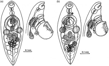
Based on seven specimens (fig. 1a, table 2). Body fusiform, smooth. Pre-oral lobe present. Oral sucker subterminal, pre-pharynx absent, pharynx spherical, oesophagus long (can reach the level of the anterior margin of the ventral sucker). Caeca end behind the posterior margin of the vitellarium, not extending to the posterior end of the body. A large ventral sucker on the border of the anterior and middle thirds of the body. Ducts of six gland cells, which are dorsally placed above the ventral sucker, pass through a thin-walled sac and open by a pore on the median line of the body between the genital pore and the ventral sucker. Testes are round, on the same level, left and right of the median line of the body, near or in contact with the posterior margin of the ventral sucker. Seminal vesicle is saccate and placed between the middle of the ventral sucker and the anterior margin of the ovary. Pars prostatica, which passes along the median line of the body dorsally to the ventral sucker, is long and lined with vesicular cells and surrounded by numerous prostatic cells. Sinus-like sac is oval and penetrated with a hermaphroditic duct. Genital pore is median and located at a small distance before the ventral sucker. A pit is connected with a thin-walled sac in the inner side and lies on the median line of the body between the genital pore and the anterior margin of the ventral sucker. Ducts of six gland cells, which are dorsally placed from the ventral sucker, pass through the sac and open in the pit.
Morphological features of adult worms of (a) Lecithaster mugilis Yamaguti, Reference Yamaguti1970 and (b) Lecithaster sudzuhensis n. sp.

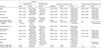
Morphometrics (in μm) of Lecithaster mugilis, L. sudzuhiensis n. sp. and L. helodes.

Ovary consists of four large lobes and is located between the posterior border of the testis and the anterior margin of the vitellarium. Seminal receptacle is round and placed on the level of the ovary and on the left of the median line of the body. Vitellarium consists of seven drop-shaped follicles and is placed at the end of the middle third and onset of the last third of the body. The uterus is located between posterior margin of the ventral sucker and posterior end of the body. Eggs small, oval and operculated. Excretory bladder is Y-shaped, excretory pore is terminal.
Molecular data
For L. mugilis collected from V. engeli, Vietnam, a total of 1821 and 1003 alignable characters, obtained using from two to six replicate sequences, were available for analysis in the 18S rRNA gene and 28S rRNA gene datasets, respectively. No variable and parsimony-informative sites for either 18S or 28S rDNA of L. mugilis were presented.
Lecithaster sudzuhensis n. sp.
Taxonomic summary
Host. Mugil cephalus Linnaeus, 1758, Mugilidae.
Type habitat. Kievka River (42°52′N, 133°39′E).
Site. Intestine.
Intensity of infection. 17 specimens.
Type deposition. Holotype no. 51-Tr., paratypes nos 52-Tr.–56-Tr., the collection of the Zoological Museum (Institute of Biology and Soil Science, Far East Branch of the Russian Academy of Sciences, Vladivostok, Russia). E-mail: petrova@ibss.dvo.ru. Deposited: 20 October 2015.
Etymology. The specific name refers to the old name of the Kievka River, Sudzuhe River.
Description
Based on six specimens (fig. 1b, table 2). Body fusiform, smooth. Pre-oral lobe present. Oral sucker subterminal, pre-pharynx absent, pharynx spherical, oesophagus long (can reach the level of the anterior margin of the ventral sucker). Caeca end at the posterior half or behind the posterior margin of the vitellarium. The large ventral sucker is on the border of the anterior and middle thirds of the body. Round testes located at the same level, on both sides of the median line of the body, near or in contact with the posterior margin of the ventral sucker. Seminal vesicle is saccate and placed between the middle of the ventral sucker and the anterior margin of the ovary. Pars prostatica, which passes along the median line of the body dorsally to the ventral sucker, is long and lined with vesicular cells and surrounded by numerous prostatic cells. Sinus-like sac is oval and penetrated with a hermaphroditic duct. Genital pore is median and located at a small distance before the ventral sucker. A pit connected with a thin-walled sac in the inner side and lies on the median line of the body between the genital pore and the anterior margin of the ventral sucker. Ducts of an uncertain number of gland cells, placed dorsally from the ventral sucker, pass through the sac and open in the pit.
Ovary consists of four large lobes and is located between the posterior border of the testis and the anterior margin of the vitellarium. Seminal receptacle is round and placed on the level of the ovary and on the left of the median line of the body. Vitellarium consists of seven drop-shaped follicles and is placed at the end of the middle and onset of the last third of the body. Uterus is located between posterior margin of the ventral sucker and posterior end of the body. Eggs small, oval and operculated. Excretory bladder is Y-shaped, excretory pore is terminal.
Molecular data
For L. sudzuhensis n. sp. collected from Mugil cephalus, Primorsky region, 1819 and 1002 alignable characters, obtained using from two to six replicate sequences, were available in the 18S rRNA gene and 28S rRNA gene datasets, respectively. No variable and parsimony-informative sites for either 18S or 28S rDNA of L. sudzuhensis n. sp. were presented.
Discussion
The adult flukes collected from mullet fish in Vietnam had identical morphology (including body and organ size) to L mugilis described by Machida (Reference Machida2003) (table 2). There are minor differences in the sizes of the ventral sucker, which are smaller in our specimens than in those described by Machida. Machida (Reference Machida2003) noted the presence of a pit with a short posterior sac, or duct, lying midway between the genital pore and ventral sucker in specimens of L. mugilis. In our material, worms had a pit connected to a thin-walled sac in the same area of the body. Ducts of glands pass through the sac (fig. 1a). Lecithaster helodes Overstreet, Reference Overstreet1973 from mullet fish that were caught in the Mississippi Sound and adjacent waters are similar to L. mugilis in most morphological features and sizes (table 2). Overstreet (Reference Overstreet1973) distinguished L. helodes and L. mugilis based on the number of lobes of the ovary (four and three lobes, respectively). However, Machida (Reference Machida2003) established that L. mugilis has a four-lobed ovary, so this morphological feature cannot be used to identify it from L. helodes. However, the differences in the sizes of the pharynx and oral sucker (table 2), the ratio between these organs (the pharynx was smaller than the oral sucker in L. mugilis and vice versa in L. helodes) and some differences in the length of the caeca (the caeca reached the zone between the ovary and posterior end of the body in L. mugilis and only the level of the posterior margin of the vitellarium in L. helodes) support the validity of L. helodes.
Among East-Asian species of Lecithaster, L. mugilis is most similar to L. confusus Odhner, 1905 and L. stellatus. In 1907, Looss (cited by Machida, Reference Machida2003) pointed out that L. confusus has a pre-acetabulum pit. Lecithaster mugilis differs from these species and other species of Lecithaster, besides L. helodes and L. sudzuhensis n. sp., by having a long oesophagus, which can reach the anterior border of the ventral sucker.
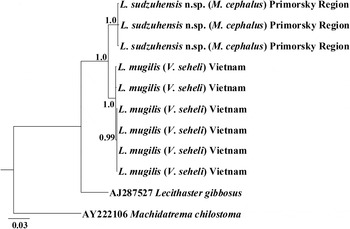
Worms found in mullet fish from the Primorsky region of Russia match the diagnostic features of the genus Lecithaster in all morphological features (Gibson, Reference Gibson, Gibson, Jones and Bray2002). When comparing morphological and metric characteristics, the newly described species has the greatest similarity to L. mugilis and L. helodes (table 2). However, L. sudzuhensis n. sp. differs from L. mugilis by having a higher mean size of the pharynx and a smaller ventral sucker, and by the ratio of suckers. The present species differs from L. helodes by having a smaller maximal and mean size of the pharynx, and smaller ventral sucker, and by the ratio of suckers (table 2). Unlike L. helodes, the pharynx of L. sudzuhensis n. sp. is smaller than the oral sucker (table 2). According to the above features, L. sudzuhensis n. sp. occupies an intermediate position between L. mugilis and L. helodes. Comparative analysis of the nucleotide sequences of L. mugilis from Vietnam and L. sudzuhensis n. sp. from the Primorsky region showed 40 (1.9%) and 28 (2.6%) variable sites in the 18S rRNA and 28S rRNA genes, respectively. The genetic differentiation between L. mugilis from Vietnam and L. sudzuhensis n. sp. from Primorsky with L. gibbosus was 5.4% and 5.5% respectively for the 18S rDNA gene sequence data, and 10% and 10.3% for the 28S rRNA gene sequence data. A phylogenetic relationship based on the combined ribosomal gene sequence data showed a clear differentiation of L. mugilis and L. sudzuhensis n. sp.; L. gibbosus appears to be a sister species to the (L. mugilis/L. sudzuhensis n. sp.) cluster (fig. 2). Thus, our molecular data support the validity of L. sudzuhensis n. sp. from M. cephalus of the Primorsky region.
Phylogenetic tree of the family Lecithasteridae based on the analysis of combined 18S rRNA (complete) and of 28S rRNA (partial) gene sequences with the Bayesian algorithm; nodal numbers indicate posterior probabilities.

The present study showed that L. mugilis, L. helodes and L. sudzuhensis n. sp. have no distinct morphometric differences. Only molecular data allowed us to distinguish L. mugilis from Vietnamese mullet fish and L. sudzuhensis n. sp. from Far Eastern fish. It is likely that L. mugilis, L. helodes and L. sudzuhensis n. sp. form a group of cryptic species. However, a final decision on the status of these species can only be made after receiving the molecular data for L. helodes.
Financial support
Grants RFBR no. 16-34-00222_mol_a and VAST.DA47.-DA 12/15-18 provided partial financial support for this research.
Conflict of interest
None.