The haemodynamic changes in patients with Fontan circulation with reduced cardiac output, chronic venous hypertension, and alterations in subdiaphragmatic venous flow pre-dispose for the development of Fontan-associated liver disease. This process is initiated by hepatic congestion with the potential for progression to liver cirrhosis, portal hypertension, and the risk of hepatocellular carcinoma.Reference Ghaferi and Hutchins1–Reference Gordon-Walker, Bove and Veldtman3
As the incidence of these complications increases with duration of follow-up, regular hepatic surveillance is recommended. Clinical examination is completed by blood chemistry, ultrasound at least annually, and CT or MRI depending on severity of Fontan-associated liver disease.Reference Kim, Yang, Jang, Yoo, Khalili and Kim4 Patients with compensated cirrhosis are generally asymptomatic; abnormal liver function test, jaundice, ascites, or variceal bleeding indicate decompensated liver disease.
Portal vein thrombosis is a relatively common complication in advanced cirrhosis but has not been mentioned in the context of Fontan-associated liver disease. The risk of portal vein thrombosis has been observed to increase with the severity of liver disease as previously described in 7–16% of patients with advanced non-cardiac cirrhosis and as a possible “surrogate marker for worsening portal hypertension”.Reference Harding, Perera, Chen, Olliff and Tripathi5
We report the previously undescribed case of a 25-year-old female with double inlet left ventricle and transposition of the great arteries, initially palliated with modified Blalock–Taussig shunt, undergoing modified Fontan surgery with a fenestrated lateral tunnel at the age of 2.5 years.
Post-operative haemodynamics at age of 18 years revealed a mean pulmonary artery pressure of 8 mmHg, left ventricular end-diastolic pressure of 3 mmHg, but effort intolerance on cardiopulmonary exercise test. Abdominal sonography described heterogeneous echogenicity of the liver with nodular surface, no splenomegaly, antegrade portal vein flow (28 cm/s). Liver enzymes were mildly elevated with further increase predominantly of gamma-glutamyl transpeptidase during the following years in the presence of normal synthetic function.
The patient remained asymptomatic with stable albeit reduced exercise capacity, and without abdominal pain or ascites. She continued on acetylsalicyclic acid but she was not treated with medical heart failure therapy.
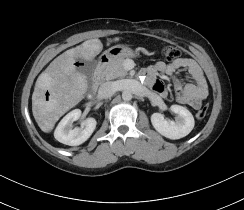
Doppler ultrasonography demonstrated absent portal vein flow with cavernous malformation as an incidental finding during follow-up in the presence of stable spleen measurements and without ascites. CT confirmed the sonographic finding of inhomogenous enhancement in the liver and multiple nodular lesions, absent portal vein flow with cavernous transformation, and portosystemic collateral vessel draining into the left-sided renal vein (Fig 1).
Axial CT image demonstrating multiple nodular hepatic lesions (black arrows). Enlarged collateral vessel draining into the left-sided renal vein (white arrowhead).

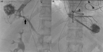
Cardiac catheterisation at age of 22 years had demonstrated mean pulmonary artery pressure of 14 mmHg, transpulmonary gradient of 5–6 mmHg, RPI 1,2 WE*m2, and arterial oxygen saturation of 89%. The angiography showed a veno-venous collateral vessel to the right upper pulmonary vein; the retrograde hepatic vein wedge angiography demonstrated only the right-sided portal vein branch, no left-sided portal vein branch (Fig 2). Recent blood tests revealed normal synthetic function, normal transaminases, stable gamma-glutamyl transpeptidase elevation (770U/l), no leucopenia, thrombocytopenia, or hyperbilirubinemia; oesophageal varices had been excluded by endoscopy.
(a) Right-sided hepatic vein wedge angiography revealed right portal vein branch (nearly 6 mm) (black arrow) and (b) left-sided hepatic vein wedge angiography could not demonstrate the left-sided portal vein branch

Comment
Late failure of the Fontan circulation may first become evident by decline in functional capacity and/or development of Fontan-associated liver disease leading to recent recommendations for a regular screening protocol. This should be initiated 5–10 years after Fontan surgery, including liver function parameters, alpha-fetoprotein, and liver imaging alongside haemodynamic evaluation.Reference Ackermann, Geerts, Van Vlierberghe, De Backer and Francois6,Reference Rahtgeber and Harris7
We report on the first documented case of portal vein thrombosis in such a patient with advanced liver disease during long-term follow-up. The patient was asymptomatic without any abdominal symptoms, and the diagnosis of portal vein thrombosis was made on routine ultrasonography with confirmation on abdominal CT scan. Delayed diagnosis is explained by the finding of an enlarged collateral vessel draining into the left-sided renal vein with decompression of the portal system. Besides the well-known pathogenetic conditions leading to Fontan-associated liver disease during long-term follow-up, this patient had some potential additional pre-disposing risk factors: advanced liver disease with multiple regenerative nodules might have caused further reduction of portal blood flow. In addition, the heterozygote state for factor V Leiden mutation in addition to well-known imbalance of pro- and anti-coagulant proteins during long-term Fontan circulation might pre-dispose to thrombotic complications.Reference Firdouse, Agarwal, Chan and Mondal8 Because of compliance problems, the patient could not be safely anticoagulated during adolescence and remained on acetylsalicyclic acid also after diagnosis of portal vein thrombosis.
The management of Fontan-associated liver disease in this case, without hypersplenism or liver dysfunction, prompts debate. With the development of a large collateral vessel and no signs of portal hypertension, the risk of variceal bleeding is not typically high. The use of transjugular intrahepatic portosystemic shunt to restore portal flow is not a suitable treatment option in Fontan patients with regard to the usually normal or reduced portosystemic gradient and development of intrahepatic collaterals; a transjugular intrahepatic portosystemic shunt procedure might only be considered as a rescue intervention in massive variceal bleeding.Reference Téllez, Rodiguez-Santiago and Albillos9,Reference Velpula, Sheron, Guha, Salon, Hacking and Veldtman10 A meso-portal vein surgical bypass (Rex shunt) is not a suitable option in this patient as only the right portal vein branch was demonstrable on angiography.
Whether portal vein thrombosis will lead to progression of the liver disease and might aggravate the “natural history of Fontan-associated liver disease” with the risk of development of variceal bleeding, ascites, intestinal ischemia, and encephalopathy is unpredictable and the patient needs close follow-up for signs and potential complications of cirrhosis, including hepatocellular carcinoma. In patients with failure of the Fontan circulation and Fontan-associated liver disease, combined heart and liver transplantation might be considered; isolated heart transplantation might allow to prevent further progression of compensated liver disease.Reference Rahtgeber and Harris7 As this patient remained compensated and presented with a good quality of life other than reduced functional capacity transplant referral was deemed unnecessary at this stage.
Financial Support
There was no grant or other funding obtained for this report.
Conflicts of Interest
None.
Ethical Standards
This case report was performed according to the local code of research conduct and data protection rules and was according to the ethical guidelines and principles of the International Declaration of Helsinki. Approval by the local ethics committee was not required.