Implications
Visceral fat deposition has been considered an important factor in cattle finishing. As maturing proceeds, large amounts of mesenteric fat can be deposited leading to production inefficiencies, which can jeopardize meat quality. Yet, no studies addressing fatty acid incorporation at the cell membrane and its outcome on membrane physical properties, including rigidity and permeability to water and solutes, are available for ruminants. This study exploits the membrane fluidity and permeability to glycerol, a key substrate involved in lipogenesis, while an underlying mechanism for differential visceral fat deposition in bovines that may be influenced by breed or diet.
Introduction
Traditional meats with Protected Designation of Origin, derived from local extensive production systems and autochthonous breeds, have the certification of European Union legislation due to their supposed quality and sensory traits, which have been associated with their specific lipid fraction properties (Council Regulation No. 2081/92 of 14 July, European Economic Community). Curiously, the scientific information available to sustain the claimed quality, mainly dependent on its lipid composition, is scarce. On the other hand, the manipulation of adipose tissue deposition in cattle has represented for many years a major breeding goal as a future guarantee for the improvement of production efficiency, visceral fat partitioning, carcass composition and meat quality (De Smet et al., Reference De Smet, Raes and Demeyer2004). The identification of key factors and molecular mechanisms responsible for the development of specific fat depots (Azain, Reference Azain2004) in autochthonous bovine breeds is necessary, in particular, mechanisms underlying visceral fat accumulation in young bulls with distinct precociousness, Alentejana and Barrosã, known as late- and early-maturing breeds, respectively (da Silva et al., Reference da Silva, Lemos, Monteiro and Portugal1998). Precociousness is intimately related to adipose tissue deposition in meat-producing animals, as early-maturing breeds deposit noticeable amounts of marbling fat before late-maturing breeds (Hocquette et al., Reference Hocquette, Gondret, Baeza, Medale, Jurie and Pethick2010).
The degree of saturation of plasma membrane acyl chains might be among the primary events in adipocyte differentiation (Stubbs and Smith, Reference Stubbs and Smith1984). Nevertheless, literature addressing bovines’ fatty acid deposition at the cell membrane level and its outcome on membrane physical properties is unavailable. Given that the membrane bilayer permeability to water and solutes is tightly related to phospholipid composition and membrane fluidity (Lande et al., Reference Lande, Donovan and Zeidel1995), it seems reasonable that distinct fat depots with specific metabolic characteristics would affect membrane physical properties. Fatty acid incorporation into cellular membranes is known to affect permeability to water and, most importantly, to glycerol (Soveral et al., Reference Soveral, Martins, Martins, Lopes, Alfaia, Prates and Moura2009; Martins et al., Reference Martins, Lopes, Martins, Madeira, Santos, Prates, Moura and Soveral2010). It is well established that glycerol is a key substrate for lipogenesis and lipolysis in adipose tissues of ruminants. The concept of membrane permeability and fluidity could therefore be critical for understanding membrane structure–function.
Specific genetic characteristics have been also described, with the purebred Alentejana breed considered phylogenetically distant from the purebred Barrosã breed (Beja-Pereira et al., Reference Beja-Pereira, Alexandrino, Bessa, Carretero, Dunner, Ferrand, Jordana, Laloe, Moazami-Goudarzi, Sanchez and Canon2003). Large differences in the levels of intramuscular fat in Alentejana and Barrosã bovines were previously reported by our group, with values of Alentejana breed (1.2%) nearly half of the other (2.3%) (Alfaia et al., Reference Alfaia, Castro, Martins, Portugal, Alves, Fontes, Bessa and Prates2007 and Reference Alfaia, Alves, Martins, Costa, Fontes, Lemos, Bessa and Prates2009). Studies in humans and mice (Field and Clandinin, Reference Field and Clandinin1984; Field et al., Reference Field, Ryan, Thomson and Clandinin1988) demonstrated that dietary fat can alter adipose cell membrane composition. Thus, the structure and physiological properties of the adipocyte membrane may be changed. The goal of the present study was to assess the effect of breed and diet on fat deposition, as well as on the lipid composition, permeability and fluidity of adipocyte membranes.
Material and methods
Experimental design: animals and diets
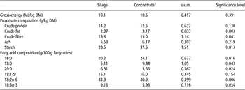
The experimental design included 24 male bovines from two phylogenetically distant autochthonous breeds, the late-maturing breed, Alentejana, and the early-maturing breed, Barrosã, allocated to silage-based or concentrate-based diets (four experimental groups of six animals each) from January to November 2009. At the beginning of the experiment, Alentejana bulls were 332 ± 10.2 days old (initial weight of 275 ± 15.6 kg) and Barrosã bulls were 268 ± 2.96 days old (initial weight of 217 ± 4.57 kg). Bulls were fed two experimental diets composed of 70/30% and 30/70% of corn silage and concentrate, respectively. The proximate composition and fatty acid profile in both experimental diets (n = 3) are shown in Table 1.
Diet composition

DM = dry matter.
n = 3; values are mean ± s.e.m.
†Silage-based diet = 30/70% of concentrate and silage, respectively.
‡Concentrate-based diet = 70/30% of concentrate and silage, respectively.
Bulls were slaughtered at 18 months of age at the INRB Experimental Abattoir by exsanguination after stunning with a cartridge-fired captive bolt stunner. The amount of subcutaneous fat was determined by dissection of the leg joint. The former has been suggested to be representative of the overall bovine carcass composition in these particular breeds (Simões and Mendes, Reference Simões and Mendes2003). Mesenteric, omental and kidney knob and channel fat (KKCF) depots were excised and weighed. Samples from the mesenteric fat were collected, flash-frozen in liquid nitrogen and stored at −80°C for subsequent analysis.
The entire experiment was conducted under the guidelines for the care and use of experimental animals in the Unidade de Produção Animal, L-INIA, INRB (Fonte Boa, Vale de Santarém, Portugal).
Preparation of membrane vesicles from bovine's mesenteric fat
Membrane vesicles from bovine's mesenteric fat were prepared by differential centrifugation with buffer without detergents, according to Martins et al. (Reference Martins, Lopes, Martins, Madeira, Santos, Prates, Moura and Soveral2010). Briefly, approximately 20 g of fat tissue from each bovine was chopped into small pieces, removing visible blood vessels, and homogenized in 200 ml of mannitol-Hepes buffer (100 mM mannitol, 10 mM Tris-Hepes, pH 7.4) in a Warring blender for 2 min. The homogenate was filtered through a 70 μm nylon mesh to separate the vascular stroma and intracellular fat retained in the filter. The filtrate was centrifuged at 46 000 × g for 45 min at 10°C to obtain a pellet of crude membranes and further washed in the same buffer. The membrane pellet was then resuspended in mannitol-Hepes buffer, transferred to a syringe and sheared by vigorously passing it 10 times through a 21-gauge needle and immediately used for transport experiments. Protein content was determined by the Bradford method (Bradford, Reference Bradford1976). The vesicle size of all membrane preparations was determined by Dynamic Light Scattering; Brookhaven Inst. BI-90.
Fatty acid composition and cholesterol concentration of adipose membranes
After membrane vesicle lyophilisation (at −60°C and 2.0 hPa), fatty acids were converted to methyl esters (FAME) (Raes et al., Reference Raes, De Smet and Demeyer2001; Christie et al., Reference Christie, Dobson and Adlof2007). The resulting FAME were then analysed by gas chromatography, using a capillary column (30 m × 0.25 mm i.d., Omegawax 250; 0.25 mm film thickness; Supelco, Bellefont, CA, USA), equipped with a flame-ionization detector. The chromatographic conditions were described in detail elsewhere (Alves and Bessa, Reference Alves and Bessa2009). The fatty acid composition was expressed as g/100 g of total fatty acids identified.
Total cholesterol was extracted from lyophilized adipose membrane vesicles through a direct saponification with saturated methanolic KOH solution (Naeemi et al., Reference Naeemi, Ahmad, al-Sharrah and Behbahani1995). Cholesterol was separated and identified using high-performance liquid chromatography (HPLC) equipment (Agilent 1100 Series, Agilent Technologies Inc., Palo Alto, CA, USA) by normal phase (Zorbax Rx-Sil column, 250 mm × 4.6 mm i.d., 5 μm particle size, Agilent Technologies Inc.). HPLC was equipped with a diode array detector set at 206 nm and the solvent (30 ml/l isopropanol in n-hexane) flowed at 1 ml/min. Total cholesterol concentration was calculated, in duplicate, based on the external standard technique, from a standard curve for peak area v. cholesterol concentration and expressed as mg/mg vesicles.
Water and glycerol permeability experiments
Stopped-flow experiments were performed on a HI-TECH Scientific PQ/SF-53 apparatus with 2 ms dead time, temperature controlled and interfaced with a PC microcomputer. Experiments were performed at temperatures ranging from 14°C to 37°C. Typically, five runs were stored and analyzed in each experimental condition. For the measurement of osmotic water permeability, membrane vesicles (0.3 mg protein/ml) resuspended in mannitol-Hepes buffer (120 mOsM) were mixed with an equal amount of isosmotic or hyperosmotic (240 mOsM) mannitol solutions to reach an inwardly directed gradient of the impermeant solute. The kinetics of vesicle shrinkage was monitored from the time course of scattered light intensity at 400 nm until a stable light scatter signal was attained. The osmotic water permeability coefficient (P f) was estimated by fitting the light scatter signal to a single exponential curve and using the linear relation between P f and the exponential time constant k (van Heeswijk and van Os, Reference van Heeswijk and van Os1986), P f = k(V o/A)(1/V w(osmout)∞), where V w is the molar volume of water, V o/A is the initial volume to area ratio of the vesicles and (osmout)∞ is the final medium osmolarity after the applied osmotic gradient. For glycerol permeability, membrane vesicles equilibrated in 120 mOsM mannitol-Hepes buffer were exposed to an external solution where the impermeant solute was partially replaced by glycerol (60 mOsM mannitol plus 180 mOsM glycerol, creating an inwardly directed glycerol gradient). After the first fast vesicle shrinkage due to water outflow, glycerol influx in response to its chemical gradient was followed by water influx with subsequent vesicle reswelling. Glycerol permeability was calculated as P gly = k(V o/A), where k is the single exponential time constant fitted to the light scatter signal of glycerol influx (Dix et al., Reference Dix, Ausiello, Jung and Verkman1985). All solution osmolarities were determined from freezing point depression on a semimicro osmometer (Knauer GmbH, Germany) using standards of 100 and 400 mOsM.
The activation energy E a of water and glycerol transport was calculated from the slope of the Arrhenius plot (ln P f or ln P gly as a function of 1/T) multiplied by the gas constant R.
Membrane fluidity measurements
Membrane fluidity was studied by a fluorescence polarization method, which measures the fluorescence anisotropy (r) of two probes incorporated in the membrane: 1,6-diphenyl-1,3,5-hexatriene (DPH), or 1-(4-(trimethylamino)phenyl)-6-phenyl-1,3,5-hexatriene (TMA–DPH; Molecular Probes, Junction City, OR, USA), as previously described (Martins et al., Reference Martins, Lopes, Martins, Madeira, Santos, Prates, Moura and Soveral2010). Membrane fluidity was assessed based on the fluorescence anisotropy values, calculated by the equation r = (I VV−GIVH)/(I VV + 2GIVH), where I VV and I VH are the fluorescence intensities and the subscripts indicate the vertical (V) or horizontal (H) orientations of the excitation and emission polarizers, and G = I HV/I HH is the instrumental factor (Lakowicz, Reference Lakowicz1999). DPH fluorescence measurements were performed with an excitation wavelength (λ exc) of 357 nm and an emission wavelength (λ em) of 428 nm. For TMA–DPH, λ exc = 343 nm and λ em = 427 nm. The fluorescence intensity data points used for calculations were the average of three replicate aliquots (after blank subtraction) measured on a Varian Cary Eclipse fluorescence spectrophotometer (Varian Scientific Instruments, Mulgrave, Victoria, Australia).
Statistics
Statistical analysis was performed using the Statistical Analysis System (SAS) software package, version 9.1 (SAS Institute, USA). Data were expressed as mean and standard error of the mean. The GLM procedure was used to perform a 2 × 2 factorial analysis to determine significant main effects of breed (Alentejana or Barrosã), diet (silage or concentrate) and their respective interaction (breed × diet). In the case of interaction, significant differences between groups were identified using Fisher's post-hoc test at P < 0.05.
Results
Mesenteric and omental fat weights are affected by diet but not by breed
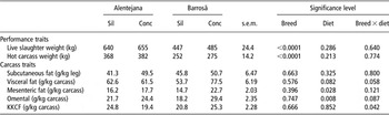
Some of the growth performance parameters, including live slaughter weight, hot carcass weight and subcutaneous and visceral fat depots weights are shown in Table 2. An effect of breed was observed for live slaughter and hot carcass weights, as Alentejana bulls had higher values of both variables in relation to Barrosã bulls (P < 0.0001).
Slaughter weight, hot carcass weight, subcutaneous and visceral adipose depots weights from Alentejana and Barrosã bulls fed silage- and concentrate-based diets

KKCF = kidney knob and channel fat.
Dietary treatments: Sil = silage diet based on 30/70% of concentrate and silage, respectively; Conc = concentrate diet based on 70/30% of concentrate and silage, respectively. Values are mean ± s.e.m.
The subcutaneous fat weight obtained through the dissection of the leg was unchanged across the experimental groups (P > 0.05). The same occurred for total visceral fat (P > 0.05). Mesenteric and omental fat weights were increased (P < 0.05 and P < 0.01, respectively), by feeding a concentrate-based diet. An interaction between breed and diet was observed for KKCF depot weight (P < 0.05). Regarding this adipose depot weight, and for concentrate-fed bovines, Alentejana bulls had lower values, whereas Barrosã bulls had the opposite.
Membrane-saturated fatty acids but not cholesterol concentration is influenced by diet and not by breed
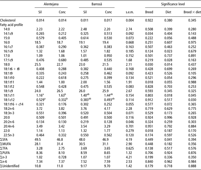
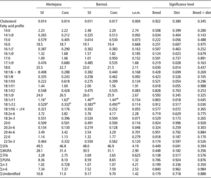
Table 3 depicts the lipid composition of membrane vesicles from mesenteric fat from the four experimental groups. Total cholesterol concentration was unaffected by breed or diet (P > 0.05). The distribution of the main fatty acid classes showed a highest occurrence of saturated fatty acids (SFA), followed by monounsaturated fatty acids (MUFA), then polyunsaturated fatty acids (PUFA) and lastly trans fatty acids (TFA). The diet affected the sum of SFA (P < 0.05), including the 18:0 fatty acid, being the values higher in silage-fed animals than in concentrate-fed bulls (P < 0.05). For this former fatty acid, a breed effect was also observed, as overall Barrosã breed showed a lower concentration (P < 0.05). In contrast, the 22:0 fatty acid was enhanced in Barrosã bulls, regardless of the diet (P < 0.05). Even if the sum of MUFA was not influenced by the factors under study (P > 0.05), 16:1c9 and 17:1c9 fatty acids were affected by diet, showing consistently higher concentrations in concentrate-fed bulls (P < 0.05). Moreover, the breed had a notorious effect in the 14:1c9 fatty acid concentration, with Barrosã bulls showing higher values than Alentejana (P < 0.05). The same effect was observed for 18:1t11 fatty acid (P < 0.05). In addition, an interaction between breed and diet was found for 18:1c11 and 18:1c12 fatty acids (P < 0.05), because significant differences on these concentrations were observed for the diet factor for Alentejana breed, but not for Barrosã. Hence, for Alentejana bulls, 18:1c11 fatty acid concentration was higher in concentrate-based than in silage-based diets; the opposite was observed for 18:1c12 fatty acid. Apart from these changes, no other significant variations regarding the sum or the individual fatty acids promoted by breed, diet or their respective interaction were observed.
Cholesterol concentration (mg/mg vesicles) and fatty acid profile (g/100 g fatty acids) in mesenteric adipose tissue membrane vesicles from Alentejana and Barrosã bulls fed silage- and concentrate-based diets
SFA = saturated fatty acids; MUFA = monounsaturated fatty acids; TFA = trans fatty acids; PUFA = polyunsaturated fatty acids.
Dietary treatments: Sil = silage diet based on 30/70% of concentrate and silage, respectively; Conc = concentrate diet based on 70/30% of concentrate and silage, respectively. Values are mean ± s.e.m.
a,bMean values within the same row with different superscript letters are significantly different (Fisher's post-hoc test, P<0.05).
FAME = fatty acid methyl esters. ΣSFA = sum of 14:0, 15:0, 16:0, 17:0, 18:0, 20:0 and 22:0; ΣMUFA = sum of 14:1c9, 16:1c7, 16:1c9, 17:1c9, 18:1c9, 18:1c11 and 18:1c12; ΣTFA = sum of 18:1t6 + t8, 18:1t9, 18:1t10, 18:1t11, 18:1t12 and 18:1t16 + c14; ΣPUFA = sum of 18:2n-6, 18:3n-3, 20:2n-6, 20:4n-6 and 22:5n-3; Σn-6 = sum of 18:2n-6, 20:2n-6 and 20:4n-6; Σn-3=sum of 18:3n-3 and 22:5n-3.
Permeability and fluidity of adipose membranes are independent of breed or diet
Membrane vesicles obtained from mesenteric fat revealed a unimodal size distribution, showing a mean diameter of 371 ± 57 nm for all tested groups. These membrane preparations were subsequently used to assess water and glycerol permeability by stopped-flow spectroscopy, as well as membrane fluidity by fluorescence anisotropy.
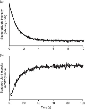
Figure 1 depicts typical stopped-flow light scatter signals of vesicle volume changes when gradients of mannitol (water permeability, panel A) and glycerol (panel B) were imposed. The time courses of vesicle volume changes are used to calculate P f and P gly, as described in the section ‘Material and Methods’.
Representative stopped flow light scatter of adipose membrane vesicles permeability to water and glycerol at 23°C. Time course of (a) vesicle shrinkage due to water outflow after an hyperosmotic shock with 120 mOsM mannitol gradient or (b) vesicle reswelling due to glycerol uptake after an inwardly directed 180 mOsM glycerol gradient.

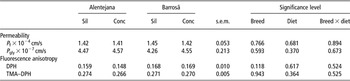
The permeability values obtained for the two bovine breeds fed silage- or concentrate-based diets are shown in Table 4. Neither breed nor diet affected the permeability of adipose membrane vesicles to water (P f) and glycerol (P gly) (P > 0.05). Accordingly, the activation energy values (E a) for water and glycerol permeation were similar among experimental groups, ranging from 14.5 ± 0.3 to 15.0 ± 0.2 kcal/mol (60.7 ± 1.3 to 62.8 ± 0.8 kJ/mol) for water (P > 0.05) and 23.7 ± 0.3 to 24.3 ± 0.2 kcal/mol (99.2 ± 1.4 to 102 ± 1.0 kJ/mol) for glycerol (P > 0.05). These relative high E a values suggest that permeation occurs mainly through the lipid bilayer with no contribution of specific protein channels for transport.
Permeability values for water (P f) and glycerol (P gly), and fluorescence anisotropy of DPH and TMA–DPH in mesenteric adipose tissue membrane vesicles from Alentejana and Barrosã bulls fed silage- and concentrate-based diets

DPH = 1,6-diphenyl-1,3,5-hexatriene; TMA–DPH = 1-(4-(trimethylamino)phenyl)-6-phenyl-1,3,5-hexatriene.
Dietary treatments: Sil = silage diet based on 30/70% of concentrate and silage, respectively; Conc = concentrate diet based on 70/30% of concentrate and silage, respectively. Values are mean ± s.e.m.
The fluorescence anisotropy of DPH and TMA–DPH in membrane vesicles from each experimental group are also shown in Table 4. Following the same pattern, no significant effects of breed or diet were detected among experimental groups (P > 0.05) for both fluorescence probes.
Discussion
The manipulation of adipose tissue growth, deposition and metabolism has important economic implications for the livestock industry, because it can improve production efficiency, carcass composition and meat quality.
Two underlying processes are responsible for increased adiposity in beef cattle: hypertrophy (larger adipocyte size) and hyperplasia (larger number of adipocytes) (Novakofski, Reference Novakofski2004), which are affected by factors such as genetics, sex, age, feeding regimen, food supply and the specific adipose tissue depot (Vernon and Houseknecht, Reference Vernon and Houseknecht1991). However, an understanding of the mechanisms of body fat deposition in farm animals and its outcome in adipocyte physiology is far from well established. Consequently, this study was designed to elucidate the contribution of breed and diet to adipose tissue membrane permeability and fluidity as possible key players on fat deposition in bovines. The morphological features between these breeds reflect differences in mature size and, consequently, fat accumulation (da Silva et al., Reference da Silva, Lemos, Monteiro and Portugal1998). Following on our previous results, in which the mesenteric fat depot had smaller adipocytes but a greater number of cells than subcutaneous fat (Costa ASH et al., unpublished data), the aforementioned visceral fat was selected for analysis due to its unique properties, regarding lipogenic activity and immune-response potential (Mukesh et al., Reference Mukesh, Bionaz, Graugnard, Drackley and Loor2010).
As expected, a clear effect of breed was observed for some growth performance parameters, with Alentejana displaying higher values for live slaughter and hot carcass weights (da Silva et al., Reference da Silva, Lemos, Monteiro and Portugal1998; Reis et al., Reference Reis, Navas, Pereira and Cravador2001).
Regarding lipid deposition, all fat depots under study were unaffected by breed. However, diet appeared to play a significant role in determining mesenteric and omental fat depots weight, which were consistently higher in concentrate-fed bovines. The chosen silage- and concentrate-based diets provided significant differences in their composition. The concentrate-based diet exhibited higher fat and starch concentrations, whereas the silage-based diet exhibited higher fiber concentration. These differences extend to the detailed fatty acid composition, in particular to the sums of SFA and PUFA classes. The proportions for SFA were higher in concentrate-based diets (silage 31.9 v. concentrate 37.2) at the expenses of 16:0, 18:0 and 20:0 fatty acids. The inverse trend was observed for PUFA (silage 53.0 v. concentrate 46.8), determined by 18:2n-6 and 18:3n-3 fatty acids.
There is convincing evidence in animal models that dietary fat influences cell membrane phospholipid composition (Clandinin et al., Reference Clandinin, Field, Hargreaves, Morson and Zsigmond1985; Jenkins, Reference Jenkins1994). Typically, ruminant diets are low in fat but high in PUFA contents. Nevertheless, unsaturated fatty acids in the diet undergo an extensive biohydrogenation in the rumen, with consequently high levels of SFA being absorbed in the intestine and deposited in the tissues (Wachira et al., Reference Wachira, Sinclair, Wilkinson, Enser, Wood and Fisher2002). In agreement, adipose membranes from visceral fat were found to be richer in SFA, displaying concomitantly a lower proportion of PUFA.
Cholesterol is a biomembrane-rigidifying component. When cholesterol is aligned in parallel array with the phospholipid fatty acyl chains, it reduces membrane fluidity (Stubbs and Smith, Reference Stubbs and Smith1984; Onuki et al., Reference Onuki, Hagiwara, Sugibayashi and Takayama2008), but inversely, increases rigidity. Our results indicate that total cholesterol concentration was unchanged by any factor under study. Accordingly, neither breed nor diet affected the permeability of adipose membranes to water or glycerol. The activation energy for both water and glycerol transport was high and relatively stable in all cases, thus indicating that permeation is not protein-mediated and occurs mainly via the lipid bilayer where permeability correlates with fluidity. Accordingly, no variations for adipose membrane fluidity were found with DPH or TMA–DPH probes, which assess fluidity at different depths in the bilayer (TMA–DPH assessing a region closer to the lipid–water interface). Altogether, these results corroborate the stability found in permeability data.
Besides cholesterol, fatty acids strongly influence membrane fluidity. With an increase in unsaturated fatty acids concentration, membrane fluidity increases because PUFA acyl chains are extremely flexible and can rapidly change conformational states. The fatty acid profile in adipose membranes from mesenteric fat of Alentejana and Barrosã bovines fed silage- and concentrate-based diets showed no variations in PUFA sum and, foremost important, included none of the n-3 fatty acids, EPA (20:5n-3) and DHA (22:6n-3), well known for their impressive range of health benefits, the latter being recognized as a potent membrane fluidizer agent (Stillwell and Wassall, Reference Stillwell and Wassall2003). These results are in accordance to Wachira et al. (Reference Wachira, Sinclair, Wilkinson, Enser, Wood and Fisher2002), who found residual concentration of both n-3 fatty acids in subcutaneous fat of sheep, even after the intake of feeding regimens enriched in linseed and fish oils. Dietary lipids do not directly affect the fatty acid composition of ruminant adipose tissues, as they do in non-ruminants (Sarkkinen et al., Reference Sarkkinen, Agren, Ahola, Ovaskainen and Uusitupa1994). Raising the PUFA content of ruminant tissues by PUFA feeding is rather complex due to the extensive hydrogenation of dietary unsaturated fatty acids by rumen microorganisms (Pond, Reference Pond1999; Jambrenghi et al., Reference Jambrenghi, Paglialonga, Gnoni, Zanotti, Giannico, Vonghia and Gnoni2007). Nevertheless, the few changes observed for the general fatty acid profile in adipose membranes appear to reflect the dietary treatment imposed, instead of a breed-related effect. The same pattern had already been observed for the fatty acid profile in mesenteric fat, with diet determining the proportions of the major fatty acids as well as their partial sums (Costa ASH et al., unpublished data). Although the sum of MUFA was kept similar across experimental groups, the 16:1c9 and 17:1c9 fatty acids were under the influence of diet, with higher concentration in concentrate-fed bulls. This is in line with previous reports stating that concentrate promotes higher expression or activity levels of delta-9 desaturase enzyme, responsible for the conversion of SFA to MUFA (Daniel et al., Reference Daniel, Wynn, Salter and Buttery2004). Nevertheless, these differences relate to residual concentration of these fatty acids, ranging from 0.48% to 1.82% and, therefore we believe, play an irrelevant physiological role. Similarly, total SFA was affected by the diet factor, being the difference observed largely determined by stearic acid (18:0) concentration in Alentejana bulls fed on silage. More 18:2n-6 fatty acid in the silage feeding regimen results in more 18:0 in adipose plasma membranes, as reported formerly (Jenkins, Reference Jenkins1994). High stearic levels are also in accordance to the reported values in the adipose tissue of lambs (Enser et al., Reference Enser, Hallett, Hewitt, Fursey and Wood1996).
Finally, a breed effect was observed for 22:0, 14:1c9 and 18:1t11. All three fatty acids concentration were higher in Barrosã bulls when compared with Alentejana bulls. The long-chain fatty acids (LCFA) occurred at very low levels in the adipose membranes of bovines, either Alentejana or Barrosã, fed any of the dietary treatments. Low levels of LCFA in ruminant's adipose tissue have already been reported and were attributed to the low incorporation of these fatty acids into the triacylglycerol fraction, as well as to the low proportion of phospholipid in the adipose tissue (Enser et al., Reference Enser, Hallett, Hewitt, Fursey and Wood1996; Wachira et al., Reference Wachira, Sinclair, Wilkinson, Enser, Wood and Fisher2002). Concerning the difference observed between breeds for the 22:0 fatty acid, it might be due to a higher elongase expression, or enzymatic activity, in the Barrosã than in the Alentejana breed. Genetic background also appeared to dictate a differential expression, or activity, of delta-9 desaturase enzyme, responsible for the conversion of 14:0 to 14:1c9 (Keating et al., Reference Keating, Kennelly and Zhao2006). These hypotheses remain to be tested. In relation to the 18:1t11 fatty acid, commonly known as vaccenic acid, it is metabolized into the c9,t11-conjugated linoleic acid (CLA) isomer (Lock et al., Reference Lock, Corl, Barbano, Bauman and Ip2004), to which numerous health claims have been attributed (Bhattacharya et al., Reference Bhattacharya, Banu, Rahman, Causey and Fernandes2006), and for this reason has been considered as beneficial or neutral.
Conclusions
This study reports that adipose membranes from ruminant's mesenteric fat depot were rich in SFA due to ruminal biohydrogenation of dietary PUFA. Membrane fluidity and permeability to glycerol were found to be independent of breed (Alentejana or Barrosã) and diet (based on 70/30% or 30/70% of corn silage and concentrate, respectively). Re-enforcing these findings, cholesterol, the main biomembrane-rigidifying component, and in particular, unsaturated fatty acid concentration were unchanged among experimental groups.
Acknowledgements
This study was supported by Fundação para a Ciência e a Tecnologia (FCT) through grant PTDC/CVT/2006/66114 and individual fellowships to Ana P. Martins (SFRH/BD/2009/65046), Ana S. H. Costa (SFRH/BD/2009/61068) and Susana V. Martins (SFRH/BPD/2009/63019). Paula A. Lopes is a researcher from the program “Ciência 2008” from FCT. We wish to express our gratitude to Rui Bessa and Susana Alves (L-INIA-REQUIMTE) for providing fatty acid profiles.