INTRODUCTION
The phylum Nemertea currently comprises around 1300 valid species names (Gibson, Reference Gibson1995; Kajihara et al., Reference Kajihara, Chernyshev, Sun, Sundberg and Crandall2008) of which maybe 30% could be dubious (Gibson, Reference Gibson1985) given the fact that their descriptions lack details that would help to securely identify them. In addition to brief and shallow descriptions, holotypes and paratypes are often either missing or non-informative. The actual number of nemertean species is, therefore, shrouded in mystery and to revise these hundreds of species would demand major efforts from the complete nemertean community. One initiative to stabilize the framework for nemertean taxonomy was provided by Sundberg et al. (Reference Sundberg, Chernyshev, Kajihara, Kånneby and Strand2009). They provided a matrix of morphological characters that can be used as a basis for descriptions of hoplonemerteans and palaeonemerteans. Several larger and smaller molecular studies published in the last decade (among others: Thollesson & Norenburg, Reference Thollesson and Norenburg2003; Sundberg et al., Reference Sundberg, Vodoti and Strand2010; Kajihara et al., Reference Kajihara, Olympia, Kobayashi, Katoh, Chen, Strand and Sundberg2011; Andrade et al., Reference Andrade, Strand, Schwartz, Chen, Kajihara, Von Döhren, Sun, Junoy, Thiel, Norenburg, Turbeville, Giribet and Sundberg2012) also improve the conditions for more pervasive and comprehensive interventions within the nemertean taxonomy.
Following tradition, much effort and time is put into nemertean histology, but the results are not always conclusive. The soft bodies, the number of preparation artefacts and the scarce number of specimens that can be investigated are all factors that may aggravate precise results (see Sundberg & Strand, Reference Sundberg and Strand2010). There are many moments of subjective interpretation, and when there are enough numbers of specimens to compare, it has been shown that intraspecific anatomical variation can be in some features that might be used as taxonomic characters (Sundberg, Reference Sundberg1979). There are others that can be consistent (Norenburg, Reference Norenburg1986). Even so, traditional morphological descriptions continue to be published with varying emphasis on different species characters. An attempt to stress and solve problems in nemertean taxonomy is the DNA-based description of Pseudomicrura afzelii (Strand & Sundberg, Reference Strand and Sundberg2011). In this publication the authors mention both the difficulties with anatomical characters, the value of studies of live specimens and some difficulties that arise when trying to apply the nomenclatural code to the results of molecular analyses. It is sometimes stated that nemertean worms lack external characters and that internal anatomical details are a must for absolute measures (e.g. Gibson, Reference Gibson1985), but there are opposing views on the subject (e.g. Sundberg, Reference Sundberg1979). Here, we make an effort to set a structure for testing the functionality of internal and external morphology in combination with a routine molecular analysis. Our results indicate obvious informative values for external characters.
MATERIALS AND METHODS
Samples
Worms were collected in May 2009 and in July 2012 in Norwegian waters with an RP-sled, from depths of more than 200 m. Specimens were observed alive, and those for histological examination were fixed in 4% paraformaldehyde in filtered seawater or Bouin's fluid, sectioned at 6 µm and stained by the Mallory trichrome technique for histological examination. Additional specimens were preserved in RNAlater and stored for subsequent DNA extraction using DNeasy (QIAgen, Inc.) following the protocol recommended by the manufacturer.
Morphological studies
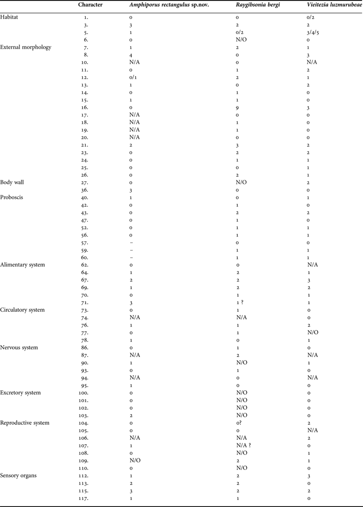
To map the morphological characters of the new species we used the character matrix from Sundberg et al. (Reference Sundberg, Chernyshev, Kajihara, Kånneby and Strand2009). Data for two species (Raygibsonia bergi Sundberg et al., Reference Sundberg, Chernyshev, Kajihara, Kånneby and Strand2009 and Vieitezia luzmurubeae Junoy et al., Reference Junoy, Andrade and Giribet2011, Appendix 2) earlier described with the same matrix were used for comparative studies (Table 1). All characters were treated as neutral.
Differences in number of characters (internal and external) between Amphiporus rectangulus sp. nov., Raygibsonia bergi and Vieitezia luzmurubeae, the three species described with character matrix.

Molecular studies and analyses
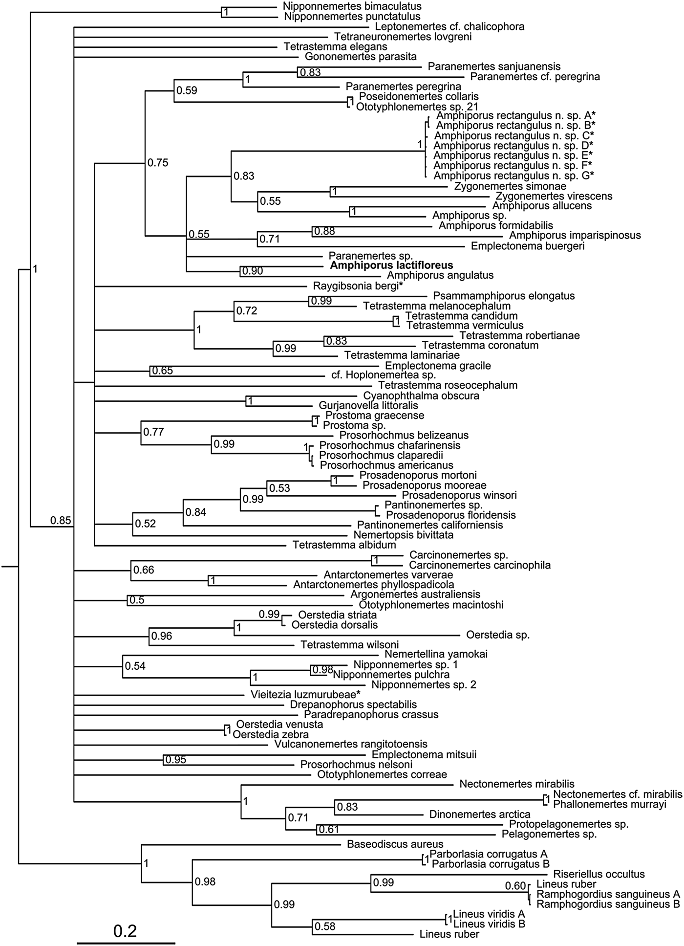
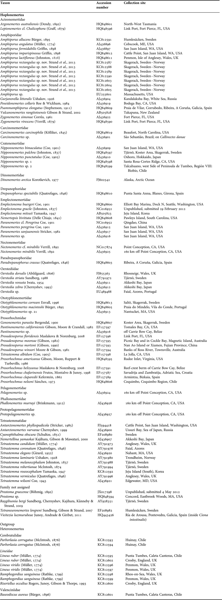
Part of the mitochondrial gene cytochrome oxidase I (COI) was used for molecular analysis (Figure 1). Data were selected with the aim to maximize hoplonemertean diversity, given available data. Outgroup consists of various heteronemertean species (Appendix 3) based on previous results of molecular analyses (Thollesson et al., Reference Thollesson and Norenburg2003; Andrade et al., Reference Andrade, Strand, Schwartz, Chen, Kajihara, Von Döhren, Sun, Junoy, Thiel, Norenburg, Turbeville, Giribet and Sundberg2012) that show clear distinction between the two clades. The third group Palaeonemertea has a more obscure relationship to both groups, which is a reason to exclude them from this analysis. The PCR was performed according to standard procedures, and sequences were obtained through Macrogen (Korea). Primers used were HCO1490 and LCO2194 (Folmer et al., Reference Folmer, Black, Hoeh, Lutz and Vrijenhoek1994).
Phylogenetic relationships of Hoplonemertea inferred from Bayesian analysis of COI mt-DNA. Numbers at nodes refer to posterior probability. The tree is rooted on outgroups represented by members of Heteronemertea. Asterisks mark species that have been coded according to the character matrix provided by Sundberg et al. (Reference Sundberg, Chernyshev, Kajihara, Kånneby and Strand2009). We have intentionally kept all branches with pp50%, partly to show the different proposed clades, but mainly to show the very weak support for existing genera.
Contigs were assembled in Geneious v.5.6.5. (Biomatters available from http://www.geneious.com). The COI was aligned straightforwardly with the reading frame for amino acids, using the general invertebrate mitochondrial genetic code with Muscle in TranslatorX (Abascal et al., Reference Abascal, Zardoya and Telford2010). Models of substitution were tested for each gene alignment with jModelTest 0.1 (Guindon & Gascuel, Reference Guindon and Gascuel2003; Posada, Reference Posada2008) under the Akaike information criterion (AIC). All datasets were analysed with MrBayes 3.1.2 (Ronquist & Huelsenbeck, Reference Ronquist and Huelsenbeck2003; Huelsenbeck & Ronquist, Reference Huelsenbeck, Ronquist and Nielsen2005) using the freely available Oslo University Bioportal Cluster (see Kumar et al., Reference Kumar, Skjæveland, Orr, Enger, Ruden, Mevik, Burki, Botnen and Shalchian-Tabrizi2009). Analyses of individual genes were run for 40 million generations. All runs were sampled at every 1000th generation under the six parameter general time reversible (GTR) model with gamma distributed rate variation across sites. Convergence was ascertained by checking the log likelihood graphs, the average standard deviation of split frequencies, and the potential scale reduction factor (PSRF). The first 25% of the sampled trees were regarded as the burn-in period. Since chain mixing was low under the default settings (four MCMC chains for each run and a heating parameter of 0.2) the dataset was analysed using eight MCMC chains in each run with a heating parameter of 0.1 to ensure sufficient chain mixing and a reliable sample from the posterior distribution. Consensus trees were built from two independent runs of 30,000 samples each for the Bayesian analyses. A maximum likelihood analysis was also run on the combined dataset using RAxML v.7.0.3 (Stamatakis, Reference Stamatakis2006) with 1000 bootstrap replicates to check for congruence with the Bayesian analysis.
TAXONOMY
SYSTEMATICS
Class ENOPLA Schultze, Reference Schultze1851
Order MONOSTILIFERA Brinkmann, Reference Brinkmann1917
Family AMPHIPORIDAE McIntosh, Reference McIntosh1874
Genus Amphiporus Ehrenberg, Reference Ehrenberg, Hemprich and Ehrenberg1831
Amphiporus rectangulus sp. nov.
(Figures 2–5)
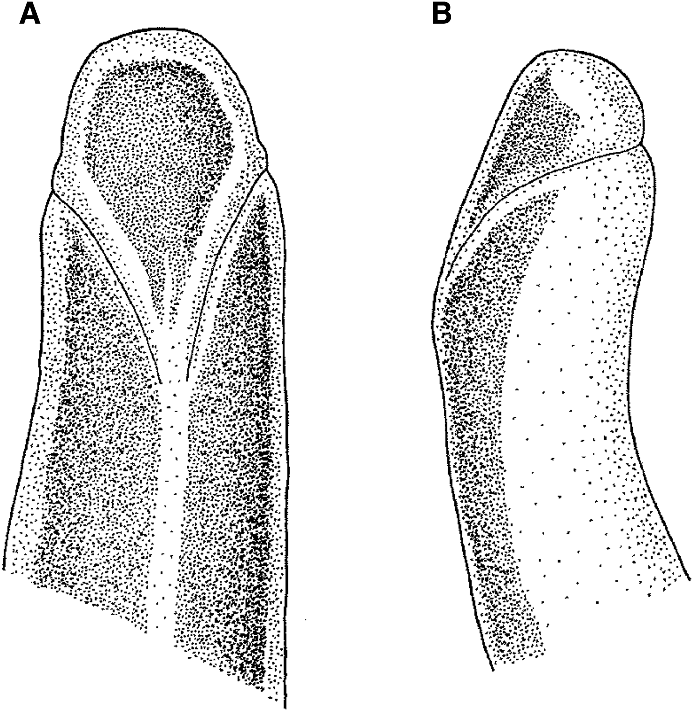
Amphiporus rectangulus sp. nov.: (A) drawing of the anterior part of the body of one specimen to show the ‘V’ pattern and the cephalic furrows in detail; (B) drawing of the lateral view of the anterior part. Type status: additional specimens. Department of Biology and Environmental Sciences, University of Gothenburg.
Amphiporus rectangulus sp. nov.: (A) photograph of a complete specimen with the characteristic ‘V’ shaped cephalic furrows; (B) photograph of a complete specimen with the papillated proboscis everted; (C) group of specimens together to compare the differences between males (♂) and females (♀). Type status: additional specimens. Department of Biology and Environmental Sciences, University of Gothenburg.
Amphiporus rectangulus sp. nov.: (A) section through pyloric region showing the details of the proboscis and its ten proboscis nerves (arrowheads); (B) microphotograph of a squeezed specimen showing the stylet and its basis. Abbreviations: CM, circular muscle layer; LM, longitudinal muscle layer. Type status: holotype. Voucher ZMBN 88199. The University Museum of Bergen, Norway.
Amphiporus rectangulus sp. nov.: (A) section through the anterior-most part of body showing the cephalic glands (arrowheads); (B) section through the pyloric region showing the parts of the alimentary system; note a mid-dorsal blood vessel (arrowhead); (C) section through the apical region of the body showing the apical organ (arrowhead); (D) detail of a section through the pyloric region showing the lateral nerve cord, the lateral blood vessel (arrow) and one excretory tubule (arrowhead); (E) detail of a section through the cephalic region showing the buccal nerves (arrowheads); (F) section through cephalic region showing the ciliated canals (arrow) that connect the cephalic furrows with the cerebral organs; note the anterior accessory pouches of the intestinal caecum (arrowheads); (G) section through the proboscis armature region of body showing the stylet apparatus (arrowhead), the deeply branched intestinal diverticula and the gonads. Abbreviations: CB, cerebral ganglion; CF, cephalic furrow; CM, circular muscle layer; CO, cerebral organ; E, epidermis; G, gonad (testis); IC, intestinal caecum; ID, intestinal diverticulum; LM, longitudinal muscle layer; LN, lateral nerve cord; P, proboscis; PY, pylorus; R, rhynchocoel; RY, rhynchodaeum; S, stomach. Type status: holotype. Voucher ZMBN 88199. The University Museum of Bergen, Norway.
TYPE MATERIAL
Holotype: sexually mature male, series of transverse sections (voucher ZMBN 88199, The University Museum of Bergen, Norway). (Skagerrak, Norway; trip ‘BIOSKAG 2’ with RP-sled; sand; coordinates: 58°57.250′N 10°34.403′E; water depth: 220 m); 18 May 2009. Partial COI mt-DNA sequence of the species has been deposited in GenBank (Accession number KC812587).
Paratype: sexually mature male, series of transverse sections (voucher ZMBN 88200, The University Museum of Bergen, Norway). (Skagerrak, Norway; trip ‘BIOSKAG 2’ with RP-sled; sand; coordinates: 58°57.250′N 10°34.403′E; water depth: 220 m); 18 May 2009. Partial COI mt-DNA sequences of the paratype and two other specimens are deposited in Genbank (Accession numbers KC812588, KC812589, KC812590).
Additional specimens: sexually mature males and females (Department of Biology and Environmental Sciences, University of Gothenburg). (Frøysjøen, Norway; RP-sled; mud; coordinates: 61°49.417′N 5°13.617′E; water depth: 450 m); 20 July 2012.
DIAGNOSIS
The genus Amphiporus is poorly defined in absolute terms and most characters are not genus specific. A compilation of available literature (Bürger, Reference Bürger1904; Gibson & Crandall, Reference Gibson and Crandall1989; Maslakova et al., Reference Maslakova, Thiel, Vásquez and Norenburg2005) renders the following list of morphological characters for the genus: proboscis well developed, rhynchocoel extends to or almost to posterior end of body, apical organ present, body wall musculature generally well developed without diagonal muscle layer between circular and longitudinal muscles, blood vascular system with three longitudinal vessels, nervous system without neurochords or neurochord cells, intestinal caecum present with a pair of anterior diverticula as well as lateral pouches, cerebral organs anterior to cerebral ganglia, excretory system positioned in the foregut region, sexes separate. None of these characters contradicts the inclusion of our new species in the genus. The rhynchocoel wall is, for many Amphiporus spp., described with separate circular and longitudinal muscle layers. This does not directly apply to Amphiporus rectangulus sp. nov., whose rhynchocoel wall musculature is difficult to define (character 43).
Earlier DNA analyses have repeatedly shown the genus Amphiporus to be non-monophyletic (see e.g. Sundberg et al., Reference Sundberg, Chernyshev, Kajihara, Kånneby and Strand2009; Kajihara et al., Reference Kajihara, Olympia, Kobayashi, Katoh, Chen, Strand and Sundberg2011). Our analysis supports these results. Amphiporus rectangulus sp. nov. nests however (Figure 3) in the same clade as the type species of the genus, Amphiporus lactifloreus (Johnston, Reference Johnston1828).
DESCRIPTION
External features
Most of specimens examined alive, before or after anaesthetization, up to 1 cm long and approximately 1 mm wide. Appearance sturdy and chubby. Body soft and movement calm. Body shape very variable, but tapering backwards. Dorsal side brownish, like nougat, with ventral side and lateral margins pale. Furrows pale, deep, and very obvious encircling head meeting in rather steep V-shape dorsally (Figure 2). In some specimens, single pale stripe present, commencing behind V-shape as longitudinal pigmentation reaching backwards full or nearly full body length. Other specimens lacking this paler stripe; coloration obviously variable intra-specifically (Figure 4). No eyes distinguishable. Body wall quite thick, through which rhynchocoel and proboscis difficult to observe.
Body wall, musculature and parenchyma
Epidermis glandular, reaching up to 130 µm in thickness (Figure 5B). Connective tissue dermal layer up to 15 µm, proximally bordering epidermis (Figure 5B). Body wall musculature comprising outer circular and inner longitudinal layers, respectively 10–15 µm and 35–40 µm in thickness (Figure 5F).
Proboscis apparatus
Proboscis pore situated antero-ventrally near tip of the head opening into rhynchodaeum whose epithelium being neither ciliated nor glandular (Figure 5A, E). Rhynchocoel reaching posterior tip of body, its wall lacking distinct musculature, appearing as simple connective membrane (Figures 4A, 5B). Proboscis epithelium well developed into papillae. In everted state, proboscis with outer circular and inner longitudinal muscle layers. Nerve supply consisting of neural ring with ten nerves situated between longitudinal muscle fibres (Figure 4A). Proboscis armature consisting of single central stylet and pair of accessory stylet pouches, containing 1–2 reserve stylets. Several specimens everted proboscis during preservation; no stylets observable in these specimens. Paired accessory stylet pouches not easily observed but clearly visible in sections. Shape of stylet bulb very difficult to determine due to artefacts in preparations (Figure 4B).
Alimentary canal
Oesophagus emerging directly from rhynchodaeum in front of brain, posteriorly reaching cephalic region. Oesophageal epithelium simple, without glands or cilia. Stomach situated under brain, developed into pyloric canal opening into dorsal wall of intestine. Intestinal caecum with two long anterior accessory pouches reaching forward to rear of brain lobes and alongside them (Figure 5F). Intestinal diverticula deeply branched and alternating with gonads in mature specimens (Figure 5G).
Circulatory system
Blood system following basic plan for enoplan nemerteans. Mid-dorsal blood vessel emerging as branch from right lateral vessel in cerebral region. For all body length, the lateral vessels running below lateral nerve cords (Figure 5D); these anastomosing anteriorly and posteriorly by cephalic and caudal loops, respectively. No vascular plug observed.
Nervous system
Brain well developed, with ventral cerebral lobes being much thicker than dorsal ones. Dorsal cerebral commissure longer and narrower than the ventral one (30 µm vs 40 µm in diameter respectively); former positioned anterior to latter. Outer neurilemma enclosing brain, but no inner neurilemma distinguished. Ventral lobes being confluent with lateral nerve cords, which throughout their length containing only single neuropile; no accessory lateral nerves (Figure 5D). Posterior junction of lateral nerve cords subintestinal. Neither neurochords nor neurochord cells distinguished in nervous system. Dorsal cephalic nerves leading forward from anterior borders of brain lobes into cephalic-gland region; two buccal nerves reaching rhynchodaeum pore (Figure 5E).
Frontal organ and cephalic glands
Small, single apical organ opening at very tip of head, 40 µm in diameter (Figure 5C). Cephalic glands scattered between muscle fibres in anterior part of head, forming quite prominent distinct lobes (Figure 5A), opening via apical organ.
Sense organs
No eyes found. Cerebral sensory organs large, located all alongside brain. 65 µm in height, 145 µm in length, each opening laterally by simple, unforked, thick-walled ciliated canal leading inwards from cephalic furrow (Figure 5F). A well-developed cap of acidophilic glands extending a short distance along upper margins of organs.
Excretory system
Well developed, confined to post-cerebral region of body, consisting of two pairs of thick-walled longitudinal collecting tubes running close to lateral nerve cords, and opening by lateral nephridiopores in pyloric region (Figure 5D). No flame cells observed.
Reproductive system
Specimens collected having mature gonads; sexes are separate. No sexual colour dimorphism. Gonads serially disposed along body from pyloric region backwards, lying from dorsal to ventro-lateral margins between intestinal diverticula (Figure 5G). Holotype mature male, with deeply bilobate testes. No gonopore found in two different males. Mature testes is 120 µm in diameter.
ECOLOGY
Specimens were found on both mud bottoms and sandy bottoms. In May 2009 most specimens seem to be sexually mature (Figure 3C). Other organism groups highly represented in the same samples where Sipuncula, Caudofoveata, Bivalvia, Gastropoda, Polychaeta.
ETYMOLOGY
The name rectangulus, a Latin adjective, refers to the shape of the live animal body in transverse section.
RESULTS
Morphology
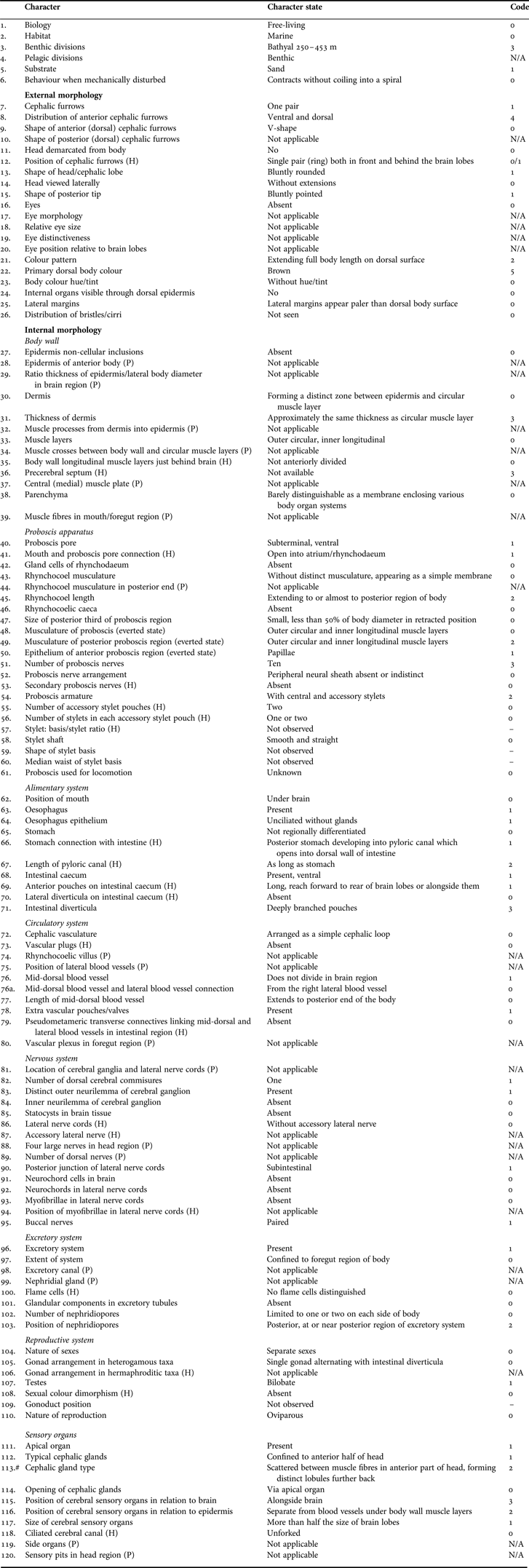
Complete matrix for Amphiporus rectangulus sp. nov. is available (Appendix 1). Between the three hoplonemertean species that have available data we find that the highest number of character differences lies within external morphology. For the comparative matrix see Appendix 2.
The character matrix (see Appendix 1) contains in total 120 characters, 16 of which are only applicable for palaeonemerteans. Of the characters relevant for hoplonemerteans, we were able to code 100, only hesitating on four (regarding gonopores and stylet basis).
Note on matrix correction: character 113 (cephalic gland type) is not only applicable for Palaeonemertea as stated in Sundberg et al. (Reference Sundberg, Chernyshev, Kajihara, Kånneby and Strand2009). It is also available for Hoplonemertea.
Also, there is a new character added to the matrix with two different states. Character 76a: mid-dorsal blood vessel and lateral blood vessel connection. State 0: from the right lateral blood vessel. State 1: from the left lateral blood vessel.
Molecular analysis
Results from the molecular analysis are consistent with previous studies using COI mt-DNA. Hoplonemerteans form a monophyletic group which inside has very little or no resolution in relation to existing genera.
DISCUSSION
The informative value of a comparative morphology matrix, in terms of relationship or species definitions, with only three hoplonemertean species, is of course extremely low. We need amounts of data of completely other dimensions if we are to evaluate the information potential with the approach. More matrix-based species descriptions will enable the testing of whether histology is helping us towards better phylogenetic resolution and/or to clarify species delimitation issues. In Table 1 we present differences in the number of characters (internal and external). Considering the fact that the three investigated species are obviously different in many aspects—genus placement, appearance, sampling localities and so on—we may expect to find differences of some obvious magnitude regarding morphological characters as well as some genetic distance between the three of them. It is, therefore, interesting to note that even though the listed number of external characters is rather low, and could be improved both in terms of numbers and resolution, most of the counted differences are to be found here. Between the 90 listed internal characters (traditionally said to be of most importance), there are only around 30 characters that show any absolute differences between the three of them. In Appendix 2 there are possibilities for readers to check whether these differing internal characters can be related to, for example, genus diagnosis. We found no such systematic or taxonomic correlations within our set of data. This is consistent with the discussion (Strand & Sundberg, Reference Strand and Sundberg2011) provided when describing Pseudomicrura afzelii with external morphology in combination with DNA. So, what are the useful characters for describing a ribbon worm species? For nemertean taxonomy, species descriptions and systematics, the difficult internal anatomical characters that we can come up with, may not be the most informative, even if they are of high biological interest. To recognize a live and fresh specimen and identify it to species level can be fairly straightforward with a description carrying a combination of notes on habitat, ecology, behaviour and external features. With a barcoding sequence attached, the species determination can be made in a more absolute sense. The molecular analyses (see Figure 1) that were performed in this study are consistent with all previous studies on COI and nemerteans. The resolution of the phylogenetic tree is in parts poor, which reflects the fact that COI is relatively unconserved, and carries many neutral mutations. More genes from both the nuclear and mitochondrial genomes would be ideal for better resolution, but it is difficult to reach the taxonomic breadth represented by the species for which COI sequences are available. However, hoplonemerteans form a monophyletic group. Within this group there are some clades that verify a generic context, but for many clades it is obvious that taxonomy does not reflect natural groups, even with poor resolution. Judging from the tree it can also be said that the barcoding approach has possibilities. For example, ‘regular’ members of the genus Tetrastemma gather in clades, while a few species of the genus separate from these. Going back to morphology, the separated species are also the ones with deviant morphological diagnosis and appearance. Our study opens new approaches for tidying up the taxonomic waste basket that most nemertean species currently dwell in. The nemertean systematics and the relationship between different taxonomic entities is a complicated issue. More than 60% of all valid nemertean genera are monotypic, while a handful of genera carry hundreds of species. To our knowledge, this has nothing to do with reality, but rather reflects historical inconsistencies and the pitfalls of taxonomic rules and traditions. Of the more species-numerous genera, very few have exclusive diagnoses (see Diagnosis part in the description above), and none of them hitherto tested with genetics have been shown monophyletic (see Results above). Resolving phylogeny, and have taxonomy reflect this, is a work of major effort in the phylum Nemertea. Our results are likely to be valid for more groups with similar complications—that is, benthic animals with soft bodies, small size and rarely found in numbers high enough to declare intraspecific variation (e.g. certain nudibranchs or sipunculans).
ACKNOWLEDGEMENT
Alfonso Herrera-Bachiller thanks Juan Junoy for his support and advice in the early revision of the specimens sections.
FINANCIAL SUPPORT
Alfonso Herrera-Bachiller received a grant from the Zoologica Scripta Travel Fellowship funded by The Norwegian Academy of Science and Letters; Malin Strand was supported by the Norwegian Taxonomy Initiative.
APPENDICES
Character matrix for Amphiporus rectangulus, sp. nov. character states and code from Sundberg et al. (Reference Sundberg, Chernyshev, Kajihara, Kånneby and Strand2009). N/A: no applicable character.
Comparative matrix for characters of Amphiporus rectangulus, sp. nov., Raygibsonia bergi and Vieitezia luzmurubeae. Character states and code from Sundberg et al. (Reference Sundberg, Chernyshev, Kajihara, Kånneby and Strand2009). N/A, no applicable character, N/O, not observed character. Characters with the same state and palaeonemertean characters are excluded.
List of nemertean species included in the phylogenetic analysis, together with collection site and GenBank Accession number. Six species of heteronemerteans are used as the outgroup.