Introduction
In radiotherapy of gynaecological malignancies, simultaneous-integrated boost intensity-modulated radiotherapy (SIB-IMRT) helps to achieve highly conformal dose distribution for the planning target volume (PTV) while minimising dose to planning risk volume and reducing treatment time and number of monitor unit.Reference Mazzola, Ricchetti and Fiorentino1–Reference Cozzi, Dinshaw and Shrivastava3 SIB-IMRT delivers different radiation doses to different areas in a single treatment session. However, daily treatment uncertainties such as internal organ motion, patient setup errors, changes in position and shape of tumour limit merits of SIB-IMRT.Reference Thornqvist, Hysing and Tuomikoski4–Reference Taylor and Powell9 Uterine cervix and pelvic tissues are adjacent to rectum and bladder. During radiotherapy of cervical cancer, changes in bladder filling and rectal content cause variation in target position and volume.
Various authors have reported a change in shape and position of the target volume due to variations in bladder and rectum volume during the course of radiotherapy of cervical cancer. Rozilawati et al.Reference Ahmad, Hoogeman and Bondar10 observed changes in uterine cervix shape and position in their investigation due to alteration in bladder volume. Georg et al.Reference Georg, Georg, Hillbrand, Kirisits and Pötter11 found differences in irradiated volumes of small and large bowel with bladder filling variations. Jhingran et al.Reference Jhingran, Salehpour, Sam, Levy and Eifel12 evaluated deformation in vaginal vault caused by rectal content and bladder filling variation during IMRT of uterine cervical carcinoma.
To detect variations in target dose and volume due to changes in bladder and rectal volumes (including air bubbles), we performed a prospective study in which CT scans were obtained once in a week during SIB-IMRT of ten cervical cancer patients. Initial planning CT scans were then fused with intra-treatment scans to determine dosimetric and volumetric variations in PTV and organs at risk (OARs).
Materials and Methods
A total of ten patients with biopsy-proven pathological diagnosis of squamous cell carcinoma of uterine cervix were recruited in this prospective analysis. The age of patients ranged from 32 to 76 years with a mean age of 48 years. According to International Federation of Gynecology and Obstetrics staging criteria, four patients had stage IB disease, three had IIB and three had IIIA disease.Reference Lee and Atri13 It was planned that all of the ten patients will undergo SIB-IMRT with 5,040 cGy dose to the tumour site and 4,500 cGy to the lymphatic nodal region.
CT simulation for treatment planning
A written instruction was given to all patients to come for CT scanning with a full bladder which pushes the small bowel away from the treatment field. All patients were instructed to drink 750 mL of water 1 hour prior to every scanning. Patients were immobilised in a supine position on the treatment table by Vac-Loc equipment with their arms up and legs relaxed. All CT scans were taken with a slice thickness of 2·5 mm from GE High Speed X/I model (General Electric Medical system, Waukesha, WI, USA). After CT scanning, target volume and OARs including rectum, bladder, femoral heads and small bowel were contoured. Tumour site was contoured as clinical target volume (CTVtumour). Tumour PTV was created by adding 0·7–1 cm margin around CTVtumour. CTVnodal was generated by contouring the connected blood vessels, then putting 0·7–1 cm vaginal cuff around the vessels. An additional 0·5 cm margin around CTVnodal was applied to generate PTVnodal.
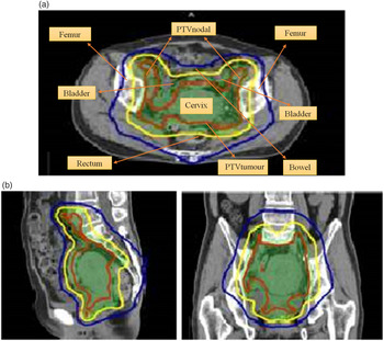
A prescription dose of 5,040 cGy to PTVtumour and 4,500 cGy to PTVnodal in 28 fractions was delivered using SIB-IMRT with double arc. Treatment plans were created, so that the ≥95% of the respective PTV be covered by ≥95% of the prescribed dose. Small bowel, bladder, rectum and femoral heads are the OARs. All SIB-IMRT plans were generated using 10 MV photon beam on treatment planning system Pinnacle3 (V8.0 d; Philips, Fitchburg, WI, USA). During radiotherapy planning, PTVtumour V95%, PTVnodal V95% and OAR volumes were measured from planning CT scans taken in 1st week. CT scans were taken once in a week over a treatment period of 5·5 weeks. Intra-treatment scans (scans taken on every Monday) were then fused with the planning scans to determine variations in the target volume and the OAR volume. Changes in radiation dose to the PTVtumour and the PTVnodal were also assessed by comparing intra-treatment scans with the planning scans (Figures 1a and 1b).
(a) CT scan of a patient undergoing simultaneous-integrated boost intensity-modulated radiotherapy: transversal view. (b) Sagittal and frontal view of a CT scan.

Results
SIB-IMRT of cervical malignancy is often made complicated by the variations in target volumes receiving tumouricidal doses due to changes in the bladder and the rectal volumes during treatment fractions.Reference Kerkhof, Raaymakers, van der Heide, van de Bunt, Jurgenliemk-Schulz and Lagendijk14
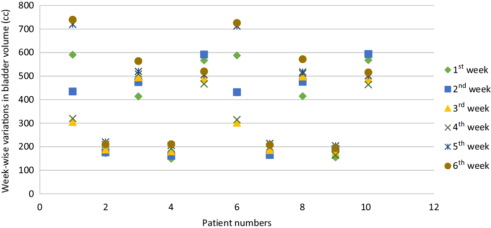
In this exploration, all patients showed inter-fraction/intra-treatment bladder volume larger than the planning bladder volume. In some cases, the bladder volume measured in last treatment week was smaller than the initial planning full bladder volume. In few patients, variations in bladder volume ranged from 153 to 220 cc. When comparing intra-treatment full bladder scans with planning full bladder scan, difference between planning bladder and inter-fraction bladder volume ranged from 4·5 to 49%. Figure 2 shows initial planning full bladder volume and subsequent inter-fraction bladder for each patient.
Representation of intra-treatment variations of bladder volumes (cc).

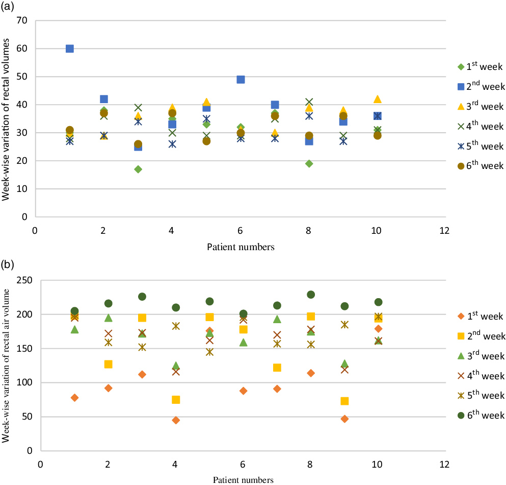
In Figure 3a, rectal volume varied from 17 to 60 cc in this exploration. Inter-fraction rectal volume of some patients showed slight variations from the planning rectal volume. In one patient, rectal volume during treatment was significantly greater than the planning rectal volume. In patients 1 and 6, rectal volume measured in 2nd week of treatment was much higher than the rectal volume measured in other weeks due to variations in rectal contents. In all cases, discrepancy between planning and intra-treatment rectal volumes was ranging from 2 to 105% during an entire course of radiotherapy. There observed no correlation between treatment week and rectal volume.
(a) Representation of intra-treatment variations in rectal volume. (b) Representation of intra-treatment variations in rectal air volume.

The presence of air bubble inside the rectum is a crucial factor which is responsible for changing the rectal volume. It expands and pushes the rectum closer to the PTV. In Figure 3b, planning rectal air volume was less than intra-treatment air volume in all patients except in one. A wide variation between planning and intra-treatment air volumes was found in most patients. This variation ranged from 78 to 229 cc. When comparing planning and intra-treatment air volumes, the difference was in a range from 2 to 367%. In one patient, rectal air variation was not large, and it ranged from 179 to 218 cc. In that patient, the maximum difference between planning and intra-treatment rectal air volumes was 21·79%. Rectal air volume measured towards the end of radiotherapy session was largest. This showed that the instructions regarding evacuation of rectal air were not followed during an entire treatment session.
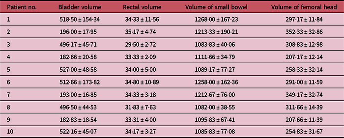
Moreover, volume of small bowel being irradiated varied with alteration in bladder filling volume. Small bowel volume varied from 958 to 1517 cc. Furthermore, irradiated volume of femoral head also varied when bladder and rectal volumes changed. Femoral head ranged from 190 to 402 cc over a treatment period of 5·5 weeks. Mean volume of OARs with standard deviation is given in Table 1.
Variations in volumes of organs at risk during radiotherapy

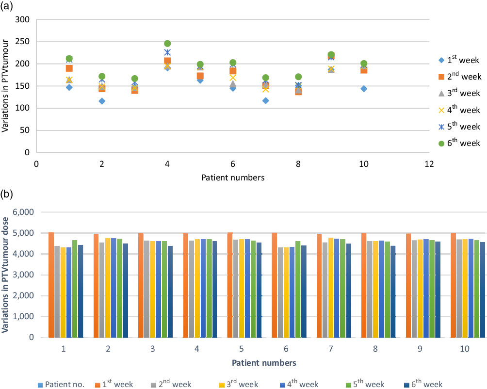
Due to bladder and rectal volume variations, PTVtumour V95% and PTVnodal V95% (volume taking 95% of the prescribed dose) did not remain constant throughout the treatment. In most of the patients, intra-treatment PTV V95% was more than the planning PTV V95%. The difference between planning and inter-fraction PTV V95% was ranging from 1·35 to 48·28%. PTVtumour V95% dose also changed with variations in target volumes. In all patients, initial planning PTVtumour V95% dose was more than 95% of the prescribed dose. Intra-treatment PTVtumour dose was less than 95% of the prescribed dose. The maximum discrepancy between intra-treatment PTVtumour dose and planning PTVtumour dose was 12·15%.
The dosimetric and volume changes in PTVtumour V95% during treatment fractions are shown in Figures 4a and 4b.
(a) Representation of intra-treatment variations in PTVtumour. (b) Representation of intra-treatment variations in PTVtumour dose. PTVtumour, planning target volume of the tumour.

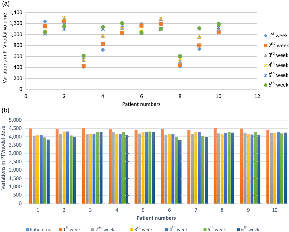
Bladder filling and rectal volume variations also brought changes in PTVnodal V95% during radiation treatment as shown in Figure 5a. Discrepancy in planning and inter-fraction PTVnodal V95% showed a range of 0·88–57·78%.
(a) Representation of variation in PTVnodal volume. (b) Representation of variation in PTVnodal dose.

Figure 5b showed that PTVnodal V95% dose also varied during SIB-IMRT. Planning PTVnodal V95% dose was more than inter-fraction PTVnodal V95% dose in all patients. About 12·87% was the maximum difference between planning and intra-treatment PTVnodal doses.
Discussion
Treatment planning and radiotherapy of cervical cancer require a comfortably full bladder and an empty rectum.Reference Eminowicz15 Full bladder pushes volumes at risk like small bowel away from the treatment field. The volume of small bowel receiving tumouricidal dose decreased when bladder volume was large reducing the probability of late bowel toxicity.Reference Sithamparam, Ahmad, Sabarudin, Othman and Ismail16,Reference Nuyttens, Robertson, Yan and Martinez17 In this analysis, inconsistent bladder filling and rectal volume variations caused changes in PTVtumour V95% and PTVnodal V95%, affecting accuracy of treatment plan by disturbing dose distribution of the target site. It is also reported in various studies.Reference Yong, Eduard, Tianfang, Chuang and Lei18–Reference Grills, Hugo, Kestin, Chao, Wloch and Yan20 To avoid this situation, Jhigran et al.Reference Jhingran, Salehpour, Sam, Levy and Eifel12 have proposed a method of adding a very large margin to CTV that includes shifts in target position to avoid inadequate dosage of the target volume. But this approach is unacceptable when a high dose of 4,500–5,040 cGy is prescribed to the target site. In another approach, they filled bladder with fixed saline volume to reduce target volume uncertainty. This is significant for a short boost treatment course for patients with high-risk features but becomes impractical for a whole period of radiotherapy.Reference Jhingran, Salehpour, Sam, Levy and Eifel12 Yamamoto et al.Reference Yamamoto, Yonesaka and Nishioka21 recommended a method to apply image-guided technique to produce alterations in field position depending on the location of fiducial markers inserted in the vagina before every fraction. This approach is applicable for target limited to the vagina and becomes unsuccessful for cases where vagina and pelvic nodes are treated simultaneously.Reference Yamamoto, Yonesaka and Nishioka21 Few studies emphasised on the use of tool for rapid evaluation of bladder volume prior to every radiotherapy fraction for accurate dose delivery.Reference Sithamparam, Ahmad, Sabarudin, Othman and Ismail16,Reference Chen, Yang, Wang and Hu22
Some patients could not achieve full bladder throughout the treatment course due to their inability to hold urine in the bladder, or they may become less attentive to bladder filling instructions. Various studies showed less variation in rectum volume than bladder volume during a course of treatment, but in this investigation presence of air bubble in the rectum increased its volume and pushed it closer to the target site.Reference Jhingran, Salehpour, Sam, Levy and Eifel12 This caused alteration in rectal and target volume dose distribution, thus influencing the accuracy of treatment plans. Chen et al.,Reference Chen, Yang, Wang and Hu22 in their study on prostate cancer showed that air bubbles in the rectum changed the shape of the rectum and causing the accuracy of dose distribution around the target site harder to achieve.
In this exploration, all patients were unable to maintain a consistent and comfortably full bladder volume over a treatment period of 5·5 weeks. In addition to this, these patients also suffered from air bubble in their rectum which produced rectal movements and disturbed the target dose distribution. In addition to this, only a small number of patients were brought under this investigation. To enhance dose distribution accuracy, it is suggested that the patients should have a constant and full bladder volume and empty rectum before every treatment fraction by strictly following departmental instructions. Furthermore, number of patients to be investigated should be increased in future studies.
Conclusion
Inconsistent bladder and rectal volumes had a significant impact on target volume and dosage during an entire course of SIB-IMRT. For radiotherapy of gynaecological malignancies, data on variations in PTV should be acquired on daily basis to target radiation dose to the tumour site with accuracy.
Acknowledgements
We are thankful to Clinical and Radiation Oncology Department, Shaukat Khanum Memorial Cancer Hospital and Research Center, Lahore and Islamia University, Bahawalpur
Conflict of interest
None.