The Wilms tumour 1 gene (WT1) was recognised as a tumour suppressor gene more than 15 years ago, when the WAGR (‘Wilms tumour predisposition, aniridia, genitourinary anomalies, and mental retardation’) syndrome was linked to germline deletion of the WT1 locus on chromosome 11. Its specific relevance to Wilms tumour was demonstrated by Housman and associates shortly thereafter, when they reported loss of heterozygosity in one WT1 allele and an intragenic deletion in the other in a sporadic Wilms tumour sample (Ref. Reference Haber1). Since those initial discoveries, the gene product of WT1 has been characterised as a nuclear transcription factor containing four C-terminal DNA-binding zinc fingers and an N-terminal transcriptional regulatory domain. WT1 is now known to be primarily expressed in cells of the developing genitourinary and haematopoietic systems. Haematopoietic expression of WT1 appears to be inversely correlated with the degree of differentiation: expression has been detected in CD34+ haematopoietic stem/progenitor cells, but not in mature leukocytes (Refs Reference Fraizer2, Reference Baird and Simmons3, Reference Maurer4, Reference Hosen5). A similar pattern is seen in renal development: initial strong expression in metanephric mesenchyme diminishes with differentiation, until in the mature kidney WT1 expression is limited to glomerular podocytes.
Although mutation and deletion of WT1 in Wilms tumour suggests a tumour suppressor function, WT1 is overexpressed in a variety of other neoplasms, including carcinomas of the lung (Refs Reference Mundlos6, Reference Oji7), colon (Ref. Reference Koesters8) and pancreas (Ref. Reference Oji9), as well as paediatric sarcomas (Ref. Reference Athale10), mesothelioma (Ref. Reference Amin11) and other mesodermal tumours (Ref. Reference Thorner12), suggesting possible oncogenic activity in these and other malignancies. With evidence supporting a tumour suppressor role in some contexts but an oncogenic function in others, the tumour-suppressor–oncogene dichotomy might not apply to a discussion of WT1, which may function differently in different types of malignancy.
Strong WT1 expression in acute leukaemias was first reported by Miwa and colleagues (Ref. Reference Miwa, Beran and Saunders13). These findings have been confirmed in numerous studies, including reports of increased expression in acute lymphoblastic leukaemia (ALL), chronic myeloid leukaemia (CML), myelodysplastic syndrome (MDS) and childhood acute myeloid leukaemia (AML). There is growing evidence that WT1 expression has prognostic importance. The first such report found that AML patients with low-level WT1 expression had a remission induction rate of 91% (20 of 22 patients) compared with 0 of 6 patients with high-level WT1 expression (Ref. Reference Inoue14). Subsequently, it was reported that patients less than 60 years old with de novo AML with high-level WT1 expression had a 3-year overall survival of only 19%, whereas patients with low-level WT1 expression had a 3-year overall survival of 64% (Ref. Reference Bergmann15). Mutations in WT1 have also been reported in myeloid leukaemias, in both diagnostic and relapse samples (Refs Reference King-Underwood and Pritchard-Jones16, Reference Miyagawa17). One report of eight such patients with acute leukaemia found that only two of the patients achieved remission and none survived (Ref. Reference King-Underwood and Pritchard-Jones16). Because the primary treatment for acute leukaemia is chemotherapy, this observation suggests the possibility that mutations in WT1 confer drug resistance to the leukaemia cells.
This review focuses on our current understanding of WT1 function in haematopoiesis and leukaemogenesis. Advances in this field are leading to the development of novel therapeutic modalities, which are also discussed.
Structure and function of WT1
The WT1 gene consists of ten exons located on chromosome 11, at locus 11p13. The gene encodes a 57 kDa protein product, comprising two major domains. The N-terminus is proline- and glutamine-rich and has transcriptional regulatory function, whereas the C-terminal DNA-binding domain is composed of four Cys2His2 zinc fingers, each encoded by a discrete exon (numbers 7–10; Fig. 1). The four zinc fingers share significant homology to those of the early growth response gene product EGR1. The zinc finger region is also required for RNA binding, particularly zinc finger 1 (Ref. Reference Caricasole18), but DNA and RNA binding are mutually exclusive (Ref. Reference Bardeesy and Pelletier19). Subnuclear localisation of WT1 was affected by treatment with RNase, but not DNase, suggesting that WT1 may function at least partly at the post-transcriptional level (Ref. Reference Caricasole18).
Schematic diagram of the WT1 transcript and its protein. (a) Alternative splicing sites. Each WT1 exon is represented by a grey box, with the codon number of the junction between exons indicated below. The alternatively spliced regions (exon 5 and the KTS insert) are represented by different colours for clarity. The truncated forms referred to in the text begin with exon 2 (codon 442), as transcription is initiated from a cryptic promoter within the first intron. (b) Protein domains. The WT1 protein has two broad domains, as indicated: an N-terminal transcriptional regulatory domain, and a C-terminal DNA-binding domain containing four zinc fingers. The relative locations of the alternatively spliced regions are shown.

Protein isoforms
The WT1 mRNA undergoes alternative splicing at two independent sites (Fig. 1). The mature mRNA may include or exclude exon 5 (Ex5), which encodes a 17 amino acid sequence whose function is as yet unknown. In addition, the use of one of two splice acceptor sites at the 5' end of exon 10 results in exclusion or inclusion of a 9 bp region encoding a three amino acid insert, designated KTS for lysine, threonine and serine. Thus, four different major protein isoforms result from the two major alternative splicing events. In the human 20-week-old kidney, the relative ratios of the isoforms Ex5−–KTS−, Ex5+–KTS−, Ex5−–KTS+ and Ex5+–KTS+ are 1.0 to 2.5 to 3.8 to 8.3, respectively; KTS-positive isoforms constitute 80% of cellular WT1 (Ref. Reference Haber20). Ex5+ isoforms were the most abundant in the human haematopoietic system, adult kidney cortex, and the testis. Mouse kidney and testis likewise showed a relative excess of Ex5+ isoforms. The ratios of isoforms containing or lacking KTS were similar between mouse and human tissues, suggesting conservation of KTS splicing across species (Ref. Reference Renshaw, King-Underwood and Pritchard-Jones21).
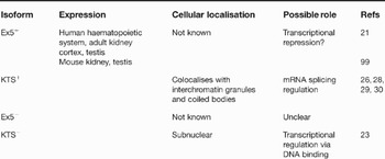
There is growing evidence that different WT1 isoforms have different cellular functions (Table 1). The KTS insert, lying between exons 9 and 10, affects the relative spacing of the third and fourth zinc fingers, which in turn determines which zinc fingers make contact with DNA, thus impacting on the DNA sequences that are bound by different WT1 isoforms. A molecular basis for this observation was demonstrated using NMR relaxation techniques; the KTS insert significantly increases the flexibility of the protein backbone. The insertion of KTS consequently prevents optimal interaction between zinc finger 4 and its cognate binding site in the DNA major groove (Ref. Reference Laity, Dyson and Wright22). KTS− isoforms localise to subnuclear compartments similar to other classical transcription factors such as Sp1 and TFIIB (Refs Reference Rauscher23, Reference Reddy, Hosono and Licht24), and are most strongly linked with transcriptional regulation, likely via a consensus DNA sequence similar to that bound by EGR1 (Ref. Reference Rauscher23) (see next section). Depending on a number of factors, including cell line and reporter promoter used, KTS− isoforms may repress or activate transcription (Refs Reference Reddy, Hosono and Licht24, Reference Reddy25). By contrast, KTS+ isoforms have a reduced ability to bind DNA (Ref. Reference Bardeesy and Pelletier19), and a consensus DNA sequence for WT1 binding for isoforms containing the KTS insert has not been defined. KTS+ isoforms colocalise with interchromatin granules and coiled bodies (Refs Reference Larsson26, Reference Englert27), and are increasingly linked with the regulation of mRNA splicing. Co-immunoprecipitation assays have revealed KTS+ isoforms strongly interact with splicing proteins U2AF65 (Ref. Reference Davies28) and RBM4 (Ref. Reference Markus29). The presence of WT1, preferentially KTS+ isoforms, in nuclear poly(A)+ ribonucleoprotein particles (Ref. Reference Ladomery30), supports the contention that WT1(KTS+) is involved in post-transcriptional processing. Specific binding of α-actinin mRNA by this isoform has also recently been demonstrated, further supporting a role in post-transcriptional regulation (Ref. Reference Morrison31).
Functions of alternatively spliced isoforms of WT1

Abbreviations: Ex5, exon 5; KTS, three amino acid insert of lysine, threonine and serine.
In addition to the four major isoforms, other novel WT1 proteins have been reported. Larger isoforms result from translation initiation at an upstream in-frame CUG codon (Ref. Reference Bruening and Pelletier32), while N-terminally truncated isoforms, measuring 36–38 kDa, result from translation initiation at a downstream, in-frame AUG located in a cryptic exon within intron 1. These truncated forms are presumed to be spliced similarly to the major isoforms. The truncated mRNA transcript was highly tissue-specific in humans, limited to fetal kidney and testis, whereas full-length WT1 was expressed in adrenal gland, fetal and adult kidney, and testis. DNA-binding capacity of the two isoforms was similar; however, the truncated WT1 isoform appeared to be more effective in the activation of SRY-driven and p21-driven reporter genes, as indicated by transient transfection assays. Furthermore, small interfering RNA (siRNA) directed against truncated WT1 inhibited K562 cell growth more substantially than did siRNA against full-length WT1. Truncated WT1 also activated genes, such as those encoding cyclin E and the insulin-like growth factor 1 receptor, that are normally repressed by the full-length isoform (Ref. Reference Hossain33). Truncated WT1 might thus exhibit oncogenic properties, in contrast to full-length WT1, although more research will be required to characterise this novel isoform fully.
WT1 transcriptional targets
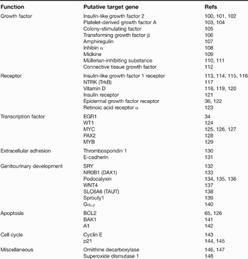
Significant effort has been expended in the identification of relevant targets of the transcriptional regulatory activity of WT1. A consensus DNA sequence (5'-GCGGGGGCG-3') bound by isoforms lacking the KTS insert has been identified (Refs Reference Rauscher23, Reference Madden34), and numerous promoters have been found to contain this sequence. Wang et al. identified a second DNA sequence that is also bound by WT1 in WT1-regulated promoters (5'-TCCTCCTCCTCCTCTCC-3') (Ref. Reference Wang35). Transient transfection assays demonstrate WT1-mediated repression of the EGR1 gene and related promoters, such as those of genes encoding insulin-like growth factor 2, insulin-like growth factor 1 receptor, platelet-derived growth factor A, PAX2, colony-stimulating factor 1 (CSF1) and transforming-growth factor β (Table 2). However, WT1 did not affect endogenous expression of many of these genes in more detailed studies (Ref. Reference Englert36). By contrast, some putative WT1 target genes identified in this fashion do appear to be affected in intact cells. Previously identified WT1 target promoters were screened in stably transfected cell lines, and CSF-1 was found to be induced in Saos2 cell lines (Ref. Reference Thate, Englert and Gessler37). Using a transgenic mouse model, deletion of the WT1-binding element abrogated nephrin promoter activation, providing in vivo evidence for nephrin as a WT1 target gene (Ref. Reference Wagner38). In vivo evaluation was similarly undertaken in wild-type and WT1-deficient transgenic mouse embryos. Transcripts of α4 integrin were shown to be decreased by 57% in the epicardium of the WT1-deficient mice by using real-time reverse transcriptase (RT)-PCR, suggesting that α4 integrin is activated by WT1 (Ref. Reference Kirschner39). The use of high-density oligonucleotide or cDNA arrays to assess expression profiles of putative targets has identified A1, the amphiregulin gene and MYC as WT1 target genes.
WT1 target genesa

aGene products are listed.
A1, BCL2 family member; BAK1, BCL2-antagonist/killer 1; EGR1, early growth response 1; MYB, v-myb myeloblastosis viral oncogene homologue (avian); NR0B1, nuclear receptor subfamily 0, group B, member 1, NTRK, neurotrophic tyrosine kinase, receptor, type 2; MYC, v-myc myelocytomatosis viral oncogene homologue (avian); PAX2, paired box gene 2; SRY, sex determining region Y; SLC6A6, solute carrier family 6 (neurotransmitter transporter, taurine), member 6; WNT4, wingless-type MMTV integration site family, member 4; WT1, Wilms tumour 1.
WT1 expression in normal haematopoiesis
Preferential expression of WT1 in the developing fetal kidney, as well as the presence of urogenital anomalies in humans and animal models with interstitial deletions of WT1, suggest that WT1 plays a role in the development of the urogenital tract. In addition to the developing kidney, WT1 expression has been localised to the mesothelial cell lining of body cavities and visceral organs (Ref. Reference Park40), thymus, brain and spleen (Ref. Reference Armstrong41). Several studies have also shown that CD34+ bone marrow progenitor cells express high levels of WT1 relative to mature haematopoietic cells (Refs Reference Fraizer2, Reference Baird and Simmons3, Reference Maurer4, Reference Hosen5). Whether the level of expression in precursor cells is comparable to that of leukaemic blasts remains controversial. Two studies did not find a difference between CD34+ and AML cell WT1 expression (Refs Reference Fraizer2, Reference Maurer42), but Inoue et al. demonstrated a tenfold increase in WT1 expression in a variety of leukaemic cell lines and patient samples compared with normal haematopoietic progenitor cells (Ref. Reference Inoue43). The discrepancy may be attributable to differences in RT-PCR conditions. However, other authors detected WT1 expression in only 1.3% of CD34+ single bone marrow cells, at levels comparable to that of K562 CML cells (Ref. Reference Hosen5). Thus, discrepancies among different studies may also reflect differences in WT1 expression among the subpopulations of cells chosen for analysis.
The finding that WT1 is expressed in normal haematopoietic stem cells but not in mature white blood cells suggests a role for WT1 in normal haematopoiesis. Steady-state WT1 levels were down-regulated in K562 cells undergoing differentiation induced by sodium butyrate or 12-O-tetradecanoylphorbol-13-acetate (Ref. Reference Phelan, Lindberg and Call44). Down-regulation of WT1 transcripts was also observed in HL60 acute promyelocytic leukaemia cells when myelomonocytic differentiation was induced (Ref. Reference Sekiya45). Furthermore, constitutive expression of WT1 in U937 cells, a human myeloid-leukaemia-derived cell line that lacks endogenous WT1, blocked differentiation in response to retinoic acid or vitamin D3 and induced G1 arrest (Ref. Reference Svedberg46). Similar results were achieved in 32D cl3 cells, an interleukin-3-dependent murine myeloid progenitor cell line. Differentiation was arrested at the promyelocyte stage, despite treatment with granulocyte colony-stimulating factor (G-CSF). G-CSF stimulation of WT1-transfected 32D cl3 cells also resulted in constitutive activation of the transcription factors STAT3α and STAT3β and continued proliferation. By contrast, control clones differentiated into mature neutrophils in response to G-CSF stimulation and demonstrated only transient activation of STAT1 (Ref. Reference Inoue47). These studies support the contention that alteration of WT1 expression might affect haematopoietic differentiation via other transcription factors, such as STAT3. In a chimaeric mouse model generated from WT1-negative embryonic stem cells and normal murine blastocysts, researchers demonstrated that WT1 is not essential for haematopoiesis but confers a competitive advantage to stem cells when present (Ref. Reference Alberta48). These findings also support the role of WT1 in regulation of proliferation or differentiation pathways as yet undefined. More investigation in nontransformed cell lines and in vivo models will be necessary to elucidate fully the role of WT1 in normal haematopoiesis.
Function of WT1 isoforms in normal and malignant haematopoiesis
There are conflicting reports regarding differential expression of WT1 isoforms in leukaemic cells. One report indicated a preponderance of Ex5+ isoforms in AML patient samples but, overall, no significant variation in WT1 expression among samples representing different myeloid leukaemia FAB subtypes (Ref. Reference Siehl49); however, in other reports M5 AML was associated with significantly lower WT1 expression, as measured by RT-PCR, in comparison with other FAB subtypes (Refs Reference Bergmann15, Reference Trka50), and M3 AML was linked to higher WT1 expression (Ref. Reference Patmasiriwat51).
In addition to differential expression of WT1 isoforms, mounting evidence supports differential functions of these isoforms in haematopoiesis and leukaemogenesis. Transfection of 32D cl3 with Ex5−–KTS− potentiated granulocytic differentiation in the presence of G-CSF, reflected in morphological changes, changes in gene expression, and expression of mature cell-surface antigens (Ref. Reference Loeb52). By contrast, 32 D cl3 cells transfected with full-length WT1 (i.e. Ex5+–KTS+) do not differentiate in response to G-CSF administration (Ref. Reference Inoue47). Expression of KTS− isoforms dramatically increased the number of U937 cells arrested in G1 phase and induced myelomonocytic differentiation in CD34+ haematopoietic progenitor cells derived from human umbilical cord blood. This was associated with an increase in activity of cyclin-dependent kinase inhibitor p21CIP1, a known WT1 transcription target. In addition, KTS− isoforms appeared to induce CD34+ precursor quiescence. Co-expression of both KTS− and KTS+ isoforms potentiated these effects, but the KTS+ isoforms alone were not sufficient to induce differentiation (Ref. Reference Ellisen53). In studies that compared frequencies of all four isoforms in various malignancies, Ex5+–KTS+ was over-represented (Refs Reference Renshaw, King-Underwood and Pritchard-Jones21, Reference Oji54, Reference Ueda55). Furthermore, constitutive expression of Ex5+–KTS+ rescued cancer cells from the growth inhibition effected by WT1 antisense oligomers (Ref. Reference Ito56). In a transgenic mouse model expressing the AML1–ETO fusion protein (encoded by the fusion gene resulting from a commonly seen reciprocal translocation between chromosomes 8 and 21 in human AML), constitutive co-expression of Ex5+–KTS+ enhanced the proliferation of bone-marrow-derived colonies and inhibited their differentiation. Mice that were subsequently transplanted with cells co-expressing WT1 and AML1–ETO quickly developed AML, while those transplanted with AML1–ETO alone demonstrated abnormal myelopoiesis but not leukaemia (Ref. Reference Nishida57). This suggests that overexpression of WT1, particularly Ex5+–KTS+, likely plays a cooperative role with other genetic hits in leukaemogenesis.
The apparent contradiction between earlier reports of differentiation being associated with down-regulation of WT1 and more-recent studies ascribing differentiation to WT1 expression may be resolved in several ways. Prior conflicting results were obtained from leukaemia-derived cell lines, arrested at a single stage of haematopoiesis, and appear to be cell-line-specific. These leukaemic cell lines are inherently resistant to differentiation by virtue of their neoplastic transformation. The use of nontransformed cell lines (such as 32D cl3) and primary bone marrow cells avoids this issue although, like leukaemia cells, 32D cl3 cells are already lineage-committed.
Alternatively, conflicting data may be attributed to the opposing roles played by the different isoforms. The earlier papers evaluated endogenous WT1 expression, which is always composed of a mixture of all isoforms, while later studies evaluated the effects of exogenous expression of individual isoforms. Transfection of haematopoietic progenitor cells, or human embryonic stem cells, with WT1 isoforms individually and in combination, and evaluation of their effect on haematopoiesis, might help resolve the controversy. In addition, the application of siRNA technology might allow silencing of individual isoforms as a complementary approach to addressing this question.
The role of WT1 mutation in leukaemias
WT1 gene mutations are seen in 10% of sporadic Wilms tumour cases. By contrast, all children with WAGR syndrome harbour interstitial deletions of chromosome 11p13, producing a hemizygous state for WT1, and have a 30–50% lifetime risk of developing Wilms tumour. Fifteen years after treatment of Wilms tumour, the cumulative incidence of secondary malignancies in all patients is 1.6%; secondary leukaemias account for up to 20% (Ref. Reference Breslow58). While many of these cases may be attributed to therapy, WT1 mutation conferring genetic predisposition could not be excluded (Ref. Reference Shearer59).
Pritchard-Jones et al. first reported the development of AML associated with a WT1 insertion mutation, in a patient with WAGR syndrome (Ref. Reference Pritchard-Jones, Renshaw and King-Underwood60). Mutations were subsequently identified in 14% of cases of sporadic AML and 20% of biphenotypic leukaemia, as well as in undifferentiated (Ref. Reference King-Underwood, Renshaw and Pritchard-Jones61) and lymphoblastic (Ref. Reference Carapeti, Goldman and Cross62) leukaemias. Heterozygous frameshift, missense and nonsense mutations were identified in exons 1, 6, 7 and 9, respectively. These alterations are expected to produce a truncated WT1 protein with absence or loss of the zinc finger region, likely impairing DNA-binding capacity. Furthermore, Summers et al. recently reported an association between FLT-3 internal tandem duplication (ITD), a known marker of poor prognosis in AML, and WT1 mutation; those patients exhibiting both gene defects had worse prognosis than patients with either mutation alone (Ref. Reference Summers, Kakkas, Smith, Smith, Macdougall, Cavenagh, Bonnet, Young, Lister and Fitzgibbon63). Resistance to induction chemotherapy and worse disease-free and overall survival have been described in patients with WT1 mutation, suggesting a role for WT1 in apoptotic response to cytotoxic agents (Ref. Reference King-Underwood and Pritchard-Jones16).
Regulation of apoptosis by WT1
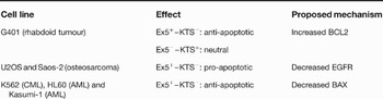
The role of WT1 in apoptosis was first suspected upon examination of WT1 knock-out mice. Kidneys were absent, and the metanephric blastema in these embryos demonstrated 10–50% apoptosis, in contrast to 1% apoptotic rate in wild-type embryos (Ref. Reference Kreidberg64). Although these findings suggested that WT1 was necessary to inhibit apoptotic cell death, subsequent studies have provided conflicting data. For example, in rhabdoid tumour cell lines that had been stably transfected, cells that expressed KTS− isoforms, but not KTS+ isoforms, demonstrated elevated expression of the anti-apoptotic factor BCL-2 and were resistant to cytotoxic therapy (Ref. Reference Mayo65). In contrast, osteosarcoma cell lines transfected with constitutively expressed Ex5+–KTS− isoform demonstrated increased apoptosis. Furthermore, activity of the epidermal growth factor receptor (EGFR) promoter was repressed fourfold in association with WT1-mediated apoptosis, identifying EGFR as a potential WT1 target. The developmental expression of EGFR in the embryonic kidney was also examined; changes in EGFR expression precede changes in WT1 expression in committed glomerular structures, supporting a role for EGFR in WT1-mediated apoptosis (Ref. Reference Englert36). Overexpression of WT1 in M1 myeloblast cells results in induction of apoptosis and G1 arrest with the Ex5+–KTS− isoform as well. WT1-induced apoptosis in this cell model functions independently of p53 and the EGFR, because M1 leukaemia cells do not express either protein (Ref. Reference Murata66). By contrast, suppression of Ex5+ isoforms with siRNA induced apoptosis in WT1-expressing leukaemia cell lines, but not in a lymphoma cell line that lacks WT1 expression. Suppression of Ex5− isoforms by siRNA did not induce apoptosis in these cell lines. In addition, the expression level of the pro-apoptotic BAX protein was significantly decreased in K562 cells transduced with the Ex5+–KTS− isoform (Ref. Reference Ito56), consistent with an anti-apoptotic function in this cell line. Thus, the impact of WT1 on apoptosis (and the mechanism by which WT1 functions) appears to be cell-line-specific. These conflicting findings are summarised in Table 3.
Cell-line-specific effects of WT1 on apoptosis
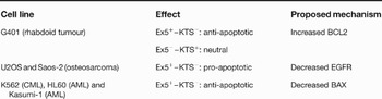
Abbreviations: AML, acute myeloid leukaemia; BAX, BCL2-associated X protein; BCL2, B-cell CLL/lymphoma 2; CML, chronic myeloid leukaemia; EGFR, epidermal growth factor receptor; Ex5, exon 5; KTS, three amino acid insert of lysine, threonine and serine; WT1, Wilms tumour 1.
Cytotoxic therapy rapidly reduced Ex5+ ratios as well as total WT1 expression in K562 and HL60 cells. Subsequently, an excess production of Ex5− transcripts was observed, suggesting increased Ex5 skipping during post-transcriptional splicing (Ref. Reference Renshaw67). To recapitulate and further examine the effects of Ex5 down-regulation, nuclease-resistant antisense oligonucleotides (ASOs) were designed, specifically targeting the 3' untranslated region (UTR) of WT1 and Ex5+ transcripts in K562 and HL60 cell lines. Both ASOs reduced K562 survival. HL60 cells were not inhibited by the UTR-derived ASO or control; however, the Ex5-derived ASO reduced cell survival to 41% of control levels. This contrasts with a previous report of HL60 resistance to ASOs (Ref. Reference Algar68). Thus, similar to its role in cellular differentiation, WT1 appears to mediate apoptosis in a cell-type- and isoform-dependent manner. Further studies are needed to elucidate its effect on programmed cell death in vivo.
Clinical implications/applications of WT1 in leukaemia patients
Prognosis
There are conflicting studies regarding the correlation between WT1 expression and prognosis in leukaemia patients. Several groups have reported that elevated expression confers poor prognosis in acute leukaemia (Refs Reference Inoue14, Reference Bergmann15, Reference Trka50, Reference Barragan69). The 3-year overall survival in adult patients with AML was 38% and 13% for low WT1 and high WT1 expression, respectively (Ref. Reference Bergmann15). Other groups have found that WT1 expression levels demonstrated no prognostic significance (Refs Reference Yanada70, Reference Schmid71, Reference Gaiger72). Still other groups found that higher WT1 expression was associated with favourable cytogenetics profiles: one group reported better outcome in children with AML (Ref. Reference Rodrigues73), while in a larger study of adults with AML, the correlation between higher WT1 expression and favourable cytogenetics was also reported, but no benefit in survival was observed (Ref. Reference Yanada70). Interpretation of these studies in toto is limited by a number of factors. The majority of these reports were retrospective and included small numbers of patients treated with a variety of therapies. The relative proportions of FAB subtypes differed among these studies as well. Thus, outcome may be attributable to factors distinct from WT1 expression. There were also technical differences related to the quantitative RT-PCR methods employed that might also have resulted in discrepant findings.
Minimal residual disease
The observation that WT1 is overexpressed in most types of leukaemia has led to the use of WT1 expression as a marker of minimal residual disease (MRD). WT1 is more attractive as an MRD marker than fusion protein markers (e.g. AML1–ETO, PML–RARα, MYH11–CBFβ), because only a minority of leukaemias are characterised by the latter. However, there initially was controversy regarding the reliability of WT1 as a means of monitoring leukaemia. In an earlier study, WT1 expression was measured via qualitative RT-PCR and was deemed to be of limited value as an MRD marker (Ref. Reference Gaiger74). With the advent of real-time quantitative RT-PCR, an increasing number of studies have validated WT1 as an MRD marker, showing concordance between WT1 expression and fusion gene transcripts in patients at diagnosis, remission and relapse (Refs Reference Kreuzer75, Reference Ogawa76, Reference Ogawa77, Reference Ostergaard78, Reference Tamaki79, Reference Cilloni80, Reference Cilloni and Saglio81). Upon examination of leukaemic cells from an acute promyelocytic leukaemia patient, RT-PCR sensitivity limits for WT1 of 10−5 for peripheral blood and 10−3 to 10−4 for bone marrow were achieved when diluted with normal cells. In comparison, PCR sensitivity for PML–RARα reached sensitivity limits of 10−4 for both peripheral blood and bone marrow (Ref. Reference Inoue14). Inoue and colleagues subsequently reported that WT1 expression increased 1 to 18 months prior to clinical relapse, suggesting that increase of WT1 relative to diagnostic levels may be a potential harbinger of relapse (Ref. Reference Inoue82). In a recent study, WT1 levels were measured at the end of induction, at 3–5 months, at 6–8 months, and at relapse. The authors reported elevated WT1 at the end of induction was an independent risk factor for relapse and death in paediatric AML patients (Ref. Reference Lapillonne83). A few groups report use of WT1 and fusion gene transcript expression to guide therapy in patients with AML and MDS (Refs Reference Miyamura84, Reference Tamura85). As of yet, there have been no randomised, controlled trials to support widespread use of MRD monitoring by WT1 expression in this manner.
Future clinical applications of WT1: immunotherapy
WT1 has been suggested as a novel tumour antigen for myriad reasons. Multiple studies have demonstrated overexpression of WT1 protein in leukaemia cells compared with normal haematopoietic cells. Measurable expression of WT1 in postnatal life is normally limited to a restricted population of cells. The viability of leukaemia cells might be, in part, maintained by WT1. Thus, it may be useful to target WT1 as other overexpressed tumour antigens, including c-erbB-2/HER-2/neu, have been (Refs Reference Fisk86, Reference Tsuboi87). The sera of patients with AML were tested for WT1 antibody; 3 of 18 patients had generated antibody directed against the N-terminus segment of WT1, suggesting that the immune system recognises WT1 as a distinct antigen (Ref. Reference Gaiger88). These findings were expanded in a subsequent review, including patients with AML, ALL, CML and MDS. The authors detected IgM antibodies against WT1 in 54.8% of patients, compared with 16.2% of healthy controls. IgG antibodies were detected to a similar extent (Ref. Reference Elisseeva89).
Several groups have evaluated the ability of selective CD8+ cytotoxic T lymphocytes (CTLs) to target WT1. CD8+ CTLs specific to a 9-mer WT1 peptide restricted to HLA class I motifs have been generated. Cytotoxic killing was demonstrated against both leukaemic cell lines and fresh cells isolated from patients, without diminishing colony formation by normal bone marrow cells (Ref. Reference Ohminami, Yasukawa and Fujita90). In a subsequent study, B6 mice were immunised with various WT1 peptides derived from the wild-type WT1 sequence. A vaccine comprising three peptides from the N-terminus of the WT1 protein elicited both proliferative T-cell responses and antibody production of the immunoglobulin IgG2a type. This group also reported CTL-mediated lysis of target cells incubated with the target peptides, as well as of malignant cells expressing WT1. Histological examination of the immunised mice 20 days after immunisation demonstrated no evidence of autoimmune toxicity (Ref. Reference Gaiger88).
A similar murine model was generated with 9-mer peptides containing HLA class I-binding motifs. Sensitised CTLs lysed WT1-expressing murine tumour cells. Furthermore, mice immunised with the most immunogenic WT1 peptide successfully rejected challenges with WT1-expressing tumour cells (Ref. Reference Sugiyama91). Vaccinated mice that had been challenged with WT1-positive tumour cells survived significantly longer than mice that had been vaccinated with control plasmid or phosphate-buffered saline (Ref. Reference Tsuboi87). HLA-A*0201-restricted (the most frequent HLA class I type in Caucasians) CTLs were generated against a 9-mer fragment of WT1, P126. Cells isolated from HLA-A*0201-negative patients with chronic phase CML were not recognised by the P126-specific CTLs. By contrast, CTLs selectively killed CD34+ cells derived from HLA-A*0201-positive patients with CML. No killing of the more mature CD34− population was observed. The authors surmised that CD34− cells escaped CTL-mediated killing because of insufficient WT1 expression by these cells, as demonstrated by RT-PCR and western blotting. Finally, anti-P126 CTLs targeted leukaemic CD34+ cells rather than normal CD34+ cells, presumably because of differences in WT1 expression (Ref. Reference Gao92).
Guo et al. reported the generation of WT1-specific CD4+ T cells against a 20-mer peptide, achieving cytotoxicity in an HLA class II-restricted manner. Thus, CD4+ WT1-specific T cells may themselves play an antileukaemic role, in addition to inducing CD8+ CTL activity (Ref. Reference Guo93).
Such in vitro findings have led to the design of human WT1 vaccine trials. In a Phase I clinical study employing HLA-A*2402-restricted (the most frequent HLA class I type in Japanese), natural or modified 9-mer WT1 peptide, patients with AML, breast cancer, lung cancer and MDS were evaluated for toxicity and clinical response. The two MDS patients were excluded early in the study, due to severe leukocytopenia after one injection. However, patients with solid tumours experienced clinically significant regression of metastatic tumours. Similarly, a decrease in residual leukaemic cells and/or WT1 expression levels was observed among de novo AML patients. This decrease was not sustained if scheduled vaccination was delayed. Toxicity in patients without MDS was limited to local erythema at the injection site (Ref. Reference Oka94). One patient with relapsed AML was treated with 15 cycles of vaccination with WT1 peptide 126–134, and showed complete haematological remission 4 months after initiation (Ref. Reference Mailander95). A recent Phase I/II study of vaccination with dendritic-like leukaemia cells (DLLCs) to stimulate WT1-specific CTL development did not produce broadly applicable results. Only a small subset of patients proceeded to vaccination; the remaining patients yielded cells that failed to differentiate to DLLCs (Ref. Reference Roddie96).
Efforts are under way to improve immunotherapy directed against WT1-positive malignancies. Mice vaccinated with Mycobacterium bovis bacillus Calmette–Guerin cell-wall skeleton (BCG-CWS) one day prior to injection with WT1 peptide survived significantly longer than nonvaccinated mice or mice vaccinated with either component alone (Ref. Reference Nakajima97). Thus, adjuvants like BCG-CWS may prove to enhance the clinical effect of WT1 vaccine for humans. Multiple epitope peptides could be incorporated into a vaccine. The use of HLA class II-restricted helper epitopes in combination with HLA class I epitopes have been shown to enhance activation of CTLs. Finally, the administration of vaccines with other drugs may optimise treatment of malignancies (Ref. Reference Oka98).
Summary and conclusions
First discovered as a causative factor in Wilms tumour, a paediatric kidney cancer, WT1 is an aberrantly overexpressed transcription factor found in many human malignancies, including acute and chronic leukaemias. An increasing volume of literature has illuminated the significance of isoform-specific roles in cellular differentiation and proliferation. The role of WT1 in leukaemogenesis has yet to be fully clarified, but is likely isoform-specific as well. In addition, a promising array of clinical uses for WT1 has emerged. The use of WT1 as an MRD marker may ultimately improve survival in leukaemia patients with poor prognosis; larger clinical trials are warranted to study the feasibility of this approach. WT1 vaccines exemplify tumour-antigen-based immunotherapy and might play an important novel role in treatment of high-risk leukaemia.
Acknowledgements and funding
We thank Greg McCarty for reviewing the manuscript and contributing the figure. We also thank our peer reviewers for their insightful comments and suggestions. Supported by the Ruth L. Kirschstein Institutional Grant at Johns Hopkins, Grant #5T32CA060441-13.