Asthma is a chronic inflammatory disease of the airways affecting approximately 300 million individuals worldwide (Ref. Reference Masoli1). The prevalence of asthma increased dramatically in many Western countries during the last 30 years of the 20th century and is now greater than 10% (Ref. Reference Marks, Correll and Williamson2). In several Western countries, the rates of asthma have reached a plateau since the 1990s, especially in adults (Ref. Reference von Hertzen and Haahtela3). Nevertheless, the high incidence of asthma represents a huge economic burden. For example, the cost to the US economy attributable to asthma was estimated to be US$12.7 billion in 1998 (Ref. Reference Weiss and Sullivan4). Furthermore, high levels of nonatopic asthma have been recognised in the developing world (Ref. Reference Mallol5).
Asthma involves recurrent episodes of airway obstruction and wheezing, ranging in severity from mild to life-threatening. The majority of asthmatics have a characteristic allergic inflammatory response in the lungs involving excessive activation of T helper 2 (Th2) cells, eosinophil infiltration, mucus overproduction, and airway hyper-reactivity. In many cases, this results from an allergic response to environmental allergens (Ref. Reference Wills-Karp6). It is important to note that asthma is highly heterogeneous, and that the asthmatic inflammatory response does not always correspond closely to classical allergy. For example, a significant number of asthmatics have an inflammatory pathology dominated largely by neutrophils rather than eosinophils (Ref. Reference Douwes7). The heterogeneity of asthma is beyond the scope of this review, and we focus here principally on the cellular and molecular mechanisms underlying allergic airway inflammation (AAI).
Animal studies have been valuable for elucidating the cellular and molecular pathways required for AAI. Murine models have been particularly useful, because of the many tools available to dissect mechanisms of allergic inflammation. Neutralising antibodies, transgenic mice (usually overexpressing selective genes) and knockout mice (lacking selective genes) are particularly powerful in assessing the role of individual molecules and cells (Ref. Reference Zosky and Sly8). The popularity of such models needs to be considered against their limitations (Ref. Reference Persson9). There are many structural and functional differences between mice and humans, and mice do not spontaneously develop asthma. Experimental AAI is usually induced artificially by parenteral administration of allergen in adjuvant. Furthermore, in a standard murine AAI model, degranulated eosinophils are detected only in the airway lumen and not in the lung tissue (Ref. Reference Clark10).
The vast majority of animal studies assess acute responses to inhalation of high concentrations of antigen, which generate vigorous cellular infiltration but are not good models of airway remodelling, a common feature of established asthma. A chronic model has been developed that has more histological similarities to clinical asthma (Ref. Reference Kumar and Foster11). In another chronic model, mice were followed for several weeks after cessation of challenge. AAI resolved, but airway hyper-responsiveness (AHR) and remodelling did not resolve during the period of observation (Ref. Reference Johnson12). There are, however, relatively few studies on chronic allergen exposure. Furthermore, the standard mouse models do not reliably demonstrate the early- and late-phase responses that are characteristic of clinical asthma (Ref. Reference Zosky13). Finally, results in mouse models may differ markedly between strains, making it difficult to extrapolate findings to clinical asthma. Despite these limitations, mouse models are widely used to identify potential targets and to assess new therapeutic agents (Refs Reference Zosky and Sly8, Reference Gelfand14). This review draws heavily on animal models of AAI. Clinical trials of novel therapeutic agents are also included, but we do not attempt a comprehensive coverage of descriptive clinical studies.
Differentiation of effector T helper cells
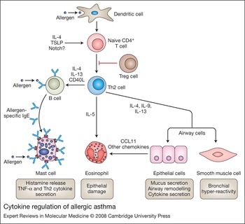
CD4+ Th cells are necessary for the pathogenesis of AAI. Depletion of murine CD4+ T cells prevents antigen-induced pulmonary eosinophilia and AHR (Ref. Reference Gavett15). The Th2 subset, which controls the allergic response through the production of cytokines such as interleukin (IL)-4, IL-5, IL-9 and IL-13, is particularly prominent in allergic asthma (Refs Reference Cohn, Elias and Chupp16, Reference Robinson17). The key role of Th2 cells is illustrated in Figure 1. Differentiation of naive CD4+ T cells is a tightly regulated process. Experiments in mice have demonstrated that the cytokine microenvironment has a major role in regulating T cell differentiation, but other factors such as antigen concentration, and expression of costimulatory molecules and Notch ligands, also contribute (Refs Reference Abbas, Murphy and Sher18, Reference Amsen19).
Cytokine regulation of allergic asthma. Pulmonary dendritic cells process and present inhaled antigens to naive CD4+ T cells. Generation of Th2 cells is enhanced by the presence of the cytokines IL-4 and TSLP, and is influenced by Notch ligands. Secretion of IL-4 by naive T cells contributes to their differentiation into Th2 cells. Th2 cells secrete IL-4 and IL-13, and express CD40L, to stimulate IgE production by B cells. IgE binds to Fc receptors on the surface of mast cells. When mast cells are activated by antigen-dependent crosslinking of IgE receptors, they degranulate, releasing histamine and TNF-α, and they secrete cytokines including IL-4 and IL-13. Eosinophil survival is supported by IL-5, and CCL11 (eotaxin-1) and other cytokines attract eosinophils to the airways. Eosinophils are toxic to epithelial cells. IL-4, -9 and -13 promote airway hyper-reactivity, mucus secretion and cytokine secretion by epithelial cells. Regulatory T cells (Treg) suppress the activation and differentiation of naive CD4+ T cells. Abbreviations: IL, interleukin; TSLP, thymic stromal lymphopoietin.

IL-4 signalling through STAT6 (signal transducer and activator of transcription 6) was originally thought to be necessary for Th2 cell commitment (Refs Reference Kaplan20, Reference Kopf21). It is now apparent that Th2 cell differentiation can occur in the absence of both IL-4 and STAT6. Nevertheless, STAT6 is essential for eosinophilic inflammation and Th2 cell tracking to the lung in murine AAI (Refs Reference Akimoto22, Reference Mathew23), and in clinical asthma, phosphorylated STAT6 is increased in peripheral blood memory CD4+ T cells, and it decreases after therapy with oral corticosteroids (Ref. Reference Gernez24). The transcription factor GATA-3 is the master regulator of Th2 cell development (Ref. Reference Zheng and Flavell25). GATA-3 regulates expression of other well-described Th2 differentiation transcription factors – c-MAF, NF-ATc (nuclear factor of activated T cells c) and STAT6 (Refs Reference Ho, Lo and Glimcher26, Reference Rengarajan, Tang and Glimcher27, Reference Rincon and Flavell28) – and controls Th2 differentiation through epigenetic transactivation of promoter regions of the genes for IL-4, IL-5 and IL-13 (Refs Reference Zheng and Flavell25, Reference Masuda29). The signals that initiate GATA-3 expression during antigen priming of a naive CD4+ T cell remain unclear. One possible mechanism may be through the Notch signal transduction pathway. This pathway regulates cell differentiation in many different tissues. Cell-surface Notch receptors can be engaged by ligands of the Delta-like or Jagged families. Expression of Delta-like or Jagged ligands on dendritic cells was elicited by Th1- and Th2-inducing stimuli, respectively (Ref. Reference Amsen19). Engagement of Notch on CD4+ T cells by Jagged on dendritic cells favoured Th2 differentiation and IL-4 production. Under these conditions, Notch induced GATA-3 and stimulated IL-4 transcription via the transcription factor RBP-Jκ (Jκ recombination signal sequence binding protein) (Refs Reference Amsen19, Reference Tu30). In the absence of GATA-3, Notch signalling promoted naive CD4+ T cells to differentiate to Th1 cells (Ref. Reference Amsen31).
For a naive T cell, an alternative pathway to Th2 commitment is polarisation into the Th1 lineage. Signalling through interferon (IFN)-γ and the T-bet transcription factor (encoded by TBX21) has been identified as the major pathway that determines Th1 differentiation, while the IL-12–STAT4 pathway also plays an important but lesser role (Refs Reference Kaplan32, Reference Seder33, Reference Szabo34). T-bet is essential for Th1 cell differentiation and commits naive T cells to this lineage by repressing GATA-3 expression (Ref. Reference Usui35). The lungs of naive T-bet-knockout (Tbx21 −/−) mice have a striking phenotype indicative of a Th2-dominated environment, including prominent perivascular and peribronchial eosinophilia, extensive airway remodelling and increased AHR (Ref. Reference Finotto36). The lung phenotype of these mice emphasises the critical role of Th2 cells in AAI. In patients with clinical asthma, reduced peripheral blood mononuclear cell T-bet mRNA expression has been reported (Ref. Reference Ko37).
While the Th1–Th2 paradigm is over two decades old, a third, distinct Th cell subset has recently been recognised. The Th17 subset is defined on the basis of secretion of the cytokine IL-17, now known as IL-17A, which has been particularly associated with neutrophilic inflammation. Th17 cells also produce IL-17F, which has a number of similar effects to IL-17A (Ref. Reference Bettelli, Korn and Kuchroo38). The transcription factors RORγt (retinoic-acid-related orphan receptor γt) and STAT3 are essential for the development of Th17 cells, and RORγt appears to be the Th17 cell counterpart of GATA-3 in Th2 cells and T-bet in Th1 cells (Refs Reference Bettelli, Korn and Kuchroo38, Reference Ivanov39). Human T cells require exposure to IL-1β and IL-6 to induce RORγt (Ref. Reference Acosta-Rodriguez40), whereas in mice the necessary factors are transforming growth factor β (TGF-β) and IL-1β (Ref. Reference Veldhoen41) or TGF-β and IL-21 (Ref. Reference Korn42). Once differentiation along the Th17 pathway has been induced, the cells expand in the presence of IL-23 (Ref. Reference Langrish43). The role of RORγt in AAI has not yet been reported. Studies on the role of IL-17A in asthma are reviewed below.
Regulatory T cells
Regulatory T cells (Treg) are another important subset of CD4+ T cells. While Treg have been most extensively studied in autoimmune diseases, there is now abundant evidence that Treg are an integral component of all immune responses, including allergic reactions (Ref. Reference Wing, Fehervari and Sakaguchi44). The best-described Treg develop in the thymus and are termed naturally occurring or nTreg. These arise from a distinct developmental pathway, which depends on the production of thymic stromal lymphopoietin (TSLP) by Hassall's corpuscles in the thymic medulla. TSLP, a cytokine related to IL-7, stimulates dendritic cells, which are necessary for nTreg development (Ref. Reference Watanabe45). nTreg are best characterised by expression of the transcription factor FOXP3 (forkhead box P3). Other populations of Treg, collectively known as inducible Treg, develop in the periphery from antigen-exposed effector T cells and may also express FOXP3 (Ref. Reference Wing, Fehervari and Sakaguchi44). The most widely used surface marker to identify Treg is the alpha chain of the IL-2 receptor (CD25), although activated effector T cells also express CD25 (Ref. Reference Sakaguchi46). In humans, Treg can be distinguished from activated effector T cells because Treg express much lower levels of the surface marker CD127. CD127loCD25+ Treg make up 6% of blood CD4+ T cells in healthy individuals (Ref. Reference Seddiki47). nTreg principally mediate immunosuppression via cell–cell contact, whereas inducible Treg may mediate inhibitory effects by secretion of the immunosuppressive cytokines IL-10 and TGF-β (Ref. Reference Wing, Fehervari and Sakaguchi44).
A role for Treg has been demonstrated in studies on mouse models of AAI. In one approach, antigen-specific CD4+CD25+ Treg cells were transferred into antigen-sensitised animals prior to challenge. The Treg reduced the resulting AHR, lung eosinophil infiltration, and Th2 cytokine production (Ref. Reference Kearley48). These effects depended on IL-10 production in the recipient, but did not require IL-10 production by the Treg themselves. In an alternative approach, depletion of Treg with an anti-CD25 antibody prior to antigen sensitisation increased AHR, eosinophil infiltration and Th2 cytokine production, and elevated the capacity of pulmonary dendritic cells to elicit a Th2 response (Ref. Reference Lewkowich49). In another study, when mice were treated with cyclophosphamide prior to sensitisation, the resulting pulmonary eosinophil infiltration and Th2 response were greatly increased, and this was associated with a reduction of FOXP3 expression in the lungs and lymphoid organs, suggesting that cyclophosphamide depletes Treg (Ref. Reference Su50). In a model of tolerance induced by respiratory exposure to allergen, pulmonary dendritic cell production of IL-10, and the resulting production of IL-10 by Treg, was required for the induction of mucosal tolerance (Ref. Reference Akbari, DeKruyff and Umetsu51). In a rat model of chronic aeroallergen exposure, Treg appeared in the respiratory mucosa and regional lymph nodes within 24 h of initial antigen exposure, and prevented further AHR (Ref. Reference Strickland52). Taken together, these animal studies indicate that Treg inhibit the extent of allergic inflammation.
There is emerging evidence that Treg can control Th2 responses in humans. CD4+CD25+ blood T cells from nonatopic donors inhibited antigen-stimulated IL-5 production by CD4+CD25− T cells, whereas inhibition by CD4+CD25+ T cells from atopic donors was much less effective (Ref. Reference Ling53). In a study on cytokine production by individual blood T cells, the dominant allergen-specific cells were IL-10-secreting in healthy individuals, resembling a type of inducible Treg known as T regulatory 1 (Tr1) cells. By contrast, in atopic individuals, the most abundant allergen-specific cells were Th2-like IL-4 secretors (Ref. Reference Akdis54). Both these papers propose that atopy may be associated with an imbalance between Th2 cells and Treg.
The effects of Treg could account for some of the observations associated with the ‘hygiene hypothesis’. This hypothesis attempts to explain the recent increased incidence of asthma in developed countries on the basis of reduced exposure to infectious agents, especially during early life. Early studies suggested that skewing of the Th response towards Th2 and away from Th1, because of reduced exposure to Th1-inducing pathogens, was a major contributor to increased asthma incidence. However, several observations argue against this. First, there is increasing evidence that in asthma in humans, and AAI in animals, Th1 cytokines contribute to pathogenesis (see section ‘Th1 cytokines’ below). Second, the incidence of several Th1-cytokine-driven inflammatory diseases such as type 1 diabetes also increased in the late 20th century (Ref. Reference Kamradt, Goggel and Erb55). Third, high parasite burdens, which are associated with strong Th2 inflammation, can protect against allergic disease (Ref. Reference Yazdanbakhsh, van den Biggelaar and Maizels56).
An alternative proposal relates the increasing incidence of asthma in developed countries to diminished Treg function (Ref. Reference Yazdanbakhsh, Kremsner and van Ree57). Infection with helminth parasites induces vigorous Th2 responses and Treg cells. In mice, infection with the gastrointestinal nematode Heligmosomoides polygyrus provided significant protection against AAI (Ref. Reference Wilson58). Remarkably, protection could be conferred following transfer of helminth-induced Tregs into uninfected allergen-sensitised mice. IL-10 is required for helminth-induced protection against AAI (Ref. Reference Wohlleben59). It is therefore possible that the low incidence of parasite infection in developed countries leads to lower generation of Treg and enhanced Th2 responses.
Dendritic cells
Antigen uptake and presentation to naive T cells is principally mediated by dendritic cells. Generation of effector T cells by respiratory dendritic cells is skewed towards a default Th2 programme (Ref. Reference Stumbles60), generating robust Th2 effector function to provoke airway and lung tissue eosinophilia (Ref. Reference Lambrecht61). In addition to activation of the T-cell receptor, costimulatory signals are required for activation of naive T cells. The best-described costimulatory pathway involves the B7 molecules CD80 and CD86 on dendritic cells, which activate CD28 on T cells. In a mouse model, CD80 and CD86 were required for efficient T-cell priming and Th2 cytokine secretion during sensitisation to antigen, but not during antigen challenge (Ref. Reference van Rijt62). However the effects of costimulation are complex, because antibody crosslinking of B7 on dendritic cells, which activates them, has been reported to inhibit airway inflammation in a mouse model (Ref. Reference Radhakrishnan63). CTLA4-Ig (abatacept), which contains a natural ligand of CD80 and CD86 that binds to these markers and blocks their costimulatory function, was trialled in active rheumatoid arthritis. CTLA4-Ig caused marked clinical and functional improvement in patients in whom at least three months of therapy with tumour necrosis factor (TNF) blockers was unsuccessful (Ref. Reference Genovese64). These findings raise the possibility that CTLA4-Ig may be beneficial in asthma, although it must be recognised that rheumatoid arthritis is not a Th2 disorder.
Priming of naive CD4+ T cells by dendritic cells towards a Th1 or Th2 phenotype is also affected by the presence of the cytokine TSLP (Ref. Reference Wang65), which not only acts directly on naive CD4+ T cells to enhance proliferation and survival (Ref. Reference Al-Shami66) but also can upregulate costimulatory molecule expression on antigen-presenting dendritic cells. Furthermore, CD4+ T cells primed from TSLP-treated dendritic cells produced significantly less IFN-γ (Ref. Reference Al-Shami67). The importance of TSLP for the development of allergic asthma was demonstrated by the attenuated inflammatory response of TLSP-receptor-knockout mice in an asthma model, and also by the protection of wild-type mice against the asthma phenotype when TSLP was neutralised in the lungs (Ref. Reference Al-Shami67). In patients with asthma, the number of airway cells expressing TSLP was increased, and correlated with airway obstruction. TSLP was expressed by epithelial cells, endothelial cells, neutrophils, mast cells and macrophages (Ref. Reference Ying68). Epithelial cells are a major source of TSLP, highlighting the important overlapping functions between innate and adaptive immunity for Th1- or Th2-directed responses (Ref. Reference Rimoldi69).
The role of dendritic cell expression of Notch ligands in Th1 and Th2 differentiation is reviewed above in ‘Differentiation of effector T cells’.
Eosinophils
Airway eosinophilia is a key feature of AAI. Eosinophils produce leukotriene (LT) C4, major basic protein and a broad range of cytokines and proallergic mediators (Ref. Reference Rothenberg and Hogan70). However, there are conflicting results on the association between eosinophils and AHR. In some studies, using IL-5-knockout mice or neutralising anti-IL-5 antibody, in which eosinophils were depleted, AHR was abolished (Refs Reference Eum71, Reference Foster72). However, other reports failed to demonstrate an association between eosinophilia and AHR. In a study on mice treated with anti-IL-5 antibody, eosinophils were depleted but AHR was not affected (Ref. Reference Corry73), and other mouse models of AAI have been reported in which eosinophils were not required for AHR (Refs Reference Tournoy74, Reference Wilder75).
Findings in recently developed eosinophil-deficient mice have also been controversial. In one strategy for generating eosinophil-deficient mice, knockout of a high-affinity GATA site in the GATA-1 promoter effectively deleted eosinophils but not other leukocyte lineages. Assessment of these ‘Δdbl GATA’ mice in AAI revealed that loss of eosinophils did not affect mucus hypersecretion or AHR, although subepithelial collagen deposition was reduced after prolonged allergen challenge (Ref. Reference Humbles76). A second eosinophil-deficient mouse strain was developed by ectopic expression of diphtheria toxin A chain under the eosinophil peroxidase promoter. When examined in a model of allergic asthma, these ‘PHIL’ mice had a modest reduction in mucus secretion and complete reversal of AHR (Ref. Reference Lee77). By contrast to evidence presented with the Δdbl GATA mice, these findings suggest a direct role for eosinophils in causing key asthma pathologies. One explanation for the discrepant results could lie in the differing genetic backgrounds of the two strains. The Δdbl GATA mice were bred on the Th2-susceptible BALB/c background while the PHIL mice originated from the Th2-resistant C57BL/6 background. These discrepant results highlight limitations in the capacity of mouse models to predict mechanisms in human asthma. The role of eosinophils in AAI remains controversial. Furthermore, human studies have failed to demonstrate a causal relationship between eosinophil infiltration and asthma (see section ‘IL-5’ below).
Mast cells
Along with eosinophils, another enigmatic cell type in asthma is the mast cell. These cells are packed with a host of preformed inflammatory molecules including histamine, TNF-α, and tryptases and chymases, which are serine proteases. Granule contents are released in response to numerous stimuli, of which the most relevant to AAI is crosslinking of the high-affinity IgE receptor (FcɛR1), and also perhaps Toll-like receptor signalling (Ref. Reference Marshall and Jawdat78). In addition to preformed mediators, mast cells also secrete de novo synthesised cytokines, chemokines and inflammatory lipids after activation (Ref. Reference Kawakami and Galli79, Reference Lorentz80). In clinical asthma, mast cells produce the key Th2 cytokines IL-4, IL-5 and IL-13 (Refs Reference Bradding81, Reference Bradding, Walls and Holgate82), although the relative contribution of mast cells versus Th2 cells to the production of these cytokines is controversial.
Investigations on the requirement for mast cells in murine AAI have given conflicting results (Refs Reference Nigo83, Reference Williams and Galli84, Reference Yu85). In clinical studies, the efficacy of an anti-IgE neutralising antibody has indirectly implicated mast cells in the pathogenesis of human allergic asthma. The humanised antibody omalizumab binds to the CH3 domain of IgE, preventing IgE from binding to both FcɛRI (high-affinity) and FcɛRII (low-affinity) IgE receptors (Ref. Reference Schulman86). Omalizumab inhibited both the early and late asthma responses to allergen, and reduced the levels of circulating IgE (Ref. Reference Fahy87). It reduced sputum and tissue eosinophilia and IgE+ and FcɛRI+ cells in the bronchial mucosa (Ref. Reference Djukanovic88). Although omalizumab is clinically very effective in the treatment of asthma, its high cost has limited its usefulness to the most severe cases (Ref. Reference Wu89). The findings suggest that mast cells are critical in the pathogenesis of clinical asthma, although the beneficial effects of omalizumab may involve other FcɛR+ cells.
Airway epithelial cells
The more sophisticated functions of airway epithelium that regulate lung inflammation are often overshadowed by its more mundane, yet important, function as a physical barrier to the external environment, and its role as a mucus secretory unit. Airway epithelial cells are responsive to many mediators involved in asthma, and activated airway epithelial cells express many chemokines, cytokines and other mediators known to promote AHR and airway inflammation and remodelling (Refs Reference Knight and Holgate90, Reference Komiya91). For example, airway epithelial cells release the neurotrophins nerve growth factor and brain-derived neurotrophic factor in response to inflammatory cytokines including TNF-α, IL-1β and IL-4 (Ref. Reference Hahn92). Nerve growth factor augments airway inflammation in a mouse model (Ref. Reference Braun93). The extensive array of pro-inflammatory mediators produced by airway epithelial cells clearly demonstrates their role in lung inflammation and remodelling, but whether stimulation of these cells can independently induce aberrant AHR is unclear.
The airway epithelium is constantly exposed to inhaled antigens and environmental pollens that may trigger an asthmatic episode. Some of these external stimuli can directly modulate cellular function, thereby contributing to the initiation and maintenance of airway inflammation. For example, the Der p1 antigen of house dust mite activates protease-activated receptors on airway epithelial cells to induce IL-6, IL-8 and granulocyte–macrophage colony-stimulating factor (GM-CSF) expression (Refs Reference Asokananthan94, Reference King95, Reference Lordan96). Interestingly, asthmatic airway epithelial cells produced more CCL20 [chemokine (C-C motif) ligand 20; also known as MIP-3α] after exposure to Der p1 than did control airway epithelial cells, indicating an intrinsic hypersensitivity of asthmatic epithelium to house dust mite allergens (Ref. Reference Pichavant97). Pollen extracts have intrinsic NADPH oxidase activity that alters the redox status in airway epithelial cells, resulting in generation of more reactive oxygen species, which ultimately augments the development of AAI in mice (Ref. Reference Boldogh98). Furthermore, protease activity in pollens such as ryegrass and Kentucky blue grass cause airway epithelial cell detachment in vitro, suggesting such proteases may disrupt airway epithelial integrity in vivo (Ref. Reference Hassim, Maronese and Kumar99).
There are few reports that specifically address the mechanisms of airway epithelial cell function in vivo in AAI. These studies have utilised the Clara cell 10 kDa secretory protein (CC10) promoter to specifically target gene expression in airway epithelial cells. Targeting of NF-κB signalling in the airway epithelium conclusively demonstrated a prominent NF-κB-dependent role for this cell type in the pathogenesis of AAI (Ref. Reference Broide100). Similarly, tissue-specific gene targeting identified a requirement for STAT6-mediated responses in airway epithelial cells for the development of IL-13-driven AHR and mucus production (Ref. Reference Kuperman101). Surfactant protein-A and -D are produced by alveolar epithelial cells, and administration of these molecules in mice led to reduced inflammation in an AAI model (Ref. Reference Madan102). IL-4 and IL-13 induced high levels of expression of the fatty-acid-binding protein aP2 in airway epithelial cells. In a model of AAI, aP2-knockout mice had markedly reduced airway eosinophils, and bone marrow chimaera studies implicated nonhaematopoietic cells, most likely airway epithelial cells, in aP2 expression (Ref. Reference Shum103). These findings suggest an intriguing link between asthma and fatty acid metabolism. Collectively, these studies demonstrate that airway epithelial cell function is vital in the pathogenesis of AAI.
Cytokines and chemokines
Cytokine regulation of AAI is complex, with a plethora of factors contributing to pathogenesis (Refs Reference Lloyd104, Reference Moffatt105, Reference Zimmermann106). Here we review the role of several key cytokines in AAI.
IL-4
IL-4 is an important cytokine for CD4+ Th2 cell differentiation (Refs Reference Kopf21, Reference Le Gros107) and IgE class switching (Ref. Reference Coffman108). IL-4 can bind to two surface receptor complexes: one formed by the IL-4Rα chain and the common γ cytokine receptor chain (γc), and the other formed by the IL-4Rα and the IL-13Rα chain. IL-13 also binds to the latter complex. Ligation of either receptor complex triggers signalling of STAT6 and other pathways (Ref. Reference Kelly-Welch109). The importance of IL-4 for Th2 cell priming during AAI was demonstrated by the reversal of allergen-induced AHR in mice treated with anti-IL-4 antibodies during both sensitisation and challenge stages, while treatment during antigen challenge alone did not confer protection (Ref. Reference Corry73). In an experimental system involving transfer of OVA-specific IL4−/− Th2 cells into sensitised mice, IL-4 regulated airway eosinophilia but not mucus hypersecretion (Ref. Reference Cohn110). Mice deficient for IL-4 displayed attenuated airway eosinophilia, although different effects on AHR were reported in different models (Refs Reference Brusselle111, Reference Hogan112), possibly reflecting the critical role on genetic background in the development of AHR in mouse models. In clinical studies, a soluble IL-4 receptor was administered weekly by nebuliser to asthma patients who discontinued inhaled steroids at the commencement of the study. The soluble IL-4 receptor demonstrated significant benefit in patient-measured FEV1 (forced expiratory volume in 1 s) (Ref. Reference Borish113), but these studies have not been followed up by publications on larger trials.
IL-5
The prominent airway and lung tissue eosinophilia in allergic asthma has led to a focus on the mediators that govern the production, survival and activation of eosinophils. IL-5 fulfils all these functions and is elevated in bronchoalveolar lavage fluid from human asthmatics and allergic mice (Refs Reference Rothenberg and Hogan70, Reference Walker114). IL-5 acts on bone marrow progenitors to accelerate the production of eosinophils (Ref. Reference Johansson115). IL-5−/− mice have a marked reduction in the number of eosinophils in the airway, lung tissue and peripheral blood in AAI (Ref. Reference Foster72), although the requirement for eosinophils in murine AHR is controversial (see section ‘Eosinophils’ above). The effects of clinical studies with neutralising anti-IL-5 antibodies were disappointing. Treatment markedly reduced the levels of blood and sputum eosinophils in asthmatic patients, but did not affect AHR. These results argue against a role for eosinophils in the pathogenesis of asthma (Ref. Reference Leckie116). Similar results have been found in other trials with anti-IL-5 antibodies (Refs Reference Flood-Page117, Reference Kips118). Furthermore, although anti-IL-5 treatment resulted in near-complete elimination of eosinophils from the blood and sputum, it reduced airway eosinophils only by 55%, suggesting that other factors may have maintained eosinophil viability in the airways (Ref. Reference Flood-Page119). These results have been discouraging for therapeutic approaches designed to target a single cytokine in allergic asthma.
IL-9
IL-9 has multiple actions similar to those of other Th2 cytokines, consistent with an effector role in AAI (Ref. Reference Soussi-Gounni, Kontolemos and Hamid120). Transgenic mice with elevated pulmonary expression of IL-9 exhibit increases in inflammatory cell influx, mucus production and mast cell numbers (Ref. Reference Temann, Ray and Flavell121). IL-9 acts via IL-13 to induce mucus production and eosinophil chemoattraction by the pulmonary epithelium (Ref. Reference Steenwinckel122). In two separate studies with mouse models of allergen-induced asthma, administration of neutralising anti-IL-9 antibodies was reported to reduce eosinophilia, AHR, airway damage and IgE (Refs Reference Cheng123, Reference Kung124). However, different findings were obtained in IL-9-knockout mice, in which AHR, eosinophilia and goblet cell hyperplasia were not impaired in a model of allergic asthma (Ref. Reference McMillan125). Although the reasons for the differences are not clear, the results in the knockout mice have discouraged attempts to assess the effects of specific blockade of IL-9 in clinical asthma.
IL-13
IL-13 binds to the IL-4Rα–IL-13Rα1 receptor complex, as does IL-4, and shares many effects with IL-4 (Refs Reference Mueller126, Reference Wills-Karp127). As mentioned above, IL-4 also signals through the receptor formed by the IL-4Rα chain and γc, and there is an additional IL-13 receptor chain, the IL-13Rα2 chain, which can act as a decoy receptor, inhibiting the effects of IL-13 (Ref. Reference Wills-Karp128). Administration of recombinant IL-13 to the airways of naive mice strongly induces mucus secretion in airway epithelial cells and is sufficient to induce both airway eosinophilia and IgE production. Neutralisation of IL-13 with a soluble fusion protein prevents AHR in an AAI model in mice (Refs Reference Wills-Karp127, Reference Grunig129). The crucial action of IL-13 acting directly at the level of the airway epithelium was demonstrated by a compelling study (Ref. Reference Kuperman101), where overexpression of IL-13 restricted to the airway epithelium was sufficient to induce AHR and mucus production. The IL-13Rα2 chain has an important role in regulating AAI. In a model of AAI, mice lacking this receptor chain exhibited enhanced AHR, mucus production and fibrosis (Ref. Reference Wilson130).
The discovery that IL-4 and IL-13 share receptors has led to the development of a receptor antagonist molecule, pitrakinra, that inhibits both cytokines. It is an IL-4 variant that competitively inhibits receptor complexes containing the IL-4Rα chain, thereby interfering with the actions of both IL-4 and IL-13. In a recent report on patients with allergic asthma, administration of pitrakinra, either subcutaneously or by inhalation, reduced the late-phase response to allergen (Ref. Reference Wenzel131). This report provides good evidence for a role for Th2 cytokines in clinical allergic asthma, and suggests that simultaneous inhibition of more than one Th2 cytokine may be necessary for beneficial effects.
Th1 cytokines
Type 1 cytokines such as IFN-γ and IL-12 inhibit Th2-cell differentiation and Th2 cytokine effector function. For this reason, Th1 cytokines were originally regarded as potentially protective against asthma, which was considered a purely Th2 response (Ref. Reference Abbas, Murphy and Sher18). Interestingly, an attempt to inhibit the Th2 response in asthmatics through recombinant IL-12 therapy reduced airway eosinophil numbers but not AHR (Ref. Reference Bryan132), which indicates a more complicated role for Th1 cytokines in asthma than is suggested by the earlier Th1–Th2 imbalance paradigm. In fact, a number of studies have reported enhanced IFN-γ levels in asthmatic patients, suggesting that IFN-γ and Th1 cells may in fact be pathogenic in asthma (Refs Reference Cho133, Reference Krug134).
In mouse asthma models, while some studies showed protective effects of Th1 cells (Refs Reference Finotto36, Reference Huang135), other experiments on transfer of Th1 cells into allergic mice failed to show Th1 suppression of Th2 cell function in vivo (Ref. Reference Hansen136). Instead of being protective, Th1 cells could potentially exacerbate inflammation (Ref. Reference Randolph137). Neutralising anti-IFN-γ antibodies in mouse models of asthma resulted in reduced AHR (Ref. Reference Hessel138, Reference Kumar139). These studies, taken together with the clinical studies described in the previous paragraph, suggest that Th1 cells and IFN-γ contribute to the pathogenesis of AAI and asthma.
IL-17A
In clinical asthma, IL-17A is overexpressed in the airways and is associated with neutrophil influx (Ref. Reference Bullens140). IL-17A induces the production by human airway smooth muscle cells of the neutrophil chemoattractant CXCL8 (IL-8) (Ref. Reference Henness141) and the eosinophil chemoattractant CCL11 (eotaxin-1) (Ref. Reference Rahman142). In a mouse model of AAI, antibodies to IL-17A inhibited neutrophil influx but enhanced eosinophil influx (Ref. Reference Hellings143). In another mouse study, IL-17A was required during the sensitisation phase for the development of allergic responses, but it attenuated the allergic response during the effector phase (Ref. Reference Schnyder-Candrian144). These findings suggest that IL-17A is more likely to be a therapeutic target in asthma associated with neutrophil influx than in classical eosinophilic inflammation.
TNF-α
TNF-α is a pro-inflammatory cytokine principally produced by monocytes and macrophages. Antagonism of TNF-α by soluble receptors or neutralising antibodies has been strikingly effective in the therapy of rheumatoid arthritis and other inflammatory diseases (Ref. Reference Feldmann and Maini145). In a mast-cell-dependent model of AAI, AHR, inflammation and Th2 cytokine production were markedly reduced in TNF−/− mice (Ref. Reference Nakae146). In severe clinical asthma, TNF was overexpressed, and a trial of soluble TNF receptor demonstrated beneficial effects on AHR, FEV1 and quality-of-life score (Ref. Reference Berry147).
Chemokines
Chemokines are a large family of cytokines with chemotactic activity. They are key regulators of leukocyte migration, both in homeostasis and in inflammation (Ref. Reference Rot and von Andrian148). Much attention has been given to the possibility of manipulating the chemokine system in the treatment of asthma. The role of chemokines in asthma has been reviewed recently (Refs Reference Palmqvist, Wardlaw and Bradding149, Reference Smit and Lukacs150) and is covered only briefly here. The complexity of the chemokine network had provided challenges in identifying suitable therapeutic targets. Many chemokines bind to multiple receptors, and many of the chemokine receptors bind multiple chemokines (Ref. Reference Palmqvist, Wardlaw and Bradding149). Despite this complexity, the chemokine receptor CCR3 is a potential target in asthma, because it is a key chemotactic receptor on eosinophils, binding the chemokines CCL5 (RANTES), CCL11 (eotaxin-1), CCL24 (eotaxin-2) and CCL26 (eotaxin-3) (Refs Reference Heath151, Reference Pope152). Knockout mice lacking CCR3 demonstrate marked reduction in AAI in asthma models, although effects on AHR have been variable (Refs Reference Pope152, Reference Humbles153, Reference Ma154). Recently, orally administered small-molecule CCR3 antagonists were shown to inhibit AAI in a mouse model (Ref. Reference Das155).
Further evidence for a role of CCR3 in AAI comes from studies on CXCL9 (MIG), CXCL10 (IP-10) and CXCL11 (I-TAC). These chemokines are agonists of the CXCR3 chemokine receptor and are induced by IFN-γ. In addition to binding to CXCR3, they function as receptor antagonists of CCR3, thereby preventing eosinophil migration (Refs Reference Loetscher156, Reference Xanthou157). These data are consistent with in vivo studies on CXCL9 and CXCL10, which inhibited airway eosinophil recruitment (Refs Reference Fulkerson158, Reference Thomas, Kunkel and Lukacs159), although expression of CXCL10 in the airways caused spontaneous AHR and airway eosinophilia (Ref. Reference Medoff160). The different findings on CXCL10 indicate the complexity of the chemokine network.
Other chemokine receptors have been identified as potential targets. A monoclonal antibody to CCR2 – a marker of macrophages, T cells, dendritic cells and neutrophils – was recently demonstrated to inhibit eosinophilic infiltration and AHR in AAI in cynomolgus monkeys (Ref. Reference Mellado161). CCR4 is another potential target, as IL-4-producing T cells in the blood and bronchoalveolar fluid of asthma patients express CCR3 and CCR4 (Ref. Reference Morgan162), and airway eosinophilia and AHR were reduced in mice lacking CCR4 (Ref. Reference Schuh163).
Given the importance of T cells to asthma, it is significant to note that airway epithelial cells produce several T cell chemokines. For example, the recruitment of CCR4-expressing Th2 CD4+ cells to the allergic lung is facilitated by airway epithelial cell production of a CCR4 ligand, CCL17 (TARC) (Ref. Reference Sekiya164). Ligation of CD40 on airway epithelium stimulates the production of CCL2 and CCL5 (Ref. Reference Propst165), further illustrating the potential for cooperation between airway epithelial cells and the adaptive immune response in directing asthma. A recent study in mice lacking the cytokine TNF-related apoptosis-inducing ligand (TRAIL) demonstrated reduced AAI and AHR. The absence of TRAIL impaired production of CCL20, thereby inhibiting the homing of dendritic cells and T cells expressing CCR6, the receptor of CCL20, to the airways (Ref. Reference Weckmann166).
Additional cytokines regulating AAI
Other cytokines have been shown to be involved in murine models and may have a role in clinical asthma. These include IL-25, which is also known as IL-17E because it is structurally a member of the IL-17 family. IL-25 is produced by Th2 cells and promotes airway inflammation and AHR in mice in an IL-13-dependent fashion (Ref. Reference Sharkhuu167). The cytokine amphiregulin, a member of the epidermal growth factor family, is produced by activated mast cells and is associated with mucus production in asthmatic airways (Ref. Reference Okumura168). Considering the preferential expression of amphiregulin in Th2 rather than Th1 cells, and the impaired nematode clearance in amphiregulin-deficient mice (Ref. Reference Zaiss169), amphiregulin may have an important role in regulating AAI. Another relevant cytokine is GM-CSF, which has a wide range of pro-inflammatory effects on granulocytes, macrophages and dendritic cells, including effects on eosinophils that overlap those of IL-5 (Ref. Reference Ritz170). Mice lacking functional GM-CSF genes had a marked reduction in bronchial eosinophilia and mucus production (Ref. Reference Su171).
It is important to note that AAI is a highly complex process, and that the activity of a vast array of cytokines and chemokines in addition to those described above is required for pathogenesis. Nevertheless, clinical experience with inhibition of TNF in rheumatoid arthritis (Ref. Reference Feldmann and Maini145) indicates that a blockade of a single critical cytokine can have potent clinically useful anti-inflammatory effects.
Concluding comments
AAI involves the interplay of multiple cell types and bioactive mediators, and is a key component of asthma pathology. Studies into the pathogenesis of AAI have informed clinical trials aimed at targeting specific components of the allergic response (Refs Reference Djukanovic88, Reference Borish113, Reference Leckie116, Reference Wenzel131, Reference Bryan132, Reference Berry147). The variable success of these trials suggests that the immunopathogenesis of asthma is very complex, involving mechanisms in addition to those suggested by mouse models of AAI. It has been suggested that AAI is most important during the early stages of asthma, prior to the development of extensive tissue damage and remodelling. This suggests that clinical trials targeting Th2 immunity might be better directed at children, rather than the adult populations that have been tested to date (Ref. Reference Holt172).
Acknowledgements and funding
The authors acknowledge support by grants from the Cooperative Research Centre for Asthma and Airways and the NHMRC of Australia. The authors thank Prof. Charles Mackay for helpful discussion, and the anonymous referees for critical appraisal of the manuscript.