I. INTRODUCTION
Suture anchors are commonly used in sports medicine and orthopedics to facilitate attachment of soft tissue to bone. Absorbable anchors are generally radiolucent and have the advantage of being absorbed over time, thus avoiding and minimizing the problems of metallic products.Reference Dejong, DeBerardino, Brooks and Judson1 The ideal absorbable fixation material would provide adequate mechanical strength and maintain sufficient strength retention in the healing period of tissues, completely degrades once no longer needed, and the space it leaves is replaced by bone.Reference Barber and Hrnack2
Polypropylene-derived polymers have been widely used in absorbable anchor manufacturing.Reference Gunja and Athanasiou3,Reference Moncal, Heo, Godzik, Sosnoski, Mrowczynski, Rizk, Ozbolat, Tucker, Gerhard, Dey, Lewis, Yang and Ozbolat4 However, most polymers do not have sufficient mechanical strength and degrade too fast to support tissue healing.Reference Ehrenfried, Patel and Cameron5 In addition, the rapid release of large degradation products during degradation can overwhelm the body’s ability to clear them, leading to an acidic environment. Many complications such as foreign-body reactions,Reference Bostman, Hirvensalo, Makinen and Rokkanen6–Reference Weiler, Helling, Kirch, Zirbes and Rehm8 systemic allergic responses,Reference Mastrokalos and Paessler9 and intra-articular inflammatory reactionsReference Kwak, Sim, Kim, Lee and Lee10 are sometimes seen.
Adding a bioceramic material to form a biodegradable composite can enhance the mechanical properties and, more importantly, delay the degradation speed of the copolymer.Reference Ehrenfried, Patel and Cameron5 Bioactive ceramic tricalcium phosphate (TCP), for instance, has been shown to buffer the pH of poly(lactic acid)–poly(glycolic acid) undergoing degradation, causing less toxicity and better biocompatibility.Reference Taylor, Daniels, Andriano and Heller11 Several studies have shown no or only a mild adverse clinical reaction to biocomposites.Reference Tecklenburg, Burkart, Hoser, Rieger and Fink12–Reference Martinek, Seil, Lattermann, Watkins and Fu14 Poly(lactide-co-glycolide)/beta-tricalcium phosphate (PLGA/β-TCP) is a composite that has been put into clinical use as a bone fixation device. PLGA/β-TCP has the highest reported osteoconductivity rates and resulted in the highest quality of new bone in the previous studies.
The addition of β-TCP can promote an appropriate degradation rate and maintain mechanical strength retention to create a stable environment for healing. A previous study has shown that increasing addition of β-TCP can successfully delay the degradation rate of PLGA/β-TCP.Reference Barber, Spenciner, Bhattacharyya and Miller15
Some scientists are concerned that an excessively long period of degradation could result in the replacement of fibrous or fatty fibrous tissueReference Bach, Carlier, Elis, Mompoint, Feydy, Judet, Beaufils and Vallee16 and could also cause complications associated with metallic anchors.Reference Ticker, Lippe, Barkin and Carroll17 Frosch et al.Reference Frosch, Sawallich, Schutze, Losch, Walde, Balcarek, Konietschke and Sturmer18 used the PLGA/β-TCP (70/30 wt%) composite anchors for anterior cruciate ligament (ACL) reconstruction in their research and found that the composite materials underwent almost no degradation in the first three months postoperatively. Other studies showed that the same osteofixation implants lost 88% of their original volume throughout a median 30-month follow-up.Reference Barber, Spenciner, Bhattacharyya and Miller15
Previous studies of the degradability of the PLGA/β-TCP focus on the biodegradable microspheres used for the sustained release of peptide drug and porous scaffolds used as bone grafts.Reference Lin, Wang, Zhou and Qian19,Reference Tracy, Ward, Firouzabadian, Wang, Dong, Qian and Zhang20 Biodegradable suture anchor as a fixation device, however, needs to maintain adequate mechanical strength at the early period of degradation, thus has a different degradation progress. The relationship between the mechanical properties and changes in the two materials of the composite during in vivo degradation still need further study. We tested clinical suture anchors (PLGA (85/15) and β-TCP: 70 wt% and 30 wt%, respectively), which are gifts given by Beijing Naton Technology Group Co., Ltd. (Beijing, China). We also set a group of pure PLGA suture anchors as a comparison to confirm the effect of the β-TCP addition on strength and degradation. Timepoints in this study followed those used in a similar experiment.Reference Nieminen, Rantala, Hiidenheimo, Keranen, Kainulainen, Wuolijoki and Kallela21 We measure the anchor’s shear strength at each time point, and its degradation properties are represented by changes in crystallinity, thermal properties, and surface observation.
II. MATERIALS AND METHODS
A. Animals and implants
The study protocol was approved by the Ethics Committee of West China School of Stomatology, Sichuan University, China. Implants were placed into the dorsal subcutaneous tissue of 8 adult beagle dogs (10–12 months of age and weighing between 10 and 18 kg). The dogs were maintained in individual cages at standard temperature and humidity, with food and water provided ad libitum.
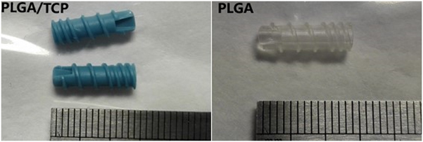
The implants were bioabsorbable PLGA/β-TCP composite anchors from Inion FixOn™ (Inion Oy, Tampere, Finland). The mass ratio of PLGA (proportion of lactic acid (LA) and glycolic acid (GA) is 85:15) and β-TCP is 70:30 (Fig. 1). According to the manufacturer, the size of β-TCP particles is under 1 μm. The control group received implants made of PLGA material, but otherwise, the parameters were consistent with the experimental group. All anchors were sterilized by epoxy ethane (EtO) and packaged in triplets in an aseptic manner.
Anchors are 4.5 mm in diameter (with thread), 14 mm in length.

B. Operation and euthanizing
The animals’ backs were shaved after general anesthesia with intramuscular injection of 3% pentobarbital sodium at a dose of 1 mL/kg body weight. After disinfection and draping the dorsal region in a sterile manner, 3 cm away from the spine, two longitudinal incisions (length: 5–7 cm) were made on both sides of the spine. After the erector spinae was exposed by dissecting the fat tissue, six muscular bags (spacing 2 cm) were prepared through blunt dissection on each side. In each animal, six PLGA/β-TCP composite anchors were put into bags on the right side, whereas six PLGA anchors were put into the left. Incisions were sutured in layers by absorbable suture material. The antibiotic (Penicillin, Potasyum, Turkey, 800000 U intramuscularly daily) was administered for 3 days after operation.
The follow-up times were 6, 12, 16, and 26 weeks, and at each time point, two animals were euthanized with a lethal injection of potassium chloride after sedation. The erector spinae muscles that contained the implanted anchors were carefully dissected. All of the anchors were removed from the soft tissue captures completely, rinsed with double distilled water, and dried in a vacuum drying cabinet for 48 h at 30 °C before further analyses. Three anchors from each group were used to measure their thermal properties [differential scanning calorimetry (DSC)]. The others were used to test their mechanical and further degradation properties.
C. Thermal analysis
A 200F3 DSC (Netzsch Co., Selb, Germany) DSC was used to determine the crystal properties and the glass transition temperature (T g) of the polymers; the temperature range was −20 to 200 °C, and the heating rate was 20 °C/min, in a nitrogen atmosphere. The DSC measurements for each sample were conducted in duplicate to establish reproducibility.
D. Microstructural characterization
Morphological characteristics of surface and longitudinal sections of the samples were monitored through scanning electron microscopy (SEM) photomicrographs (Model JSM-7500F, JEOL, Tokyo, Japan). Before examination, samples were coated with a layer of gold–palladium ∼30 nm thick.
E. XRD
Frozen samples were pulverized in liquid nitrogen, and flat block pieces were evaluated by powder X-ray diffraction (XRD). A PANalytical X-ray diffractometer (PANalytical, Almelo, The Netherlands) was used with CuKa radiation (40 kV and 30 mA), a step size of 0.02° (2θ), and a step time of 1 s. The percentage of crystallinity was calculated using the area of identified crystalline peak divided by the total area of diffraction pattern. Crystallographic measurements were performed in a range of 2θ = 10°–70°, and the area of the identified peak was used for the quantitative analysis by JADE software (MDI, Livermore, California).
F. Mechanical strength
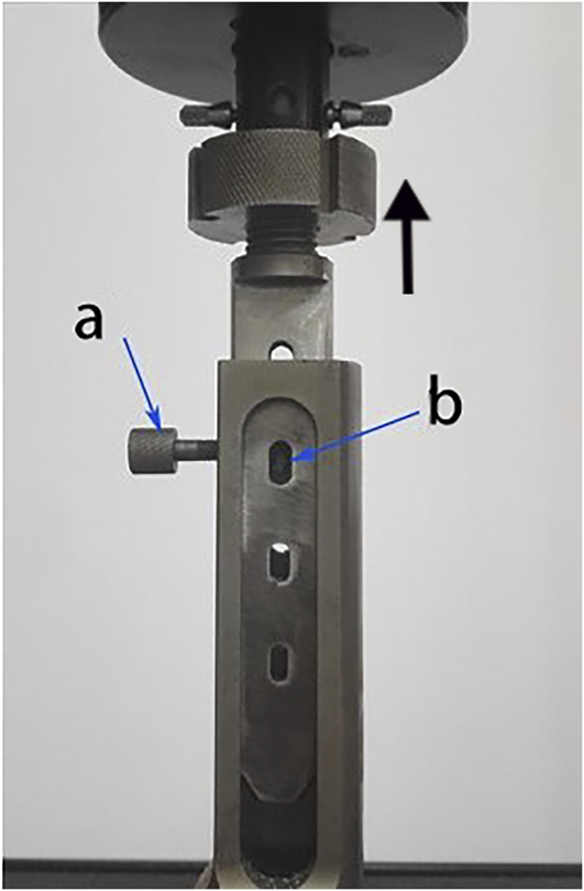
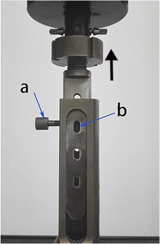
According to the guidelines of the standard ASTM F543, the anchors were subjected to shear strength testing using an Instron 5565 universal material testing machine (INSTRON Co., Norwood, Massachusetts). Anchors were bolted to the testing machine with a pair of forces in opposite axial directions acting on it (Fig. 2). The shear test measured the maximum shear load of the anchors with a constant speed of 5 mm/min until fracture. The maximum load (in newtons, N) was calculated in the form of mean ± SD, and this equation was used to convert the load value (N) into strength (MPa):  $\tau = {{{F_{\max }}} \over S}$ (shear strength τ [MPa]).
Shear strength test using a Instron 5565 universal material testing machine. A retainer bolt (a) is used to fix the anchors (b). The testing speed was 5 mm/min.
G. Statistical analysis
The data obtained from the degradation and mechanical tests were analyzed using statistics software SPSS 19.0. The comparison of differences between the samples in the PLGA/β-TCP and PLGA groups was conducted through one-way analysis of variance (ANOVA), and the comparison of samples of different timepoints in the same group was conducted through S–N–K (Student–Newman–Keuls) test, with statistical significance set at P < 0.05.
III. RESULTS
A. PLGA/β-TCP showed more stable shear strength retention and sharply declined after 12 weeks
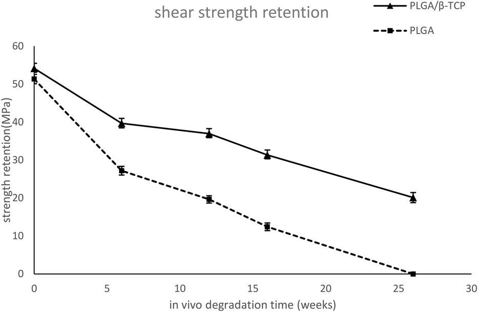
The shear strength retention of the materials at different follow-up times is shown in Fig. 3. The PLGA anchors at 26 weeks in vivo had disintegrated and their shear properties could no longer be measured. The strength retention of PLGA/β-TCP group (54.15 ± 1.05 MPa) was 5.49% higher than the PLGA group (51.33 ± 1.23 MPa) at 0 week. The difference became much more significant at later timepoints; at 16th week, the shear strength of PLGA/β-TCP (31.32 ± 1.00 MPa) was 152.04% higher than the 12.42 ± 1.00 MPa of PLGA, suggesting that the addition of β-TCP enhanced the mechanical strength of the composite; thus, the decreasing tendency of the pure PLGA group was larger than that of the PLGA/β-TCP group. For the PLGA/β-TCP anchors, the strength retention at the 12th week did not reduce significantly compared with the 6th week, suggesting that PLGA/β-TCP were more stable. The decline of strength of PLGA/β-TCP accelerated after 12 weeks; we hypothesized that this change was caused by the disappearance of the enhanced effect of β-TCP.
Shear strength retention of the PLGA/β-TCP and the plain PLGA anchors: strength retention of the PLGA/β-TCP groups was significantly higher than the PLGA group; the decreasing tendency of the pure PLGA group was larger than that of the PLGA/β-TCP group; and reduction of shear strength retention stayed stable from 6th to 12th week and then accelerated at 16th week.

B. XRD, DSC, and SEM: After 12th week, the connection between two materials in the composite became less compact
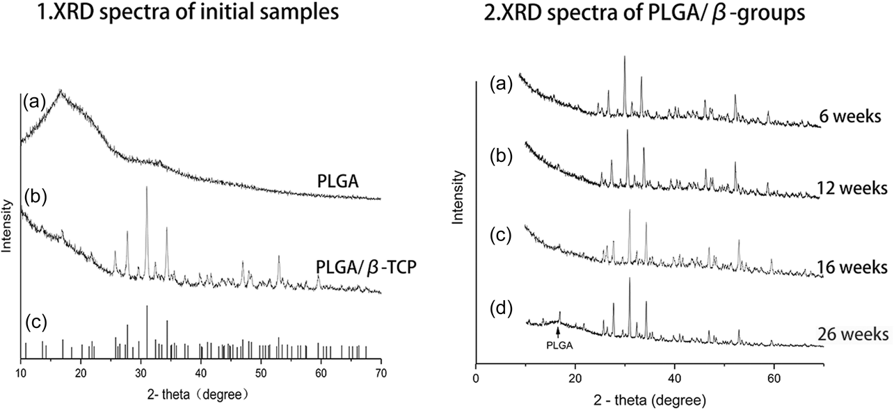
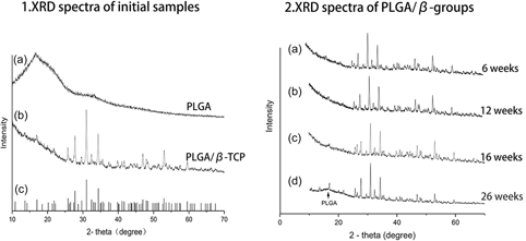
The XRD spectrum of the initial PLGA sample [Fig. 4(1a)] shows a typical broad signal of an amorphous substance in the range of 10–28° (2θ), indicating that PLGA is in a totally amorphous state. Spectra of all PLGA/β-TCP samples showed high and sharp XRD peaks, indicating that β-TCP had a perfect crystallinity [Fig. 4(1b)]. The main diffraction peak can be assigned to the structure of Ca3(PO4)2 (PDF card No. 09-0169).
XRD analysis: 1. (a) XRD spectrum of the initial PLGA sample: a typical broad signal in a range of 10–28°, the peak of PLGA was at Theta 17°. (2θ) showed PLGA was in a totally amorphous state. (b) XRD spectrum of the initial PLGA/β-TCP sample. (c) The main diffraction peak could be assigned to the hkl of Ca3(PO4)2 (PDF card No. 09-0169). 2. XRD spectra of the PLGA/β-TCP samples at various timepoints: peaks of PLGA (2θ = ∼17°) were becoming significant at 26th week [2(d) arrow], and the intensity of the β-TCP peak (2θ = 25–35°) also increased.

In the comparison of the XRD curves at 6, 12, 16, and 26 weeks, broad signal of amorphous PLGA (at Theta 17°) was becoming more significant at 26 weeks [Fig. 4(2d)], and the intensity of the β-TCP peak (at Theta 25–35°) also increased [Fig. 4(2)]. This phenomenon may infer that the connection between β-TCP and PLGA became less compact during the degradation progress.
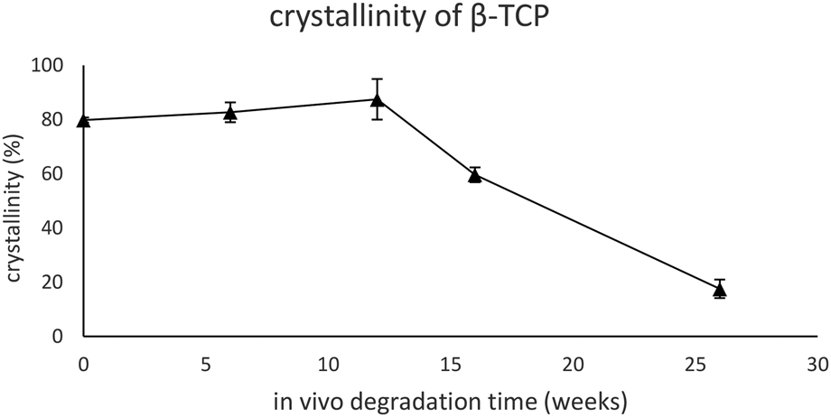
The crystallinity of β-TCP slowly increased from 79.8 ± 0.95% to 87.47 ± 7.48% during the first 12 weeks without significant differences, but decreased to 20% in the following 14 weeks (Fig. 5).
Crystallinity value of β-TCP at various timepoints: the crystallinity had slightly increased during the first 12 weeks and sharply declined from 12th week to 26th week.

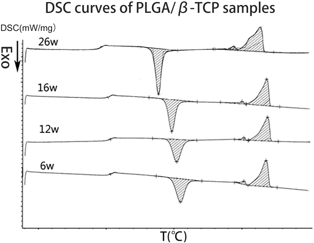
At each time point, the glass transition temperature (T g) of the PLGA/β-TCP was significantly higher compared with that of the PLGA. All the DSC spectra had a exothermic peak before melting; considering that the temperature of DSC analysis is far lower than β-TCP phase transition temperature, we attributed the exothermic peak to the cold crystallization of the amorphous PLGA. The cold crystallization peak of the initial PLGA/β-TCP composite was broader and shorter compared with that of the plain PLGA, suggesting that the β-TCP particles in the composite had slow down the speed of crystallization of PLGA. Crystallinity of PLGA of the composite is increased from 0.17 to 2.16%. At all timepoints, the areas of the cold crystallinity peak of the PLGA/β-TCP had no significant difference, suggesting that during degradation, the crystallinity of PLGA in the composite could be ignored. At 26 weeks, the cold crystallization peak became significantly sharper and higher (Fig. 6), which was similar with the pure PLGA group, indicating that the remaining PLGA became easier to crystallize, which further proved that the connection between β-TCP and PLGA became less compact during the degradation progress at later timepoints.
DSC curves of PLGA/β-TCP anchors at 6, 12, 16, and 26 weeks: at 26 weeks, the cold crystallization peak became significantly sharper and higher during degradation.

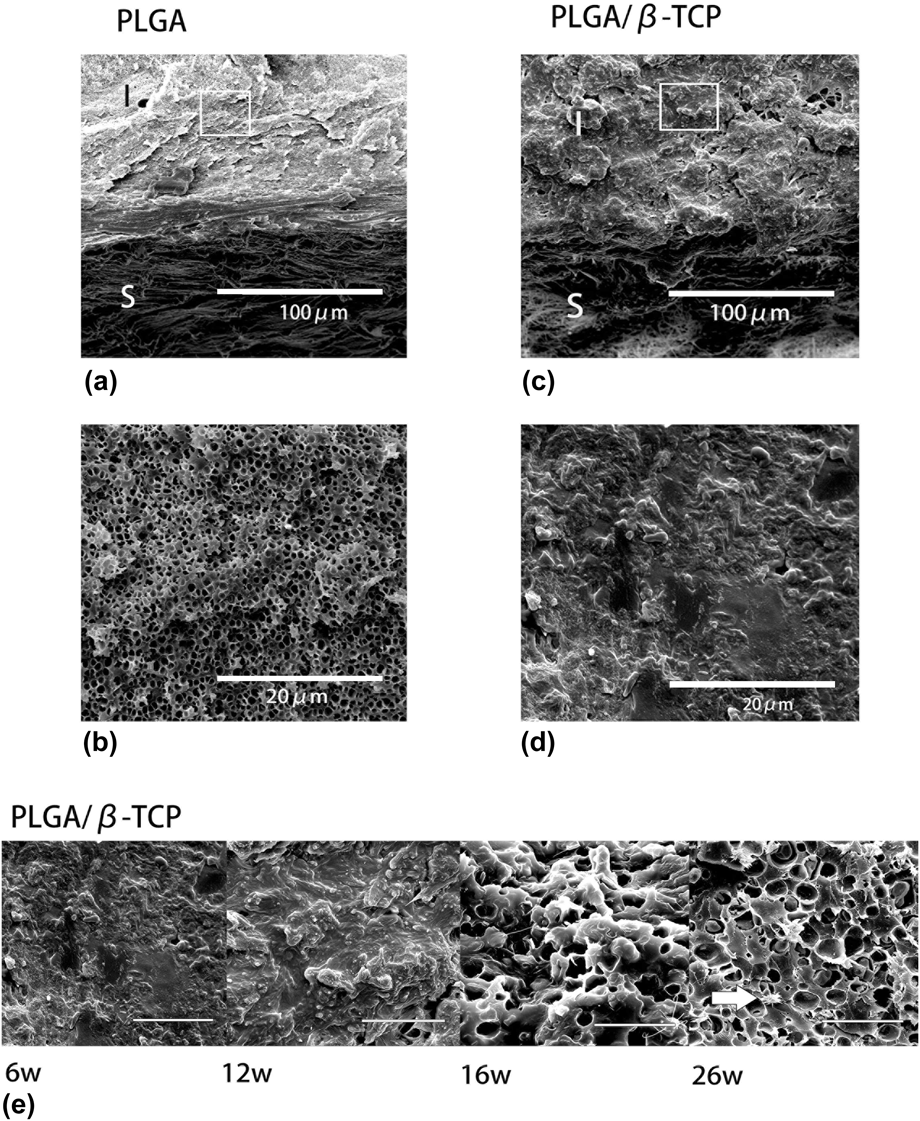
According to the SEM analysis of the surface and longitudinal section of the PLGA/β-TCP composite and plain PLGA samples, the initial structures of both materials were similar. In the composite anchors, β-TCP particles have a dispersed distribution in amorphous PLGA. Six weeks after implantation, we observed the formation of micropores in the interior of the plain PLGA sample [Figs. 7(a) and 7(b)]. Surface center segregation can be observed in the longitudinal section of the PLGA after 12 weeks in vivo. We did not observe similar micropores in the interior of PLGA/β-TCP composite samples at 6 and 12 weeks in vivo [Figs. 7(c)–7(e)]; these samples were eroded from the outside toward the center. Micropores began to appear in the inner part of the PLGA/β-TCP composite until 16 weeks in vivo. Hydroxyapatite (HAP) crystals, degradation products of TCP, were observed on the surface of the β-TCP particles at 16 weeks, confirming that the β-TCP particles in the interior of anchor began to degrade. Micropores in the composite were larger than that in the plain PLGA. After 26 weeks, more micropores appeared inside the composite and fewer β-TCP particles could be seen, and the structure seemed much like the early degradation structure inside plain PLGA [Fig. 7(e)]; this outcome may be explained because the β-TCP particles are dissolved and released into the body fluids once they lose the packaging of PLGA. The observation also agreed with the result of XRD and DSC analysis that in the later period of degradation, the content of β-TCP in the composite declined.
(a and b) Longitudinal section of the PLGA anchor at 6 weeks: at 6 weeks in vivo degradation, the formation of micropores in the interior of the plain PLGA sample was observed. (S: surface, I: interior); (c and d) Longitudinal section of the PLGA/β-TCP anchor at 6 weeks: we did not observe similar micropores in the interior of the PLGA/β-TCP composite samples at 6 weeks in vivo, the samples were eroded from the outside toward the center. (e) Longitudinal sections of the PLGA/β-TCP anchor at 6, 12, 16, and 26 weeks, interior parts: similar micropores were not observed in the PLGA/β-TCP composite samples at 6 and 12 weeks in vivo. Micropores began to appear in the inner part of the PLGA/β-TCP composite until 16 weeks in vivo. The remaining matrix at 26 weeks was similar to the interior part of the plain PLGA; HAP crystals were observed on the surface of the β-TCP particles (arrow). Scale bar, 20 μm.

IV. DISCUSSION
The purpose of this study is to investigate the degradation mechanism of the PLGA/β-TCP and to discuss the potential relationship between the β-TCP enhancement effect and the reduction of mechanical strength.
The addition of β-TCP can significantly enhance the mechanical strength of PLGA. Lin et al.Reference Lin, Wang, Zhou and Qian19 reported that an increasing in bending strength for poly-DL-lactide (PDLLA) rods with TCP adds up to 50% and they attributed the increase of strength to the effect of crack deviation by TCP particles. In terms of shear strength in this study, the strength retentions of the PLGA/β-TCP anchors show no significance between 6 and 12 weeks, and the decreasing rate accelerates after 12 weeks. The overall results indicate that after 12 weeks of in vivo degradation, the β-TCP particles begin to detach from the PLGA matrix, so that the reduction of mechanical strength becomes more obvious at the same period. After being placed subcutaneously for 26 weeks, the PLGA anchors became fragmented and there was practically no shear strength left, whereas the PLGA/β-TCP anchors kept their strength at a relatively high level of 31.31 Mpa (57.82% of the initial shear strength). The lower degradation rate can prolong the duration of the material with high mechanical properties. The gradually declining shear strength of PLGA/β-TCP is also ideal for preventing stress shielding effects because of the gradual shift of the force onto the healing bone.
XRD results showed that PLGA is in a totally amorphous state, and DSC results of composite indicated that the crystallinity of PLGA in the composite could be ignored. Thus, the crystallinity of composite is contributed only by β-TCP. The crystallinity of the composite can represent β-TCP content relatively. Although β-TCP is resorbed much faster in vivo than other calcium phosphates,Reference Jensen, Bornstein, Dard, Bosshardt and Buser22–Reference Nandi, Fielding, Banerjee, Bandyopadhyay and Bose24 the crystallinity of β-TCP was slightly increasing during the first 12 weeks without significance, and this may be explained by the fact the amorphous part of β-TCP dissolves first. Once the β-TCP particles start to degrade, the physical connection between the β-TCP particles and PLGA break apart; in addition, some particles are released from the polymer matrix into the body fluids so that the crystallinity significantly declines from 12 to 26 weeks. In the comparison of the XRD curves at 12, 16, and 26 weeks, peaks of PLGA (at Theta 17°) were becoming more significant over time. It may cause by an increasing of PLGA content in the composite.
Some polymers are reported to have two progresses occurring simultaneously during degradation: recrystallization and fracture of crystallinity. As generally accepted, degradation reactions of semicrystalline polymers proceed predominantly in amorphous regions.Reference Obadal, Čermák, Raab, Verney, Commereuc and Fraïsse25 In this study, XRD and DSC were used to measure the crystallinity of initial pure PLGA samples and show a complete amorphous state (crystallinity: 0.13%). DSC was also used to measure the crystallization of PLGA in the PLGA/β-TCP composite during degradation and found it increased to 2% at 26 weeks, but we consider that it did not show a significant difference and contributed very limit impact to the degradation. The DSC results suggested that the addition of β-TCP particles slows down the crystallization speed of amorphous PLGA in the initial composite. At the 26-week time point, the cold crystallization peak became significantly sharper and higher whereas its area did not change much, showing that the cold crystallization speed was significantly increasing, which suggests an easier tendency to crystallize PLGA in the composite during DSC test. This tendency may also be caused by a declination of the β-TCP particles, which have detached from the PLGA matrix and dissolved into body fluid.
Results of XRD and DSC both indicated that after 12 weeks, β-TCP particles have detached from the composite and remain a structure basically consist of PLGA matrix. We used SEM to observe the visualized changes in the composite and pure PLGA during degradation.
According to the SEM observation of the surface and longitudinal section of the PLGA/β-TCP composite and plain PLGA samples, the initial structures of both materials were similar. In composite anchors, micron-sized β-TCP particles had a dispersed distribution in amorphous PLGA. Six weeks after implantation, we observed the formation of micropores in the interior of the plain PLGA sample. This structural change during degradation agreed with the conclusion of a previous study that in the hydrolysis of an amorphous polymer, water penetrates the whole structure and begins to shorten the molecular chains all over the material.Reference Nieminen, Rantala, Hiidenheimo, Keranen, Kainulainen, Wuolijoki and Kallela21 For the degradation of aliphatic polyesters, degradation products are formed both at the surface and in the inner part of the material. Those carboxylic end groups localized inside the specimens are easy to concentrate, and thus, they catalyzed ester degradation, resulting in a surface center segregation.Reference Bostman, Hirvensalo, Makinen and Rokkanen6,Reference Therin, Christel, Li, Garreau and Vert26 This segregation can be observed in the longitudinal section of pure PLGA after 12 weeks.
We did not observe similar micropores in the PLGA/β-TCP composite samples at 6 or 12 weeks in vivo (Fig. 7); the samples were eroded from the outside toward the center. There are two factors to be considered for the delayed degradation of the PLGA/β-TCP composite. At the initial stage of degradation, water is blocked by the β-TCP particles and could only penetrate into the surface, and therefore the degradation was also limited to the surface.Reference Lin, Chen, Lin and Lee27 This agrees with the nonmicropores structure observed in the inner part of the PLGA/β-TCP composite at 6 and 12 weeks. The second reason is that when the TCP began to dissolve, the basic products of β-TCP compensated the acidity degradation products of the PLGA, preventing the inner autocatalytic effect of PLGA degradation.Reference Taylor, Daniels, Andriano and Heller11
Micropores begin to appear in the inner part of the PLGA/β-TCP composite at 16th week. HAP crystals were observed on the surface of the β-TCP particles [Fig. 7(e)]. When the β-TCP particles were dissolved, the Ca2+ and PO43− diffused out, HAP crystals are formed on the surface of β-TCP when the two ions reach a saturation concentration and recrystallize.Reference van Blitterswijk, Hesseling, Grote, Koerten and de Groot28 Micropores in the composite were larger than that in the plain PLGA; these may be caused by the water block effect of the β-TCP particles. The water blocked by β-TCP and hydrolysis reactions are limited to one area, resulting in larger pores. Another explanation is that the pores are caused by the separation of micron-sized β-TCP particles. β-TCP is resorbed much faster in vivo than other calcium phosphates.Reference Jensen, Bornstein, Dard, Bosshardt and Buser22,Reference Jensen, Yeo, Dard, Hunziker, Schenk and Buser23,Reference Velu and Singamneni29,Reference Velu, Kamarajan, Ananthasubramanian, Ngo and Singamneni30 When β-TCP particles contact the body fluid and begin to dissolve, the connection between β-TCP particles and amorphous PLGA is broken, and thus, some β-TCP particles can be released into the fluid. In the PLGA/β-TCP composite after 26 weeks in vivo, the structure of the longitudinal section was similar to that of the plain PLGA at 6 weeks; the pores were more intense, and the β-TCP particles were sparser. This demonstrated that more β-TCP particles were dissolved or released from the polymer matrix, and it agrees with the decline of crystallization at 26 weeks evaluated by XRD.
When the β-TCP detached from the matrix polymer, the delay degradation effect will also weaken. SEM observation at 26 weeks showed that a very open and porous structure was left so that water could easily access. We hypothesize that the degradation rate after 26 weeks will become even faster. Subsequent long-term experiments are ongoing to detect the complete degradation of the composite anchors.
V. CONCLUSIONS
The addition of β-TCP particles efficiently enhances the mechanical properties and thermal characteristics and delays the degradation rate of the anchor. After analysis of the results by XRD, DCS, and SEM observation, we can infer that in the first phase of degradation, hydrolysis is blocked by the β-TCP particles and can only occur at the very surface, and the basic products of TCP prevent the inner autocatalytic effect of PLGA degradation.Reference Taylor, Daniels, Andriano and Heller11 Once the β-TCP particles start to degrade, the physical connection between the β-TCP particles and PLGA breaks apart, and some particles may be released from the polymer matrix into the body fluid. The loss of β-TCP particles and the acceleration of strength reduction both onset at 16 weeks. We hypothesize that the degradation rate after 26 weeks will accelerate. Long-term in vivo experiments are ongoing to detect the complete degradation of the PLGA/β-TCP anchors.
ACKNOWLEDGMENTS
This work was supported by the National High-tech Research and Development Program (863 Program), No. 2015AA033701. Samples were gifts given by Beijing Naton Technology Group Co., Ltd., Beijing.