I. INTRODUCTION
Discovery of ferromagnetism (FM) in Mn-doped gallium arsenide (GaAs),Reference Ohno, Shen, Matsukura, Oiwa, Endo, Katsumoto and Iye1 has drawn considerable interest in dilute magnetic semiconductors (DMSs) for the development of spintronics devices.Reference Prinz2–Reference Wolf, Awschalom, Buhrman, Daughton, Molnar, Roukes, Chtchelkanova and Treger4 However, the observed Curie temperature (T C) ∼ 172 K in Mn-doped GaAs system was far below the room temperature value (required for any practical device application). Later, the theoretical prediction of room temperature ferromagnetism (RTFM) in Mn-doped zinc oxide (ZnO) systemReference Dietl, Ohno, Matsukur, Cibert and Ferrad5 and the experimental observation of RTFM in cobalt doped TiO2 filmReference Matsumoto, Murakami, Shono, Hasegawa, Fukumura, Kawasaki, Ahmet, Chikyow, Koshihara and Koinuma6 have boosted the hope that wide band-gap oxide system doped with a few atomic percent of transition metal (TM) such as TM (Co,Mn):TiO2Reference Bhattacharyya, Pucci, Zitoun and Gedanken7, Reference Tian, Yuan, He, Wang, Li, Xie, Liu and Yin8 would be the potential candidate for the spintronic devices. The reports from literature so far suggest that the RTFM in TM:TiO2 may originate from precipitation of magnetic metallic clusters or due to the presence of secondary magnetic phases.Reference Pinto, Cruz, da Silva, Alves and Godinhoa9, Reference Kim, Park, Park, Noh, Oh, Yang, Kim, Bu, Noh, Lin, Hsieh and Chen10 On the contrary, some other reports support the idea that the observed RTFM in TM:TiO2 is an intrinsic property.Reference Griffin, Pakhomov, Wang, Heald and Krishnan11–Reference Li, Wu, Hu, Xing, Liu, Yu, Yang, Lu, Li and Liu13 Thus the controversy over the origin/nature of observed FM in TM:TiO2 system is still persisting. Further, the direct exchange or superexchange interaction is attributed for magnetic coupling in DMS that is insulating in nature.Reference Janish, Gopal and Spaldin14 In fact, the different microstructures may be responsible for different mechanisms of FM ordering even in the same material. Kasper et al.Reference Kaspar, Heald, Wang, Bryan, Droubay, Shutthanandan, Thevuthasan, McCready, Kellock, Gamelin and Chambers15 have reported the presence of distinct ferromagnetism in chromium-doped titania (Cr:TiO2) films that were of bad quality, whereas the films exhibiting excellent structural quality have shown negligible ferromagnetism. Kim et al.Reference Kim, Park, Lee, Choia, Lee, Kim and Park16 have recently reported that the existence of oxygen vacancy is necessary for RTFM in Mn-doped rutile TiO2 thin films. Doping of Mn was also found not to play any key role in inducing the FM attribute in TiO2 thin film.Reference Hong, Sakai, Ruyter and Brize17 Furthermore, it has been reported that the FM depends on oxygen vacancies and/or defects in the TM:TiO2 system and the FM ordering is explained in terms of bound magnetic polarons (BMPs).Reference Coey, Venkateshan and Fitzgerald18 This inconsistency over the origin of FM ordering in DMSs might be due to a sensitive dependence of sample preparation conditions, and there is still no definite agreement so far about the nature of magnetic properties of DMSs prepared by different methods and different groups.
In the present work, highly resistive thin films of Ti1−xMnxO2 (0 ≤ x ≤ 0.15) were deposited on quartz substrates by spray pyrolysis technique, and an attempt has been made to correlate the structural, electronic structure and optical properties of these films with their magnetic properties. It is worth mentioning here that the technique used for the deposition of the Mn-doped film is simple (not requiring any vacuum) and cost-effective as compared with the other techniques, and the observed ferromagnetism was found to be comparable in terms of the values in other reports.
II. EXPERIMENTAL DETAILS
Thin films of Ti1−xMnxO2 (x = 0.00, 0.05, 0.10, and 0.15) were deposited by a simple and cost-effective spray pyrolysis technique. For the deposition of undoped TiO2 film titanyl acetylacetonate (C10H14O5Ti) precursor was first dissolved in ethanol. The solution was then sprayed using N2 as a carrier gas at a pressure of 0.4 kg/cm2 on the ultrasonically cleaned fused-quartz substrates maintained at ∼450 °C. Likewise, doped samples of Ti1−xMnxO2were deposited using a solution containing prerequisite amounts of manganese acetate tetrahydrate along with titanyl acetylacetonate. Every time 80 mL solution was sprayed over the substrate with a flow rate of 0.25 mL/s.
Spectroscopic ellipsometer (Model M-2000F, M/S J. A. Woolam Co., Inc., Lincoln, NE) was used to estimate the thickness of the films. The films were characterized for their phase identification and particle size by glancing angle x-ray diffraction (GAXRD) with GA = 1° using a Philips x-ray diffractometer (Model X’pert Pro, PANalytical, Almelo, Netherlands). An electrometer (Model 617, Keithley Instruments, Inc., Cleveland, OH) was used to estimate the electrical resistivity of these films at room temperature using two-probe mode of resistivity measurement. Silver paste was used to make electrical contacts between the fine copper wires and the films for the resistivity measurement. The magnetic measurements were carried out by a superconducting quantum interference device (SQUID) (MPMS XL 7, Quantum Design, San Diego, CA) in RSO (reciprocating sample option) mode with sensitivity of 10−8 emu. Measurement of normal transmittance (T) as a function of incident wave length varying from 1000 to 200 nm was carried out at room temperature using a double beam spectrophotometer (Model Lambda 900, PerkinElmer, Waltham, MA) having a resolution of 0.05 and 0.2 nm in the ultraviolet-visible (UV–VIS) and near infrared (NIR) ranges, respectively. Compositional analysis and oxidation states of Mn in Ti1−xMnxO2 films were estimated by x-ray photoelectron spectroscopy (XPS; Model SPECS) equipped with Mg-Kα(1253.6 eV) x-ray source. High-resolution transmission electron microscope (HRTEM) (Technai G20-stwin, FEI, Eindhoven, Netherlands) was used to study the particle size distribution and for microstructural analysis of the films.
III. RESULTS AND DISCUSSION
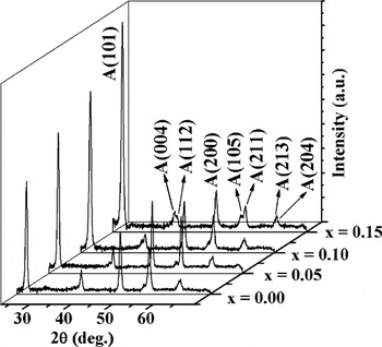
Thickness of Ti1−xMnxO2 thin films with x = 0.00, 0.05, 0.10, and 0.15 was estimated to be in the range of 300–500 nm. Figure 1 shows the x-ray diffractograms recorded on these films. It is observed that all the XRD peaks correspond to phase pure tetragonal anatase structure of TiO2 [matched with the Joint Committee on Powder Diffraction Standards (JCPDS) card No. 21-1272]. No peak related to Mn clusters or any other secondary phase including MnO2, etc., could be seen in any of the diffractograms suggesting the incorporation of Mn in TiO2 film matrix. Incorporation of Mn in TiO2 matrix is further supported by the decrease in lattice parameter and unit cell volume. As the Mn concentration (x) in Ti1−xMnxO2 thin films is raised from 0.0 to 0.15 the decrement in both, lattice parameter “c” and unit cell volume (V), was estimated to lie in the range of 9.470–9.424 Å and 135.240–134.440 Å3, respectively. The estimated lattice parameter “a,” however, did not show any noticeable change with increase in “x.” Contraction in the lattice of TiO2 due to the incorporation of Mn in film matrix can be explained in terms of ionic radii of Mn and Ti ions. For a coordination number of 6 the radius of Mn+3 ion of 0.58 Å (the radius for Mn+4 ion is 0.53 Å and for Mn+2 ion is 0.82 Å) is smaller than the ionic radius of Ti+4, i.e., 0.61Å.Reference Lide19 Thus, the estimation of lattice parameters from x-ray diffractograms suggests that Mn+3 is the most probable ionic form of Mn substituting Ti in TiO2 matrix. However, the presence of Mn+2 and Mn+4ions in these films cannot be ruled out merely on the basis of XRD.
X-ray diffractograms of Ti1−xMnxO2 films for different concentration of Mn (x = 0.00, 0.05, 0.10, and 0.15).

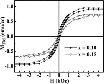
As-recorded isothermal magnetic moment versus field (m–H) curves at room temperature for Mn-doped TiO2 films with “x” = 0.10 and 0.15 exhibited nonlinear m–H behavior in low-field region suggesting the presence of ferromagnetism in these films.Reference Sharma, Chaudhary, Kashyap and Sharma20 Corresponding magnetization versus field plots (in units of emu/cc) for the extracted FM fraction of 10 and 15 at.% Mn-doped TiO2 films at room temperature (within ±4 kOe range) are shown in the Fig. 2. On the other hand, isothermal m–H measurement of substrate and Ti1−xMnxO2 thin films with x = 0.00 and 0.05 exhibited only diamagnetic signal at room temperature even in the low-field region. It is worthwhile to note here that none of the oxide phases of Mn or Ti/Mn oxide (such as MnTiO3, Mn2TiO4, and MnTi2O4) can account for the observed FM fraction of Mn-doped TiO2 films with “x” = 0.10 and 0.15, since none of these is ferromagnetic except Mn3O4 and some Mn–Ti phases.Reference Wang, Tang, Chen, Spinu, Zhou and Tung21 Mn3O4 and Mn-Ti phases are reported to have Curie Temperature T C ∼ 40 K.Reference Wang, Tang, Chen, Spinu, Zhou and Tung21 So all these Mn–Ti related phases, including all the oxides of Mn or Mn/Ti cannot be responsible for observed FM ordering at room temperature. Coercivity values of around 85 and 140 Oe, estimated for Mn-doped films with “x” = 0.10 and 0.15, respectively, further confirm the presence of FM ordering at room temperature in these samples. However, the saturation magnetization (MSFM) tends to decrease from 0.938 to 0.716 emu/cc (or 0.035 to 0.017 μB/Mn) as the concentration of Mn is increased from “x” = 0.10 to 0.15 suggesting partial or reduced incorporation of Mn ions in TiO2 lattice above “x” = 0.10. There might be two possible causes for the observed decrease in magnetization for x = 0.15. First, the solid solubility limit of Mn in TiO2 matrix is limited and is somewhat below 15 at.% and second, the increased antiferromagnetic superexchange interaction between Mn moments at higher quantum of incorporation of Mn, which could explain the observed decrease of saturation magnetization.Reference Ding, Liu, Yang and Li22 Hence, it is proposed that some kind of magnetic exchange interaction among the Mn ions incorporated in TiO2 lattice might be responsible for observed ferromagnetic ordering in these Mn-doped TiO2 thin films. Absence of ferromagnetism in low Mn concentration x = 0.05 suggests that either the interaction between Mn moments is not strong enough to initiate FM ordering even if Mn ions substitute for Ti in TiO2 matrix, or there is some kind of percolation threshold required for Mn moments to order ferromagnetically. The observed values of MSFM in these films are quite comparable to the value (0.019 μB/Mn ion) observed by Tian et al.Reference Tian, Yuan, Wang, He, Yin, Liu, Yuan and Liu23 in Mn-doped TiO2 bulk samples prepared by solid-state reaction method albeit lower than the value (5.7 emu/cc) recently reported by Sellers et al.Reference Sellers and Seebauer24 in Mn-doped anatase TiO2 film prepared by atomic layer deposition (ALD) technique. It may be mentioned here that, according to a previous report,Reference Hong, Sakai, Ruyter and Brize17 the Mn doping does not play any key role in inducing FM ordering in TiO2thin films. However, these authors found oxygen vacancies and/or defects mediated FM ordering in undoped film, which is contrary to the result observed for undoped film in present study.
Extracted MFM–H curves of Ti1−xMnxO2 with “x” = 0.10 and 0.15 at 300 K.

In a previously reported detailed magnetization investigations of these series of samples,Reference Sharma, Chaudhary, Kashyap and Sharma20 it was found that Ti1−xMnxO2 film having x = 0.10 exhibited (i) distinct thermomagnetic irreversibility (TMI), i.e., the signature of FM ordering and (ii) small kink around 43 K in zero field cooling (ZFC) curve possibly due to the formation of ferrimagnetic Mn3O4 (that could not be detected by XRD either due to its small volume-fraction or it being amorphous). Presence of Mn3O4 (in which Mn is present in mixed oxidation states of +3 and +2) in doped films is further supported by the XPS measurement of Ti1−xMnxO2 thin films (shown in Ref. Reference Sharma, Chaudhary, Kashyap and Sharma20) whereas no signature of elemental Mn or Mn+4could be evidenced in the present study.
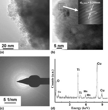
To further probe the origin of observed RTFM in Mn-doped TiO2 film, HRTEM and TEM measurements were performed on Ti1−xMnxO2 film with x = 0.10. For the TEM analysis we have deposited the sample directly on the carbon-coated copper grids. Figure 3(a) shows the TEM micrograph of the x = 0.10 film. It can be seen from the figure that the particles in the film are of irregular shape, and have size of ∼25 nm. Dark-shaded region in the micrograph is due to the relatively thick portion of the sample loaded on the TEM grid. Careful and numerous HRTEM observations [as shown by the squares in Fig. 3(b)] taken on different places on the grid did not reveal any evidence of the presence of metallic clusters of Mn excepting the anatase phase of TiO2. This is in agreement with the XRD results of Mn-doped TiO2 films. Thus all the XRD, XPS, and TEM investigations suggest that observed RTFM could not have originated either due to extrinsic reasons or due to any other secondary phase. The diffused spotty SAED pattern [Fig. 3(c)] indicates the polycrystalline nature of the film. To identify the Mn ions in the film, energy-dispersive analysis of x-rays (EDAX) attached with TEM was also performed and the recorded pattern is shown in Fig. 3(d). Quantitative analysis of the pattern revealed 8.58 at.% concentration of Mn ions in the film, which is in close proximity to the added concentration (10 at.%) via Mn precursor.
TEM analysis of the Mn-doped TiO2 polycrystalline thin film having “x” = 0.10; (a) overall particle morphology of the film, (b) lattice fringes of anatase phase structure, (c) SAED pattern, and (d) corresponding EDAX spectrum of the film.

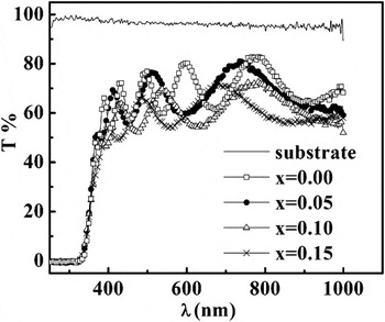
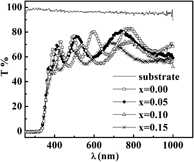
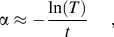
Figure 4 shows the optical transmittance (T) spectra of Ti1−xMnxO2 thin films along with the transmittance of substrate used. It can be seen from the spectra that all the films are highly transparent in the visible region and the maximum transmittance decreases as the concentration of Mn is increased. Transmittance decreases rapidly as the wave length reaches the ultraviolet region and a sharp absorption edge is observed around ∼360 nm for all the films. To estimate the optical band gap E gof the films, absorption coefficient α is calculated near the absorption edge according to the relation:
 $${\rm{\alpha }} \approx - {{\ln (T)} \over t},$$
Optical transmittance spectra of Ti1−xMnxO2 films for different concentrations of Mn (x = 0.00, 0.05, 0.10, and 0.15).

where “t” is the thickness of the film. Optical band gap E gcan be derived from the expression as
 $${\rm{\alpha }}(h\nu ) = B(h\nu - E_{\rm{g}} )^n ,$$
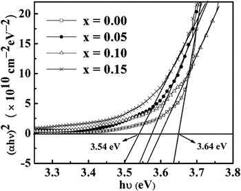
where  $h\nu $ is the incident photon energy, B is a constant, and n = ½ for direct allowed transition. The value of E g can be estimated by extrapolating the linear portion of Tauc’s plot to the photon energy axis as shown in Fig. 5. The estimated value of E g varies from 3.64 to 3.54 eV as the concentration of Mn is increased from “x” = 0.00 to 0.15, however, the variation in E g is not much dependent on Mn concentration incorporated in TiO2films.
Tauc’s plots of Ti1−xMnxO2 films for various Mn concentrations “x.”

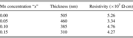
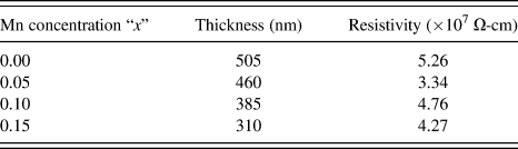
Electrical resistivity measurement performed at room temperature only (through the two-probe instrument) revealed very high resistivity (ρ ∼ 107 Ohm-cm) of all these samples. The data is summarized in Table I for all the samples along with the thicknesses of various films. Nearly unchanged high electrical resistivity of these films in the present case suggests that there are very few free carriers for conduction as compared with the Mn ions. We know that the oxygen vacancies are created in TiO2 lattice when lower valence Mn ions are substituted for higher valence Ti+4 ions for the sake of charge neutrality within the lattice. It is reported that a majority of oxygen vacancies, which are created by the incorporation of lower valence Mn ions at higher valence Ti+4 ion sites, might remain localized near substitutional dopant ion sites. As a result of above, unlike the nonlocalized oxygen vacancies, these localized oxygen vacancies do not provide the free electrons for conduction in the TiO2 lattice.Reference Griffin, Pakhomov, Wang, Heald and Krishnan11, Reference Ogale25 and hence the lattice remains highly resistive even after the creation of oxygen vacancy due to the substitution of Mn ion at Ti ion site.
Electrical resistivity data of Ti1−xMnxO2films measured at room temperature along with the thickness of the films.
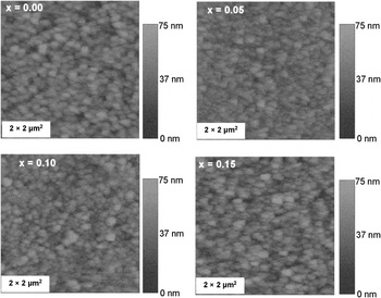
Surface morphological study of Ti1−xMnxO2 (x = 0.00, 0.05, 0.10, and 0.15) thin films suggest that all the films have densely packed microstructure (Fig. 6). The morphology and microstructure of the films do not change significantly with Mn incorporation in TiO2matrix. This is understandable since all the deposition parameters such as temperature of substrate, pressure of carrier gas, and flow rate of the solution were kept nearly identical over the same substrate (quartz), so no significant change in morphology and microstructure with Mn addition could be seen. However, the root mean square (RMS) value of roughness (7 nm) of the pristine film is found to register a small increase to 11 nm with increase in Mn concentration.
Surface morphological micrographs of Ti1−xMnxO2 films for various Mn concentrations “x.”

Now, we switch the focus of our discussions to the possible mechanism of FM ordering in these films. No unique theory has so far been developed to satisfactorily explain the observed FM ordering in all DMS oxides. The best description of observed RTFM behavior in the present case of highly insulating (ρ ∼ 107 Ohm-cm) Ti1−xMnxO2 films is given by BMP model.Reference Kaminski and Sarma26 A BMP is constituted when an electron is trapped in oxygen vacancy/defects and these vacancy/defects are coupled with the moments of Mn within their hydrogen-like orbit around the vacancy/defects. Now, there might be two kinds of possible contribution to magnetic behavior of Ti1−xMnxO2 films: (i) probability of overlapping of two BMPs leading to FM ordering in the oxide lattice and (ii) paramagnetic contribution arising due to noninteracting Mn moments. Thus the net magnetization of the sample will be the result of competition between these two factors. Dominating paramagnetic contribution from noninteracting Mn moments at higher concentrations could be responsible for the observed decrease in saturation magnetization of Ti1−xMnxO2 films as the concentration of Mn is increased from “x” = 0.10 to 0.15.
IV. CONCLUSIONS
Thin films of Ti1−xMnxO2 (x = 0.00, 0.05, 0.10, and 0.15) have been deposited on quartz substrates by spray pyrolysis technique. X-ray diffraction patterns established phase pure anatase structure for all the films along with Mn ion substituting Ti ions, and no trace of elemental Mn clusters or any other secondary phases such as oxides of Mn were seen in any of the diffractograms. This was further confirmed by HRTEM measurement. However, magnetic measurement suggests the formation of ferrimagnetic Mn3O4 (T C ∼ 40 K) indicating partial incorporation of Mn in TiO2 lattice. Further, weak FM ordering at room temperature is observed in Ti1−xMnxO2 thin films with x > 0.05. The observed ferromagnetism in these highly insulating samples is best described within the scope of the BMP model.
ACKNOWLEDGMENTS
The present work was supported by Department of Science and Technology, New Delhi via funding for the SQUID facilities (available at IIT Delhi). One of the authors (S.S.) would like to thank the University Grant Commission (UGC) for providing the Senior Research Fellowship.