Pulmonary artery sling is a malformation that occurs due to an anomalous origin of the left pulmonary artery from the right pulmonary artery. Typically, in the setting of pulmonary sling, the aberrant left pulmonary artery crosses the trachea posteriorly and courses between the trachea and the oesophagus. In effect, the tracheo-bronchial tree may be compressed.
As already described in the literature, the anatomy of the pulmonary artery branches can vary in different forms,Reference Erickson, Cocalis and George 1 sometimes giving rise to more than one left pulmonary artery, with the aberrant left pulmonary artery arising from right pulmonary artery and passing anterior to the trachea, thus avoiding airway compression. A rare case reported from our unit demonstrated partial pulmonary sling of the upper left pulmonary artery with the upper lobe branch in sling position and the lower lobe in normal position. This patient presented with airway symptoms and required repair of sling and slide tracheoplasty for significant tracheal stenosis.Reference Abelardo, Hewitt, Elliott and Muthialu 2 A vascular sling is by definition formed when the left pulmonary artery arises anomalously from the right pulmonary artery and courses between the trachea and the oesophagus. The embryology of the anomalous left pulmonary artery from right pulmonary artery is uncertain, but it has been described as a collateral artery derived from the pulmonary branches of the right sixth aortic arch.Reference Digilio, Marino, Toscano, Giannotti and Dallapiccola 3 In the setting of a pulmonary sling, the anomalous course of the aberrant left pulmonary artery has been reported to be typically posterior to the trachea, although Jue et al theorised that a left pulmonary artery might arise from the right pulmonary artery and pass anterior to the tracheo-bronchial tree, and such a case was not reported until 1991.Reference Jue, Raghib, Amplatz, Adams and Ewards 4
Another theory for the anomalous left pulmonary artery position is the “space-available theory”. According to this theory, a complexity of primitive structures, including the growing respiratory diverticulum, bronchial buds, lung buds, sixth branchial arch, and left common cardinal vein, all compete for the same space and, therefore, are at greater risk of becoming maligned or mal-connected with respect to one another, and this is unique to the left side alone, explaining why pulmonary artery slings are seen on the left side and not on the right side. Accordingly, a right pulmonary artery sling is seen only in a setting of visceral isomerism,Reference Landing 5 and we had a similar case of right pulmonary artery sling in the presence of visceral isomerism recently at our unit.
Kabuki syndrome, also known as Nikawa–Kuroki syndrome, is a rare congenital anomaly whose pathogenesis still remains unknown, and was first described in 1981 by Nikawa et al and Kuroki et al, independently of each other.Reference Nikawa, Matsuura and Fukushima 6 , Reference Kuroki, Suzuki and Chyo 7 Children affected by this syndrome present with five cardinal malformations: characteristic facies (100%), skeletal anomalies (92%), dermatoglyphic abnormalities (93%), mild-to-moderate mental retardation (92%), and postnatal growth deficiency (83%). The association between Kabuki syndrome and congenital heart disease is well-established,Reference Digilio, Marino, Toscano, Giannotti and Dallapiccola 3 and several congenital heart defects have been described, the most common being coarctation of the aorta (23–31%), atrial septal defect (16–20%), and ventricular septal defect (17–31%).Reference Ohdo, Madokoro and Sonoda 8
Partial anomalous origin of the left pulmonary artery and Kabuki syndrome in association is very rare, and to our knowledge there are only two cases reported in the literature so far.Reference Bhat, Davenport and Cocalis 9 , Reference Collins, Weinberg and Goldmuntz 10 In both the previously reported cases, in the setting of Kabuki syndrome, the aberrant left pulmonary artery did not contribute to cause a complete pulmonary sling, as its course was anterior to the trachea.
Case 1
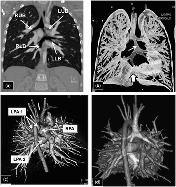
This patient was referred to our tracheal multi-disciplinary team for assessment of the airway passage in the context of a congenital cardiac malformation. Following a history of recurrent chest infections and persistent wet cough, cardiac and respiratory investigations were performed. Echocardiogram confirmed usual atrioventricular and ventriculo-arterial connections with levocardia. There was a large secundum atrial septal defect with volume overloaded right heart. There was a left aortic arch with confluent pulmonary branches; however, high parasternal short-axis view revealed the second left pulmonary artery arising from the right pulmonary artery, having an appearance typical of a pulmonary arterial sling (Fig 1). The patient underwent bronchoscopy and contrast CT angiogram that revealed atrial septal communication and the presence of two left pulmonary arteries. The upper left pulmonary artery with a narrowed origin, which was in the upper surface of the main pulmonary artery, supplied the left upper lobe. The distal left pulmonary artery, showing pulmonary sling configuration, originated from the proximal right pulmonary artery and passed between the oesophagus and the trachea. In addition, the tracheal branching pattern had a “Christmas tree” morphology, with the upper lobe bronchi originating from the same level as the distal trachea, with a narrow bridging bronchus passing inferiorly and dividing into two lower-lobe bronchi. There was no right middle lobe. The medial basal aspects of the lower lobes appeared fused, with the “horseshoe” isthmus running between the oesophagus and the pericardium (Fig 2).
Two-dimensional echocardiogram with colour flow mapping from high parasternal short-axis view in patient with “Christmas tree” bronchial arrangement and duplicated left pulmonary artery. Typical picture consistent with a pulmonary sling ( a ) showed left pulmonary artery (LPA 2) originating from right pulmonary artery (RPA) and running behind the trachea (asterisk). More anteriorly ( b ), there was, however, another left pulmonary artery (LPA 1) seen originating from the usual pulmonary bifurcation area and with anterior course to the trachea. AO=aorta; PA=pulmonary trunk.

Images from CT angiogram of the patient with “Christmas tree” bronchial arrangement and duplicated left pulmonary artery. ( a ) Coronal 1-mm reformat showing right (RUB) and left (LUB) upper bronchi and right (RLB) and left (LLB) lower bronchi, connected via narrowed trachea on CT angiogram. ( b ) Volume-rendered image of the airway and lungs shows bronchial arrangement, narrowed tracheal segment (arrow), and horseshoe lung segment (block arrow). ( c ) Volume-rendered image, posterior view, illustrating duplicated left pulmonary artery (LPA 1, LPA 2); LPA 2 originates from the Right Pulmonary Artery (RPA).( d ) Volume-rendered image, posterior view, illustrating relationship of airway (blue) to vessels ( d ).

The patient underwent surgical closure of the atrial septal defect and relief of vascular ring, and remains well clinically. In our experience, this is the second case with such a complex tracheal malformation in the setting of a duplicated left pulmonary artery.
Case 2
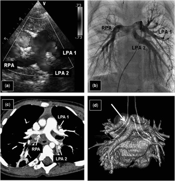
The second patient with Kabuki syndrome, pulmonary sling, global developmental delay with hypotonia, microcephaly, and cleft palate was referred from another tertiary institution for further investigation of pulmonary hypertension based on recurrent chest infections. This patient was also known to have restrictive-outlet ventricular septal defect. The chest X-ray revealed prominent pulmonary arteries; subsequent echocardiogram confirmed small ventricular septal defect and duplicated left pulmonary artery, suggestive of partial pulmonary sling. Contrast CT angiogram showed unusual branching pattern of the pulmonary arteries, with two left pulmonary arteries: the first of the left pulmonary arteries originated in the expected normal position of the left pulmonary artery, directly from the main pulmonary artery; and the second left pulmonary artery arose from the origin of the right pulmonary artery and passed between the oesophagus and the left main bronchus in a sling-like manner to reach the left lung. There was also some mild circumferential narrowing of the left main bronchus, not directly compressed by the vessels (Fig 3).
Composite cross-sectional imaging of the patient with duplicated left pulmonary artery and Kabuki syndrome. Two-dimensional echocardiogram with colour flow mapping from high parasternal short-axis view, ( a ) pulmonary angiogram and ( b ) demonstrating two left pulmonary artery branches of similar size. ( c ) 4 mm maximal intensity projection (MIP) images illustrating left anterior (LPA 1) and left posterior pulmonary artery (LPA 2); the left main bronchus runs between the two pulmonary arteries. The right pulmonary artery (RPA) is normal. ( d ) Volume-rendered image, posterior view demonstrating the relationship of airway to vessels (the aorta has been removed). The proximal left main bronchus is narrow (arrow).

Cardiac catheterisation confirmed pulmonary arterial hypertension with a pulmonary vascular resistance index of 20 Wood units×m2 reducing down to 13.1 Wood units×m2 when pulmonary vasoreactivity tested using pulmonary vasodilators. The patient remained clinically stable on bosentan and sildenafil and under regular follow-up by the pulmonary hypertension team.
Case 3
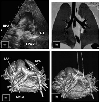
The third patient was diagnosed with Kabuki syndrome, systemic lupus erythematosus, small left kidney, hypothyroidism, and gastro-oesophageal reflux. The patient never presented with central cyanosis, heart failure, or upper airway obstruction. The echocardiogram was performed because of the presence of a cardiac murmur and revealed a mild mitral valve stenosis – restricted posterior mitral leaflet – likely due to systemic lupus erythematosus rather than structural abnormality of the valve. Apart from mitral stenosis, a complete segmental study revealed dual pulmonary artery supply to the left lung, with a duplicated left pulmonary artery arising from the right pulmonary artery posteriorly in addition to normal origin of the anterior left pulmonary artery. Contrast CT angiogram was subsequently performed to further delineate the pulmonary arterial anomaly and possible tracheal compression from the partial left pulmonary artery sling. A complete CT angiogram showed abnormal pulmonary branching anatomy with the right pulmonary artery giving rise to the aberrant left lower lobe branch, which was positioned below the left main bronchus and supplied the left lower lobe. The left lower pole pulmonary artery had a conventional origin passing superiorly to the left bronchus, which did not appear compressed (Fig 4). Cardiovascularly, this patient remains completely asymptomatic and continues to be seen for the mitral stenosis by cardiologists on a regular basis.
Composite cross-sectional imaging of the second patient with duplicated left pulmonary artery associated with Kabuki syndrome. Two-dimensional echocardiogram with colour flow mapping in high parasternal short-axis view ( a ) demonstrates two left pulmonary arteries (LPA 1, LPA 2); however, echocardiogram was unable to correctly identify the trachea. ( b ) Volume-rendered image of the pulmonary artery branches, posterior view demonstrating duplicated LPA branches (LPA1, LPA2), with LPA 2 originating from RPA. ( c ) Volume-rendered image showing relationship of airway to vessels; the tracheo-bronchial anatomy is normal, with no airway narrowing.

Discussion
The pulmonary sling is an anomaly whose embryology is still unknown and is subject to hypotheses based on observations in patients who visit for evaluation. The normal pulmonary arteries are thought to be part of the arterial buds from the sixth aortic arch. It was proposed that the pulmonary sling arises when the left lung bud fails to connect with the left sixth arch and forms instead an anastomosis with the right sixth aortic arch.Reference Jue, Raghib, Amplatz, Adams and Ewards 4 If this connection is dorsal to the developing lung bud, it will result in a pulmonary artery that arises from the right pulmonary artery passing posterior to the trachea, as it is in the case of the usual pulmonary sling reported. Embryologically, it may also be related to the “space-available” theory from the embryonic foregut mesoderm, thus making it unique to the left side alone. The developmental hypothesis of Jue et al proposed that such a connection might occur ventral to the developing bud, and in this case the pulmonary arteries would both pass anterior to the bronchi and not cause any compression to the airways. In the setting of a normal pulmonary artery branch configuration, the anatomical arrangement will be difficult to distinguish from normal, but in the presence of two co-existing left pulmonary arteries, using the normal left pulmonary artery as a landmark, the anomalous component would be expected to pass behind the trachea in the case of a dorsal connection and anterior to the trachea in the case of the predicted ventral connection.
To the best of our knowledge, there is only a single previous case report on a patient with partial anomalous left pulmonary artery with the anomalous component passing behind the trachea,Reference Bamman, Ward and Woodrum 11 suggesting a connection dorsal to the lung bud, and one from our unit with similar features of partial anomalous left pulmonary artery bud with airway anomaly.Reference Abelardo, Hewitt, Elliott and Muthialu 2 Since the initial description of the possible variants in pulmonary artery branching, there have been few cases in the literature with the aberrant left pulmonary artery coursing anterior to the trachea without causing airway compression.Reference Erickson, Cocalis and George 1 , Reference Fountain-Dommer, Shirali, Wiles and Larsen 12 , Reference Ge, DeGroff, Knudson, Strain and Chan 13 Erickson et al,Reference Erickson, Cocalis and George 1 in 1996, were the first group to report a case with duplicated left pulmonary artery in a 1-year-old patient with associated ventricular septal defect and hypospadias. Surgery was not performed as the ventricular septal defect closed spontaneously and the patient remained asymptomatic from the respiratory point of view. Abelardo et al reported a 3-month-old child with two anomalous left pulmonary arteries to individual lobes, with one being in a sling position while the other in an anatomically normal position, with associated complex tracheo-bronchial tree anatomy, imperforate anus, and recto-vaginal fistula, but with no other intra-cardiac anomalies.Reference Abelardo, Hewitt, Elliott and Muthialu 2 In this case report, we document a very similar anatomy as in case 1, where a complex tracheo-bronchial anatomy, the so-called “Christmas tree”, was associated with the presence of a duplicated left pulmonary artery with the usual branching in the upper surface of the main pulmonary artery, supplying the left upper lobe. The second branching, arising more distally, showed pulmonary sling configuration, originating from the proximal right pulmonary artery and passing between the oesophagus and the trachea.
In the other two cases, the anomaly of pulmonary branching was incidentally discovered. In case 2, the boy was under follow-up for pulmonary hypertension, whereas in case 3 the girl was under follow-up for mild mitral stenosis. Interestingly, both these patients were affected with Kabuki syndrome. To the best of our knowledge, in the literature, only two similar cases have been described in patients with Kabuki syndrome: Collins et alReference Collins, Weinberg and Goldmuntz 10 reported the case of a 4-year-old patient with Kabuki syndrome, atrial septal defect, and dual pulmonary artery supply to the left lung with a partial anomalous left pulmonary artery from the right pulmonary artery, coursing anterior to the bronchi and, therefore, not causing any compression to the airways. The second case was reported in 2012 by Bhat et alReference Bhat, Davenport and Cocalis 9 in a newborn patient presenting with anomalous left pulmonary artery and coarctation of the aorta for which he underwent successful aortic coarctation repair. Neither of these cases required any intervention on the pulmonary artery branches, as none of these patients presented with significant bronchial compression.
Published cases and our own experience are suggestive of a strong relationship between Kabuki syndrome and a duplicated left pulmonary artery. In other words, if we talk about the embryologic hypothesis, which was raised upon the development of an anomalous pulmonary artery branching pattern, it seems that these findings support the association of Kabuki syndrome with the “ventral hypothesis” proposed by Jue et al. Regarding the support for Jue’s hypothesis, there are a few other cases described in the literature with the anterior course of the anomalous vessel; we think that it is of extreme interest to underline that in all these other cases it was not clearly reported whether the suspicion of a genetic syndrome (i.e. Kabuki syndrome) was raised at that time. In three of the reported cases in the literature, the anomalous left pulmonary artery was associated with other cardiac abnormalities; in the case described by Ge et al, the patient had mitral valve hypoplasia and hypoplastic left ventricle;Reference Ge, DeGroff, Knudson, Strain and Chan 13 in one of the cases described by Fountain–Dommer et al,Reference Fountain-Dommer, Shirali, Wiles and Larsen 12 the patient presented with double-outlet right ventricle, hypoplastic left ventricle, and hypoplasia of the mitral and aortic valves, whereas the second case showed this anomaly associated with coarctation of the aorta. It appears that obstructive left heart lesions are more frequently associated with Kabuki syndrome than with other abnormalities.
In all the reported cases, no further explanations regarding the possible underlying genetic syndrome were given, predominantly due to the fact that one of the reported patients died after stage-one palliation for hypoplastic left heart, and the other two patients underwent successful pulmonary artery banding and coarctation repair, respectively, and no further detailed investigations were undertaken. Furthermore, it seems very likely that in abnormalities affecting skeletal systems, the typical syndromic features may be revealed at a later age, and/or in some cases it can be detected incidentally due to the absence of clinical symptoms in association with duplicated left pulmonary artery, as in two of our cases.
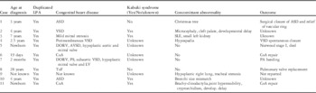
Our three case reports and review of the literature underline the importance of evaluating the relationship between the vascular (arterial) and tracheo-bronchial systems, as detailed information gained may contribute to understanding the natural history of the disease as well as to general surgical and intensive care unit management of symptomatic patients, including intubation and mechanical ventilation. The existence of these reported lesions supports the prediction of Jue et al and broadens the spectrum of known pulmonary artery malformations, giving more knowledge to our understanding about embryological development and also about a possible strong association with a genetic syndrome, which is of clinical interest (Table 1).
All reported patients and our three patients with duplicated LPA (patients 1–3 from GOSH).

ASD=atrial septal defect; AVSD=atrioventricular defect; CoA=coarctation; DORV=double-outlet right ventricle; GOSH=Great Ormond Street Hospital; LPA=left posterior pulmonary artery; LV=left ventricle; PA=pulmonary artery; PS=pulmonary stenosis; SLE=systemic lupus erythematosus; ToF=tetralogy of Fallot; VSD=ventricular septal defect
Acknowledgements
Dr Giudici contributed in collecting the literature data for the review. Dr Giudici, Dr Kanani, Dr Muthialu, Dr Carr, Dr Calder, Dr Owens, Dr Cook, and Prof. Marek contributed to the planning and writing the work. Dr Calder, Dr Owens, and Prof. Marek contributed to select the best quality images. Prof. Marek is responsible for the overall content as guarantor.
Financial Support
This research received no specific grant from any funding agency, commercial, or not-for-profit sectors.
Conflicts of Interest
None.