Neonatal myocardial infarction is a rare event. Mortality has been estimated at up to 90%, despite early diagnosis and management in tertiary centres.Reference De Vetten, Bergman, Elzenga, Van Melle, Timmer and Bartelds 1 The most common causes are enteroviral myocarditis,Reference Murugan, Gnanapragasam and Vettukattil 2 coagulopathy,Reference Clark, Stokes, Krous and Carbine 3 maternal diabetes,Reference Abbal, Paranon, Brierre, Dulac, Casper and Acar 4 thromboembolism,Reference Poonai, Kornecki, Buffo and Pepelassis 5 anomalous coronary artery, and birth asphxya.Reference Farooqi, Sutton, Weinstein, Menegus, Spindola-Franco and Pass 6 We describe two different cases of neonatal myocardial infarction with structurally normal hearts and normal coronary arteries. In both cases, the mothers had routine prenatal care during pregnancy.
Brief report
Baby 1
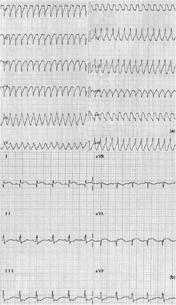
A full-term male newborn presented to the neonatal intensive care unit after severe asphyxia during caesarean delivery. His Apgar scores were 4 and 6 at 3 and 5 min, respectively. The asphyxia was felt to be due to maternal anaesthesia and/or prolonged labour. On admission, he was pale, hypotonic, and had a high respiratory rate. His vital signs showed a heart rate of 180 bpm and a blood pressure of 40/30 mmHg. Nasal positive pressure ventilation was started, and hypoglycaemia and metabolic acidosis were corrected. After 1 h of admission, a short run of wide complex tachycardia with a heart rate of 250 bpm was noted. Electrocardiogram showed ventricular tachycardia with a right ventricular outflow tract morphology (Fig 1a). Transthoracic echocardiogram showed a structurally normal heart, normal origin and flow in the coronary arteries, severe global hypokinesia of the left ventricle with an ejection fraction of 10%, moderate mitral regurgitation, and mild tricuspid regurgitation with an estimated right ventricular systolic pressure of 50 mmHg. The systemic flow was maintained by right-to-left shunting through the patent ductus arteriosus. Cardiac catheterisation was not done given good visualisation of the coronaries by transthoracic echocardiogram. The diagnosis of myocardial infarction was based on ST-T segment elevation (Fig 1b) and marked ST segment depression from V1 to V6 on electrocardiogram along with significant elevation of myocardial enzymes. Circulatory support consisted of inotropes, diuretics, and levosimendan. Thrombolysis was not felt to be necessary. The ejection fraction gradually increased to 35% by day 4 and the duct closed spontaneously.
( a ) Ventricular tachycardia; ( b ) elevation of ST–T segment in I and aVL.

The patient was discharged from the intensive care unit on day 5 of life and out of the hospital, with normal ventricular function, on day 10 of life. At 4 years of age, the patient is asymptomatic with normal ventricular function and normal neurological development.
Baby 2
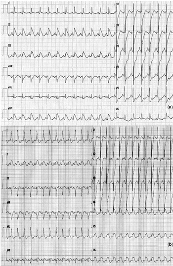
A full-term female newborn was born by normal vaginal delivery 24 h after rupture of membranes. Her Apgar scores were 8 and 10, respectively, at 3 and 5 min. Her birth weight was 2.880 kg. The baby was referred at 72 h of life because of evidence of myocardial ischaemia on electrocardiogram. On admission, she was noted to be hypotonic, with red and dry skin. Her vital signs showed a heart rate of 150 bpm and a blood pressure of 50/30 mmHg. Her haematocrit was 80%. The high haematocrit level was felt to be due to maternal smoking during pregnancy, about 15 cigarettes a day. Electrocardiogram was indicative of diffuse myocardial ischaemia with marked inferior acute myocardial infarction (Fig 2a). Echo showed global moderate to severe hypocontractility of the left ventricle and moderate to severe mitral regurgitation. Contractility was markedly affected in the inferolateral region of the left ventricle. Specific myocardial enzymes (troponin I) reached 16 time normal value. Coronary angiography was normal. The patient was admitted to the neonatal intensive care unit. The elevated haematocrit was reversed with adequate rehydration, and low cardiac output syndrome was treated with intravenous inotropes, diuretics, and vasodilatators; systemic heparinisation was performed. The diagnosis of acute myocardial infarction rather than myocarditis was made on the basis of electrocardiogram findings, elevation of specific myocardial enzymes, laboratory investigations, and clinical evaluation. Specifically, laboratory investigations ruled out the presence of infection, metabolic and bleeding disorders. Plasminogen activator inhibitor 1 has not been investigated. Over time, the ventricular function gradually improved with rapid progression of myocardial hypertrophy and impaired diastolic function. Myocardial ischaemia evolved into lateral myocardial infarction (Fig 2b). The patient was discharged from the intensive care unit on day 3 of life, and home from the hospital in good cardiovascular condition on day 30 of life. Discharge therapy included captopril, propranolol, clopidogrel, and aspirin. After 4 months of follow-up, the patient is asymptomatic with appropriate neurological development for age. The electrocardiogram continues to show signs of stabile myocardial infarction and biventricular hypertrophy. Echo shows altered left ventricle diastolic function due to persistence of severe biventricular hypertrophy of the posterior wall and interventricular septum.
( a ) Diffuse myocardial ischaemia. ST segment elevation in II, II, aVF; ST segment depression in precordial leads; ( b ) ST segment elevation in I and aVL, V4, V5, V6.

Discussion
Neonatal myocardial infarction is an extremely rare neonatal event in newborns with a structurally normal heart. In this report, we describe the cases of two newborns who survived neonatal myocardial infarction. Both patients presented with unique features. The first one presented at birth with severe asphyxia, and systemic cardiac output was maintained during the acute phase via a patent ductus arteriosus. This may have allowed time for recovery of the left ventricular systolic function. The second case presented with acute coronary syndrome associated with marked erythrocytosis, with a haematocrit of 80%. Severe polycythaemia has already been reported as a cause of acute coronary thrombosis.Reference Rossi, Randi, Zerbinati, Rinaldi and Girolami 7 To the best of our knowledge, however, this is the first reported case of severe polycythaemia associated with myocardial infarction in a neonate. Both patients were treated medically and survived, most likely due to early diagnosis and treatment. Mortality has been reported to be as high as 90% in the early series but it has been decreasing since the introduction of extracorporeal membrane oxygenationReference Farooqi, Sutton, Weinstein, Menegus, Spindola-Franco and Pass 6 indicating that temporary circulatory support may lead to recovery of contractile function, allowing survival even in patients with cardiogenic shock. Interestingly, in both of our cases, serial echocardiograms showed myocardial hypertrophy during the recovery phase. This could represent ventricular remodelling potentially through myogenic proliferation, and hyperplasia rather than cellular hypertrophy.Reference Holtz 8 Hyperplastic remodelling, active in foetal and neonatal life, may explain the reported full recovery of ventricular function in patients who survived the acute phase.Reference Samson, Bonnet and Heimburger 9 , Reference de Almeida, McQuinn and Sedmera 10 The potential for full recovery categorises neonatal myocardial infarction as reversible myocardial dysfunction and warrants aggressive treatment of low cardiac output syndrome including extracorporeal membrane oxygenation when necessary.
Conclusion
We describe two cases of reversible newborn myocardial infarction: the first representing a rare case in terms of treatment and the second with a previously undescribed aetiology in the newborn population. Both children are still alive: the first with 48 months of follow-up, and the second with 4 months of follow-up.
Acknowledgement
Special thanks to Jeferey A. Feinstein, MD, MPH, Associate Professor, Lucile Packard Children's Hospital/Stanford University Medical Center.